. 2009 Nov 5;28(44):3892-902.
doi: 10.1038/onc.2009.247. Epub 2009 Aug 24.

Macrophage-derived IL-1beta stimulates Wnt signaling and growth of colon cancer cells: a crosstalk interrupted by vitamin D3

Affiliations

Macrophage-derived IL-1beta stimulates Wnt signaling and growth of colon cancer cells: a crosstalk interrupted by vitamin D3

P Kaler et al. Oncogene. .

Abstract

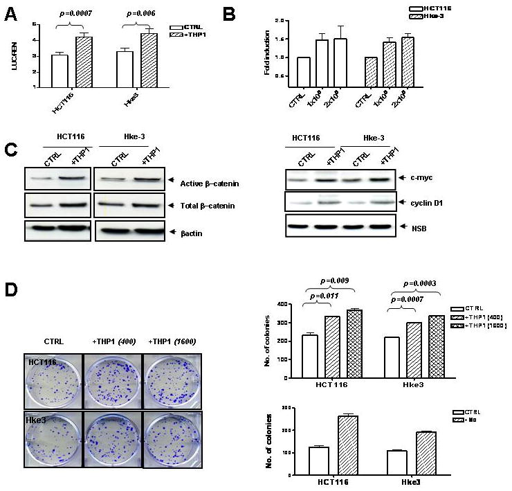
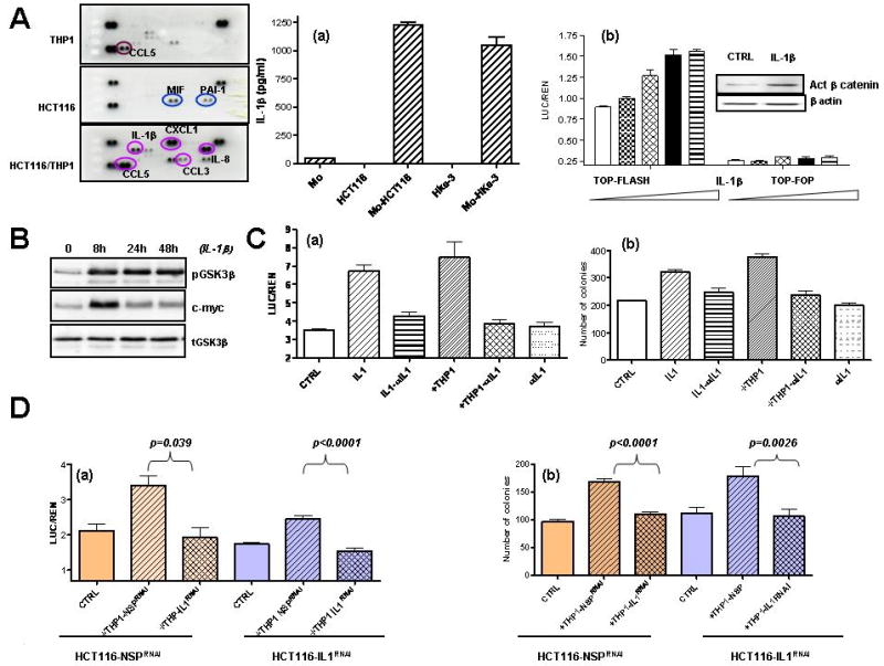
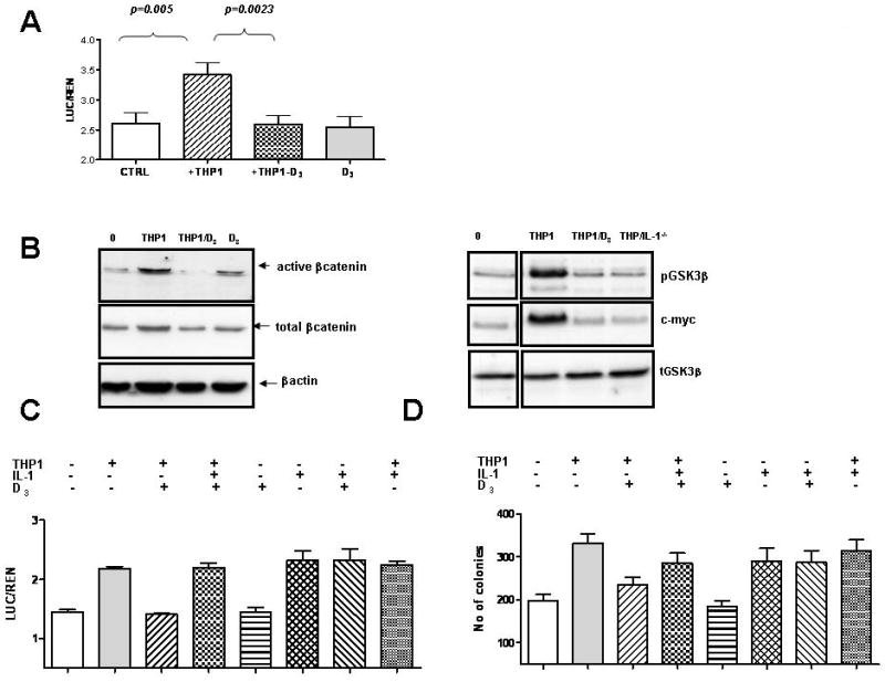
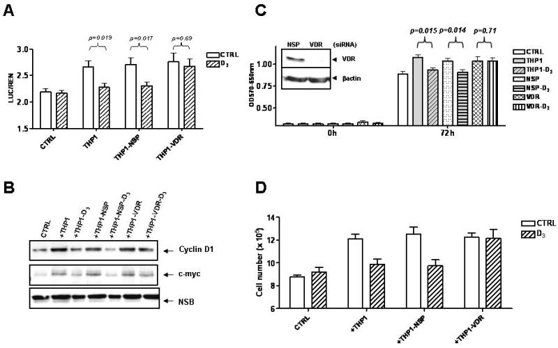
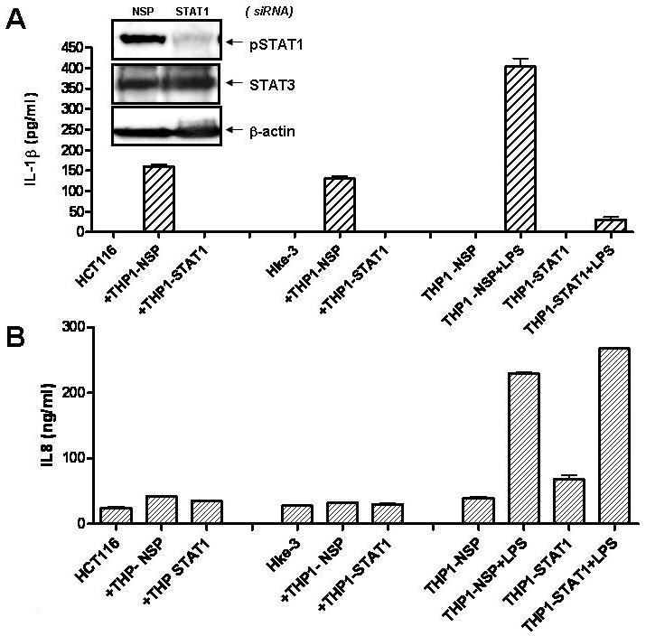
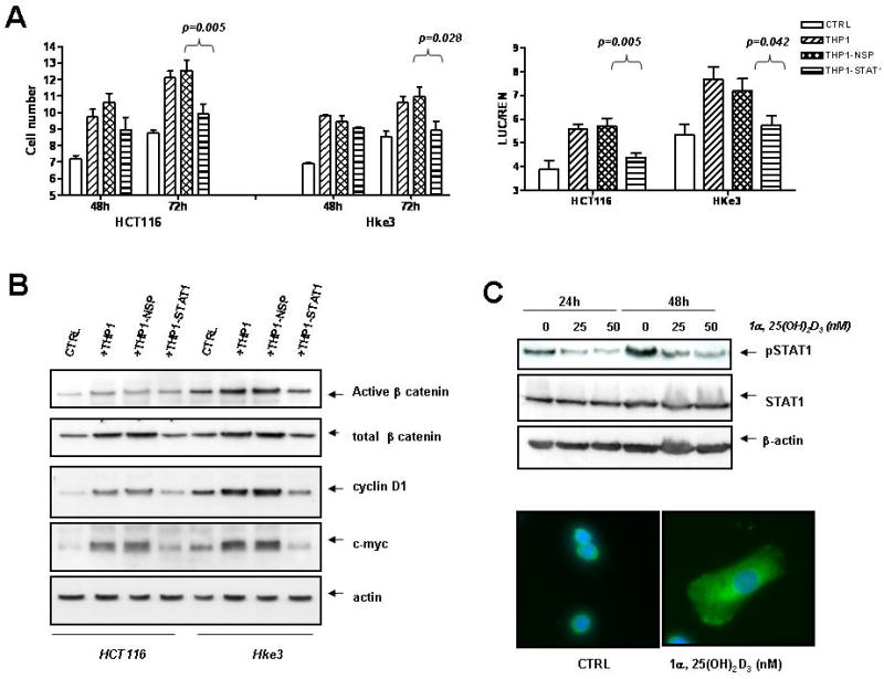
Tumor-associated macrophages mediate the link between inflammation and cancer progression. Here, we showed that macrophage-derived soluble factors induce canonical Wnt signaling in colon cancer cells and promote their growth. Tumor cells induced the release of interleukin (IL)-1beta from macrophages, which induced phosphorylation of GSK3beta, stabilized beta-catenin, enhanced T-cell factor (TCF)-dependent gene activation and induced the expression of Wnt target genes in tumor cells. Neutralization experiments using anti-IL-1beta-specific antibodies, or silencing of IL-1beta in THP1 macrophages, showed that IL-1beta was required for macrophages to induce Wnt signaling and to support the growth of tumor cells. Constitutive activation of signal transducer and activator of transcription (STAT)1 in THP1 macrophages was essential for the induction of IL-1beta and thus for the activation of beta-catenin signaling in tumor cells. Vitamin D3, an effective chemopreventive agent, interrupted this crosstalk by blocking the constitutive activation of STAT1 and the production of IL-1beta in macrophages, and therefore-in a vitamin D receptor-dependent manner-inhibited the ability of macrophages to activate Wnt signaling in colon carcinoma cells. Our data therefore established that vitamin D3 exerts its chemopreventive activity by interrupting a crosstalk between tumor epithelial cells and the tumor microenvironment.


Figures

Fig. 1
Fig. 1. Macrophages activate Wnt signaling in colon cancer cells and promote their clonogenic growth
HCT116 and Hke-3 cells were transfected with TOP-FLASH reporter plasmid and were cultured in the absence or the presence of THP1 macrophages (A) or normal human peripheral blood monocytes (B). C: The levels of active β–catenin, total β–catenin, c-Myc and cyclin D1 were determined in HCT116 and Hke-3 cells that were grown alone, or were co-cultured with THP1 macrophages. D: HCT116 and Hke-3 cells were plated alone or together with THP1 macrophages or peripheral blood monocytes (Mo) as described in Material and Methods.
Figure 2
Figure 2. IL-1β is sufficient and required to activate Wnt signaling
A: HCT116 and THP1 cells were cultured alone or together as indicated for 48 hours, and the expression of soluble mediators was determined by Human cytokine array (R&D Systems). (a): The amount of IL-1β was determined by ELISA in supernatants of HCT116 and Hke-3 cells, normal human monocytes (Mo) and in co-cultures. (b): HCT116 cells were transfected with TOP-FLASH or TOP-FOP reporter genes and were treated with increasing concentrations of IL-1β (0.5-10 ng/ml). Inset: The amount of active β-catenin in IL-1β treated cells. B:The levels of pGSK3 (Ser9), total GSK3 and c-myc in control and IL-1β treated HCT116 cells. C: (a) HCT116 cells were transfected with TOP-FLASH reporter gene and were treated as indicated. (b) HCT116 cells were treated as indicated and the clonogenic assay was performed as described in Material and methods. D: HCT116 were transfected with NSP (nontargeting) siRNA or siRNA specific for IL-1β. They were cultured with nontransfected THP1 cells or THP1 cells transfected with NSP siRNA or siRNA specific for IL-1β. Wnt signaling (a) and clonogenic assay were (b) were performed as indicated.
Fig. 3
Fig. 3. Vitamin D3 inhibits macrophage-mediated increase in Wnt signaling
A: HCT116 cells transfected with TOP-FLASH reporter plasmid were incubated with THP1 cells as indicated. B: The levels of active β–catenin, total β–catenin, pGSK3β, total GSK3β and c-Myc were determined in colon cancer cells cultured alone or together with THP1. HCT116 cells were also cultured with THP1 cells transfected with IL-1β specific siRNA (THP1/IL1-/-) as indicated. C: HCT116 cells were transfected with TOP-FLASH reporter and were cultured in the presence of THP1 cells, IL-1β and vitamin D3 as indicated. E: The effect of vitamin D3 and IL-1β on clonogenic potential of HCT116 cells.
Fig. 4
Fig. 4. Inhibition of Wnt signaling by vitamin D3 requires VDR expression on macrophages
A: HCT116 cells transfected with TOP-FLASH reporter gene were cultured with nontransfected THP1 cells, or THP1 macrophages transfected with nontargeting (NSP) siRNA or VDR specific siRNA. B: Proteins were isolated from HCT116 cells co-cultured with THP1 cells as described above and the expression of cyclin D1 and c-myc was determined by immunoblotting. C: Growth of HCT116 cells with conditioned medium collected from untreated or vitamin D3 treated THP1 cells transfected with nontrageting siRNA or VDR specific siRNA, as determined by MTT assay, D: HCT116 and THP1 cells were co-cultured as indicated and the number of HCT116 cells determined after 48 hours.
Fig. 5
Fig. 5. Secretion of IL-1β, but not IL-8, from THP1 macrophages, requires STAT1
HCT116 and Hke-3 cells were cultured with THP1 cells transfected with nontargeting siRNA (THP1-NSP) or siRNA specific for STAT1 (THP1-STAT1) (A, inset). Cultures were treated with LPS (1 μg/ml) and the levels of IL-1β (A) and IL-8 (B) were determined by ELISA.
Fig. 6
Fig. 6. Activated STAT1 is required for pro-tumorigenic activity of THP1 macrophages
A: The number of tumor cells was determined upon coculture with nontransfected THP1 cells, THP1 cells transfected with NSP (nontargeting) siRNA or STAT1 specific siRNA. Right panel: HCT116 and Hke-3 cells were transfected with TOP-FLASH and cultured with THP1 cells as indicated. B: The levels of active β-catenin, total β-catenin, cyclin D1 and c-myc were determined by immunoblotting, C: THP1 cells were treated with vitamin D3 as indicated and the levels of pSTAT1 and total STAT1 were determined by immunoblotting. Subcellular localization of STAT1 was determined by immunofluorescence (green); nuclei were stained by DAPI. THP1 cells were left untreated or were treated with vitamin D3 (25nM) for 16 hours.

Similar articles

Cited by

References

    1. Albini A, Sporn MB. The tumour microenvironment as a target for chemoprevention. Nat Rev Cancer. 2007;7:139–47. - PubMed
    1. Allavena P, Sica A, Solinas G, Porta C, Mantovani A. The inflammatory micro-environment in tumor progression: the role of tumor-associated macrophages. Crit Rev Oncol Hematol. 2008;66:1–9. - PubMed
    1. Bissell MJ, Labarge MA. Context, tissue plasticity, and cancer: are tumor stem cells also regulated by the microenvironment? Cancer Cell. 2005;7:17–23. - PMC - PubMed
    1. Biswas SK, Gangi L, Paul S, Schioppa T, Saccani A, Sironi M, et al. A distinct and unique transcriptional program expressed by tumor-associated macrophages (defective NF-kappaB and enhanced IRF-3/STAT1 activation) Blood. 2006;107:2112–22. - PubMed
    1. Brabletz T, Jung A, Hermann K, Gunther K, Hohenberger W, Kirchner T. Nuclear overexpression of the oncoprotein beta-catenin in colorectal cancer is localized predominantly at the invasion front. Pathol Res Pract. 1998;194:701–4. - PubMed

Publication types

MeSH terms