. 2009 Oct 29;28(43):3837-46.
doi: 10.1038/onc.2009.244. Epub 2009 Aug 24.

Dithiolethione compounds inhibit Akt signaling in human breast and lung cancer cells by increasing PP2A activity

Affiliations

Dithiolethione compounds inhibit Akt signaling in human breast and lung cancer cells by increasing PP2A activity

C H Switzer et al. Oncogene. .

Abstract

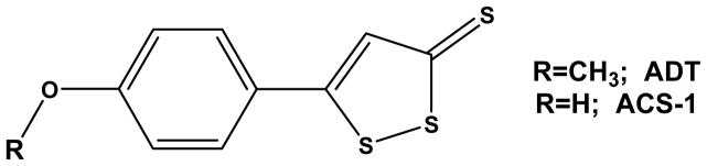
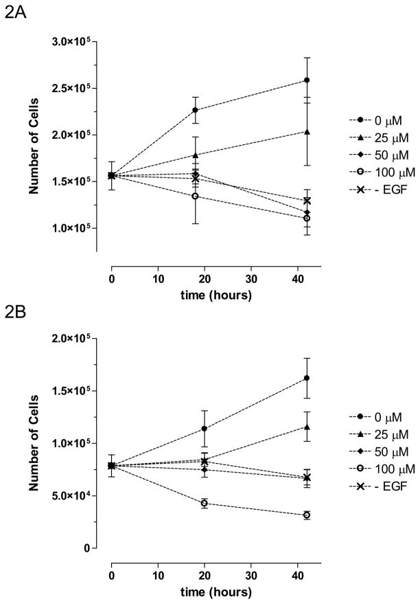
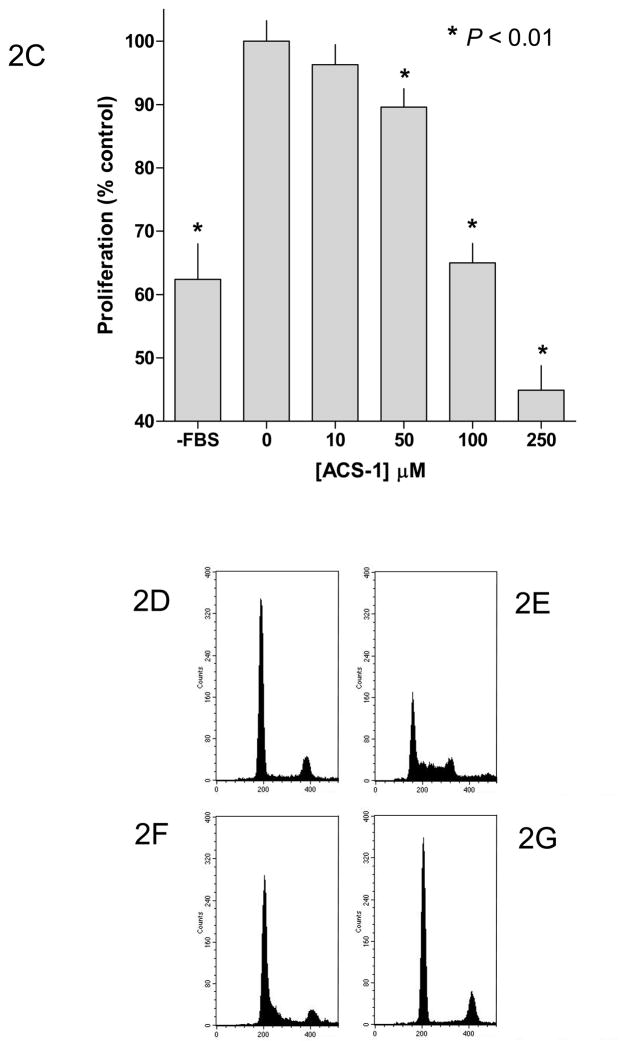
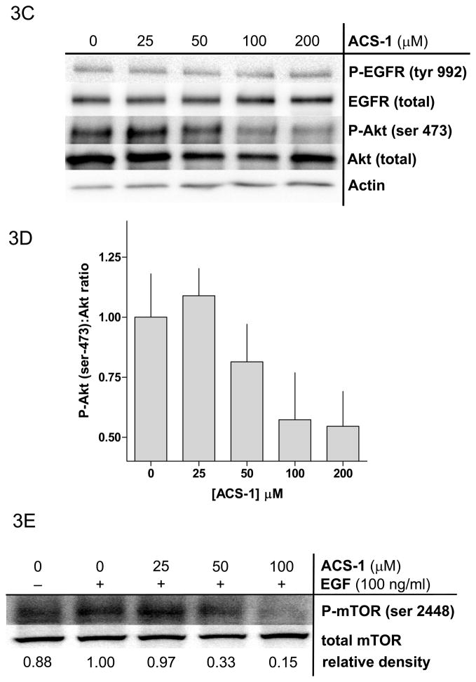
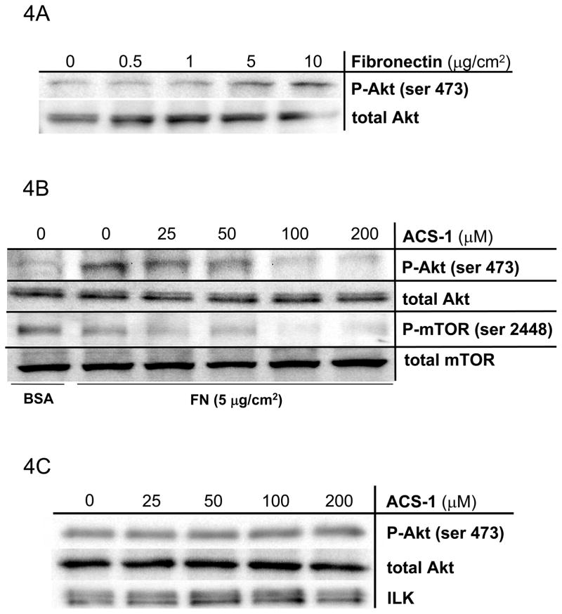
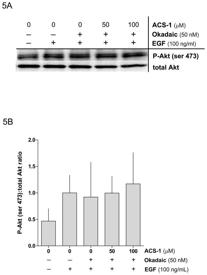
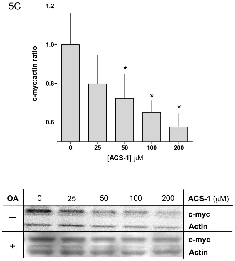
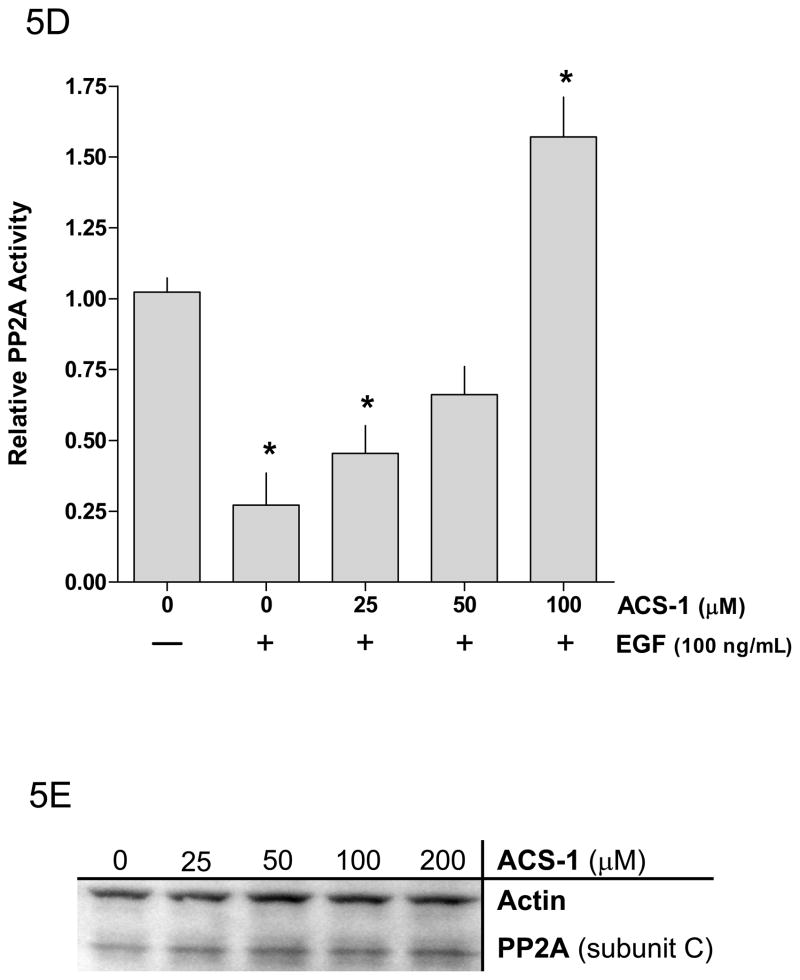
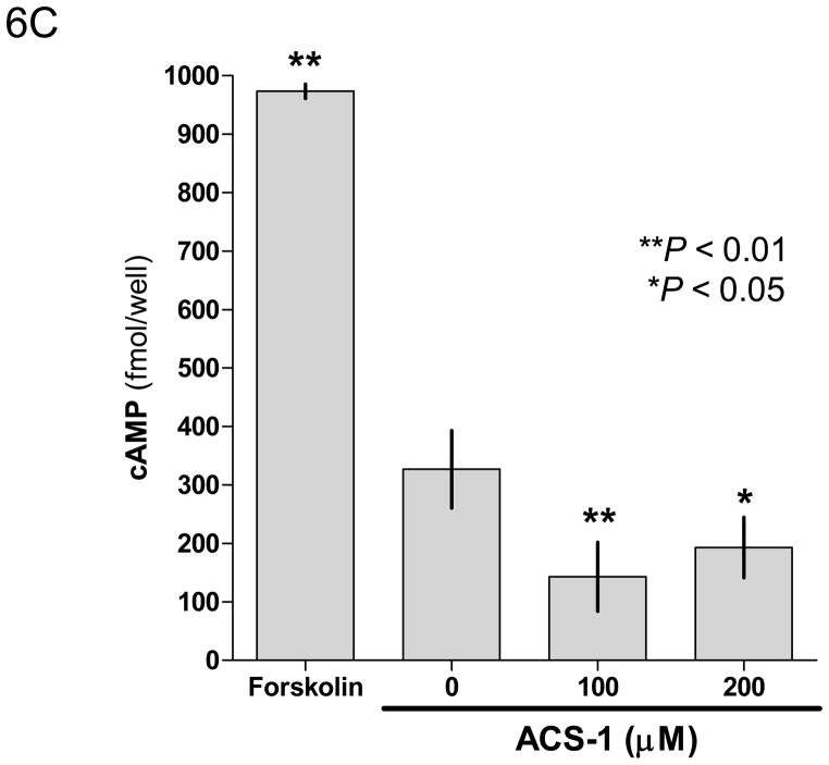
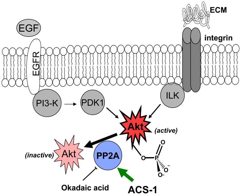
The chemopreventative effects of dithiolethione compounds are attributed to their activation of antioxidant response elements (AREs) by reacting with the Nrf2/Keap1 protein complex. In this study, we show antiproliferative effects of the dithiolethione compound ACS-1 in human cancer cell lines (A549 and MDA-MB-231) by increasing the activity of the tumor suppressor protein phoshatase 2A (PP2A). ACS-1 inhibited epidermal growth factor (EGF)-induced cellular proliferation in a concentration- and time-dependent manner. Akt activation, as determined by serine-473 phosphorylation, was inhibited by ACS-1 in cells stimulated with either EGF or fibronectin. Furthermore, ACS-1 inhibited mammalian target of rapamycin signaling and decreased c-myc protein levels. ACS-1 did not proximally alter EGF receptor or integrin signaling, but caused a concentration-dependent increase in PP2A activity. The effect of ACS-1 on Akt activation was not observed in the presence of the PP2A inhibitor okadaic acid. ACS-1 effects on PP2A activity were independent of ARE activation and cAMP formation. In addition to ACS-1, other dithiolethione compounds showed similar effects in reducing Akt activation, suggesting that this class of compounds may have other effects beyond chemoprevention.


Conflict of interest statement

Conflict of interest

Piero Del Soldato and Anna Sparatore are shareholders of Sulfidris, Milan, Italy. This company has patent rights on some reagents used in this study.

Figures

Figure 1
Figure 1
Chemical structure of ADT and ACS-1.
Figure 2
Figure 2
ACS-1 inhibits cellular proliferation in EGF-stimulated A549 and MB231 cells. (A) A549 and (B) MB231 cells were incubated with ACS-1 in serum free DMEM +0.1% BSA + EGF (100 ng/mL) for the indicated times are counted. Values represent the mean ± SD (n=6). (C) MTS assay showing ACS-1 mediated (24 hour exposure) inhibition of A549 cellular proliferation in complete growth media. (D–G) Cell cycle analysis of synchronized MB231 cells incubated for 24 hours in serum free media (D), serum replete media with ACS-1 (0, 25 and 100 μM) (E, F and G, respectively).
Figure 2
Figure 2
ACS-1 inhibits cellular proliferation in EGF-stimulated A549 and MB231 cells. (A) A549 and (B) MB231 cells were incubated with ACS-1 in serum free DMEM +0.1% BSA + EGF (100 ng/mL) for the indicated times are counted. Values represent the mean ± SD (n=6). (C) MTS assay showing ACS-1 mediated (24 hour exposure) inhibition of A549 cellular proliferation in complete growth media. (D–G) Cell cycle analysis of synchronized MB231 cells incubated for 24 hours in serum free media (D), serum replete media with ACS-1 (0, 25 and 100 μM) (E, F and G, respectively).
Figure 3
Figure 3
ACS-1 inhibits EGF activation of Akt and mTOR in MB231 and A549 cells. Serum starved cells were treated with ACS-1 for 2 hours and stimulated with EGF. (A) Western blot analysis of EGF signaling pathway activation in response to EGF and/or ACS-1. (B) Graphical representation of Akt activation in MB231 cells; ratios of P-Akt-(ser 473):total Akt are presented as percent of EGF control (mean ± SD). (C) Western blot analysis of ACS-1 effects on EGF signaling in A549 cells. (D) Graphical representation of Akt activation in A549 cells; ratios of P-Akt-(ser 473):total Akt are presented as percent of EGF control (mean ± SD). (E) Representative western blot analysis of P-mTOR-(ser 2448) in MB231 cells treated with ACS-1 and stimulated with EGF.
Figure 3
Figure 3
ACS-1 inhibits EGF activation of Akt and mTOR in MB231 and A549 cells. Serum starved cells were treated with ACS-1 for 2 hours and stimulated with EGF. (A) Western blot analysis of EGF signaling pathway activation in response to EGF and/or ACS-1. (B) Graphical representation of Akt activation in MB231 cells; ratios of P-Akt-(ser 473):total Akt are presented as percent of EGF control (mean ± SD). (C) Western blot analysis of ACS-1 effects on EGF signaling in A549 cells. (D) Graphical representation of Akt activation in A549 cells; ratios of P-Akt-(ser 473):total Akt are presented as percent of EGF control (mean ± SD). (E) Representative western blot analysis of P-mTOR-(ser 2448) in MB231 cells treated with ACS-1 and stimulated with EGF.
Figure 4
Figure 4
ACS-1 inhibits Akt activation in FN-stimulated A549 cells. (A) FN, in a concentration-dependent manner, activates Akt in A549 cells. Serum starved A549 cells were plated into Petri dishes coated with differing amounts of FN and incubated for 2 hours. (B) ACS-1 inhibits FN-induced Akt and mTOR activation. ACS-1 treated A549 cells seeded onto FN-coated dishes and incubated for 2 hours. (C) ILK was immunoprecipitated from cells plated on FN in the presence of ACS-1 and ILK kinase activity was determined by recombinant Akt phosphorylation (ser 473).
Figure 5
Figure 5
Okadaic acid abates the effect of ACS-1 on Akt activation. (A) Western blot and (B) densitometry analysis of Akt-(ser 473) phosphorylation in A549 cells incubated with ACS-1 and/or Okadaic acid for 2 hours and stimulated with EGF for 30 minutes. (C) ACS-1 increases PP2A activity as determined by total c-myc protein expression in MB231 cells treated with ACS-1 for 2 hours. C-myc protein levels are not altered in the presence of okadaic acid (OA). (D) Serum starved MB231 cells were treated with ACS-1 for 2 hours stimulated with EGF and lysed. PP2A (subunit C) was immunoprecipitated from whole cell extracts, washed with assay buffer and PP2A phosphatase activity measured. (E) ACS-1 does not affect the level of PP2A expression. Serum starved MB231 cells were treated with ACS-1 for 2 hours and stimulated with EGF for 30 minutes. PP2A and actin protein levels were determined by Western blot analysis.
Figure 5
Figure 5
Okadaic acid abates the effect of ACS-1 on Akt activation. (A) Western blot and (B) densitometry analysis of Akt-(ser 473) phosphorylation in A549 cells incubated with ACS-1 and/or Okadaic acid for 2 hours and stimulated with EGF for 30 minutes. (C) ACS-1 increases PP2A activity as determined by total c-myc protein expression in MB231 cells treated with ACS-1 for 2 hours. C-myc protein levels are not altered in the presence of okadaic acid (OA). (D) Serum starved MB231 cells were treated with ACS-1 for 2 hours stimulated with EGF and lysed. PP2A (subunit C) was immunoprecipitated from whole cell extracts, washed with assay buffer and PP2A phosphatase activity measured. (E) ACS-1 does not affect the level of PP2A expression. Serum starved MB231 cells were treated with ACS-1 for 2 hours and stimulated with EGF for 30 minutes. PP2A and actin protein levels were determined by Western blot analysis.
Figure 6
Figure 6
ACS-1 effects PP2A activity independent of protein production or phase II activation. Serum starved MB231 cells were treated with ACS-1 for 2 hours. (A) Western blot analysis of HO-1 and actin indicate that ARE controlled genes are not upregulated within the time of PP2A activation by ACS-1. (B) MB231 cells were exposed to 50 μM ACS-1 for indicated times and the induction of phase II genes (UGTA1, GSTP1, GCT1, and GCLM) were measured by RT-PCR. (C) Effect of ACS-1 on cellular cAMP levels. MB231 cells were exposed to forskolin or ACS-1 for 2 hours and cAMP was measured via ELISA.
Figure 6
Figure 6
ACS-1 effects PP2A activity independent of protein production or phase II activation. Serum starved MB231 cells were treated with ACS-1 for 2 hours. (A) Western blot analysis of HO-1 and actin indicate that ARE controlled genes are not upregulated within the time of PP2A activation by ACS-1. (B) MB231 cells were exposed to 50 μM ACS-1 for indicated times and the induction of phase II genes (UGTA1, GSTP1, GCT1, and GCLM) were measured by RT-PCR. (C) Effect of ACS-1 on cellular cAMP levels. MB231 cells were exposed to forskolin or ACS-1 for 2 hours and cAMP was measured via ELISA.
Figure 7
Figure 7
Scheme of protein interactions. Akt is activated by convergent pathways in human cancers. ACS-1 inhibits Akt activation by increasing the activity of PP2A and not by altering EGFR or integrin signaling.

References

    1. Andjelkovic M, Jakubowicz T, Cron P, Ming XF, Han JW, Hemmings BA. Activation and phosphorylation of a pleckstrin homology domain containing protein kinase (RAC-PK/PKB) promoted by serum and protein phosphatase inhibitors. Proceedings of the National Academy of Sciences of the United States of America. 1996;93:5699–5704. - PMC - PubMed
    1. Arnold H, Sears R. A tumor suppressor role for PP2A-B56α through negative regulation of c-Myc and other key oncoproteins. Cancer and Metastasis Reviews. 2008;27:147–158. - PMC - PubMed
    1. Arnold HK, Sears RC. Protein Phosphatase 2A Regulatory Subunit B56{alpha} Associates with c-Myc and Negatively Regulates c-Myc Accumulation. Mol Cell Biol. 2006;26:2832–2844. - PMC - PubMed
    1. Bae EJ, Yang Yoon Mee, Kim Jin Wan, Kim Sang Geon. Identification of a novel class of dithiolethiones that prevent hepatic insulin resistance via the adenosine monophosphate-activated protein kinase-p70 ribosomal S6 kinase-1 pathway. Hepatology. 2007;46:730–739. - PubMed
    1. Busserolles J, Megías J, Terencio MC, Alcaraz MJ. Heme oxygenase-1 inhibits apoptosis in Caco-2 cells via activation of Akt pathway. The International Journal of Biochemistry & Cell Biology. 2006;38:1510–1517. - PubMed

MeSH terms

Substances