. 2008 Aug 21;594(1):102-110.
doi: 10.1016/j.nima.2008.05.061.

A small-animal imaging system capable of multipinhole circular/helical SPECT and parallel-hole SPECT

Affiliations

A small-animal imaging system capable of multipinhole circular/helical SPECT and parallel-hole SPECT

Jianguo Qian et al. Nucl Instrum Methods Phys Res A. .

Abstract

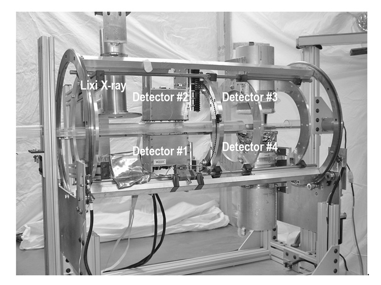
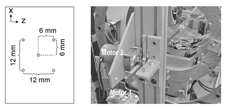
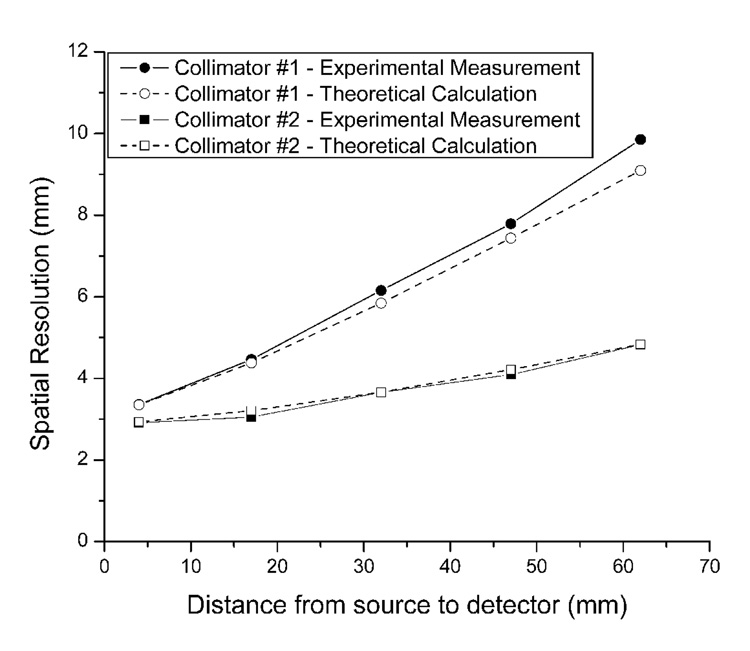
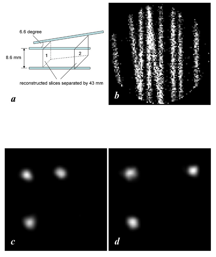
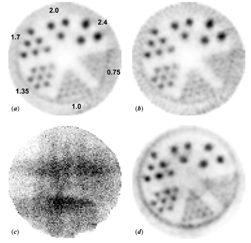
We have designed and built a small animal single photon emission computed tomography (SPECT) imaging system equipped with parallel-hole and multipinhole collimators and capable of circular or helical SPECT. Copper-beryllium parallel-hole collimators suitable for imaging the ~35 keV photons from the decay of (125)I have been built and installed to achieve useful spatial resolution over a range of object-detector distances and to reduce imaging time on our dual-detector array. To address the resolution limitations in the parallel-hole SPECT and the sensitivity and limited field of view of single-pinhole SPECT, we have incorporated multipinhole circular and helical SPECT in addition to expanding the parallel-hole SPECT capabilities. The pinhole SPECT system is based on a 110 mm diameter circular detector equipped with a pixellated NaI(Tl) scintillator array (1x1x5 mm(3)/pixel). The helical trajectory is accomplished by two stepping motors controlling the rotation of the detector-support gantry and displacement of the animal bed along the axis of rotation of the gantry. Results obtained in SPECT studies of various phantoms show an enlarged field of view, very good resolution and improved sensitivity using multipinhole circular or helical SPECT. Collimators with one, three and five 1 mm diameter pinholes have been implemented and compared in these tests. Our objective is to develop a system on which one may readily select a suitable mode of either parallel-hole SPECT or pinhole circular or helical SPECT for a variety of small animal imaging applications.


Figures

Fig. 1
Fig. 1
Photograph of the compact parallel-hole and pinhole circular/helical SPECT imaging system. Signal processing and storage instruments are not shown in this picture. All detectors are incorporated in a cylindrical gantry capable of rotating 360 degrees. Detectors #1–2 are “mouse-sized” compact gamma cameras and detectors #3–4 are 110 mm diameter circular detectors.
Fig. 2
Fig. 2
(Left) Diagram of the 5-pinhole collimator. (Right) Photograph of the setup to accomplish step-and-shoot helical orbit. Motor 1 controls the rotation of the cylindrical gantry round the AOR. Motor 2 controls the displacement along the AOR of the rack with a mouse bed fixed to one end.
Fig. 3
Fig. 3
Comparison of spatial resolution with the newly constructed detector equipped with 5 mm thick high-sensitivity collimator #1 and 6 mm thick high-resolution collimator #2 respectively. Relatively high resolution was persevered over a useful range with the 6 mm thick collimator while the efficiency was lessened by a factor about 6.
Fig. 4
Fig. 4
A 2 MBq three-capillary phantom study of 3-pinhole helical SPECT. (a) Setup of the phantom. (b) A 3-min projection. (c) Reconstructed slice 1. (d) Reconstructed slice 2
Fig. 5
Fig. 5
Reconstruction resolution as a function of ML-EM iteration number for various modes of pinhole SPECT/HSPECT
Fig. 6
Fig. 6
Six profiles (a) – (f) of the capillary in the transverse reconstructed slices after 13 iterations for 1-, 3- 5-pinhole helical SPECT (a, c, e) and circular SPECT (b, d, f)
Fig. 7
Fig. 7
(a) A transaxial view of the phantom made with 6 capillary tubes placed side by side. Each tube contained ~0.37 MBq Na125I in 8 cm length. (b) A transaxial image of a 3-pinhole helical SPECT scan of the phantom. The step increment along AOR is 0.5 mm. (c) The profile of the six capillaries in the reconstructed image (b).
Fig. 8
Fig. 8
(a) A transaxial view of the hot-rod phantom using single-pinhole helical SPECT with 3° angular increments and 0.1 mm step increments along the AOR. Each of the 120 projections was 3 min/projection. The hot-rod phantom contained 10 MBq 125I with a concentration of 2 MBq/ml. The hot rod diameters of the six wedge-shaped regions in the phantom are 0.75, 1.0, 1.35, 1.7, 2.0, and 2.4 mm, respectively. (b) An image reconstructed from 60 slices out of those 120 projections to represent three-hour 1pHSPECT (c) One 3-min projection of the hot-rod phantom of a three-pinhole helical SPECT scan with the same imaging parameters as the 1pHSPECT scan. (d) A transverse image reconstructed from the 120 projections of the 3pHSPECT scan. Each reconstructed image presented here is 4.4 mm in thickness.

References

    1. Schramm NU, Lackas C, Hoppin JW, Schurrat T, Behe M, Engeland U, Behr TM. 2003 IEEE Nucl. Sci. Symp. Conf. Rec; 2004. pp. 2823–2824.
    1. Schramm NU, Schipper M, Schurrat T, Behe M, Alfke H, Engeland U, Ebel G, Behr TM. 2003 IEEE Nucl. Sci. Symp. Conf. Rec; 2004. pp. 2077–2079.
    1. Schramm NU, Ebel G, Engeland U, Schurrat T, Behe M, Behr TM. IEEE Trans. Nucl. Sci. 2003;NS-50(3):315–320.
    1. Schramm NU, Ebel G, Engeland U, Schurrat T, Behe M, Behr TM. 2002 IEEE Nucl. Sci. Symp. Conf. Rec; 2003. pp. 774–777.
    1. Schramm NU, Wirrwar A, Halling H. 2001 IEEE Nucl. Sci. Symp. Conf. Rec; 2002. pp. 1585–1586.

LinkOut - more resources