

Jejunal small ectopic pancreas developing into jejunojejunal intussusception: a rare cause of ileus
- PMID: 19701981
- PMCID: PMC2731263
- DOI: 10.3748/wjg.15.3954
Jejunal small ectopic pancreas developing into jejunojejunal intussusception: a rare cause of ileus
Abstract
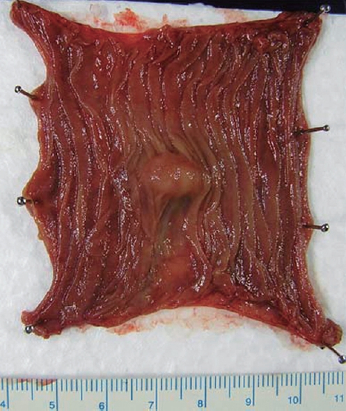
Intussusception is rare in adults. We describe a 62-year-old man with jejunal ectopic pancreas that led to jejunojejunal intussusception and ileus. The patient was admitted to our hospital because of intermittent abdominal pain. Plain abdominal radiography showed some intestinal gas and fluid levels. Abdominal CT scan demonstrated a target sign suggesting bowel intussusception. Jejunography using a naso-jejunal tube showed an oval-shaped mass about 15 mm in diameter with a smooth surface in the jejunum, which suggested a submucosal tumor (SMT), and edematous mucosa around the mass. Partial jejunal resection was carried out and the resected oval-shaped tumor, 14 mm x 11 mm in size, was found to be covered with normal jejunal mucosa. The tumor was histologically diagnosed as type III ectopic pancreas according to the classification proposed by Heinrich. Abdominal pain resolved postoperatively. This case reminds us that jejunal ectopic pancreas should be included in the differential diagnosis of intussusception caused by an SMT in the intestine.
Figures


Similar articles
-
Jejunal Intussusception Due to Heterotopic Pancreas: A Case Report.Cureus. 2021 Apr 20;13(4):e14586. doi: 10.7759/cureus.14586. Cureus. 2021. PMID: 34036004 Free PMC article.
-
Ectopic pancreas, intussusception, and a ruptured mesenteric band: an unusual association.Clin Anat. 2011 Jan;24(1):128-32. doi: 10.1002/ca.21052. Clin Anat. 2011. PMID: 20949486
-
A case of jejunal cancer arising from ectopic pancreas.Clin J Gastroenterol. 2025 Aug;18(4):610-614. doi: 10.1007/s12328-025-02148-5. Epub 2025 Jun 16. Clin J Gastroenterol. 2025. PMID: 40522573 Free PMC article.
-
Intussusception caused by a heterotopic pancreas. Case report and literature review.JOP. 2004 Nov 10;5(6):476-9. JOP. 2004. PMID: 15536284 Review.
-
Intussusception Caused by Heterotopic Pancreas: A Tunisian Case Series of 5 Pediatric Patients.Arch Iran Med. 2022 Dec 1;25(12):844-846. doi: 10.34172/aim.2022.131. Arch Iran Med. 2022. PMID: 37543913 Free PMC article. Review.
Cited by
-
A case report of incidental ectopic pancreatic tissue during laparoscopic appendicectomy.Int J Surg Case Rep. 2018;45:77-78. doi: 10.1016/j.ijscr.2018.03.023. Epub 2018 Mar 19. Int J Surg Case Rep. 2018. PMID: 29574401 Free PMC article.
-
Jejunal Intussusception Due to Heterotopic Pancreas: A Case Report.Cureus. 2021 Apr 20;13(4):e14586. doi: 10.7759/cureus.14586. Cureus. 2021. PMID: 34036004 Free PMC article.
-
Cystic jejunal duplication with Heinrich's type I ectopic pancreas, incidentally discovered in a patient with pancreatic tail neoplasm.World J Clin Cases. 2016 Sep 16;4(9):281-4. doi: 10.12998/wjcc.v4.i9.281. World J Clin Cases. 2016. PMID: 27672644 Free PMC article.
-
The Role of Laparoscopy in the Management of a Diagnostic Dilemma: Jejunal Ectopic Pancreas Developing into Jejunojejunal Intussusception.Case Rep Surg. 2017;2017:8452947. doi: 10.1155/2017/8452947. Epub 2017 Jul 27. Case Rep Surg. 2017. PMID: 28819577 Free PMC article.
-
Ectopic pancreatic tissue in the small intestine: An uncommon finding following trauma.Int J Surg Case Rep. 2024 Nov;124:110461. doi: 10.1016/j.ijscr.2024.110461. Epub 2024 Oct 16. Int J Surg Case Rep. 2024. PMID: 39418989 Free PMC article.
References
-
- Ishikawa O, Ishiguro S, Ohhigashi H, Sasaki Y, Yasuda T, Imaoka S, Iwanaga T, Nakaizumi A, Fujita M, Wada A. Solid and papillary neoplasm arising from an ectopic pancreas in the mesocolon. Am J Gastroenterol. 1990;85:597–601. - PubMed
-
- Tanaka K, Tsunoda T, Eto T, Yamada M, Tajima Y, Shimogama H, Yamaguchi T, Matsuo S, Izawa K. Diagnosis and management of heterotopic pancreas. Int Surg. 1993;78:32–35. - PubMed
-
- Hirasaki S, Tanimizu M, Moriwaki T, Nasu J. Acute pancreatitis occurring in gastric aberrant pancreas treated with surgery and proved by histological examination. Intern Med. 2005;44:1169–1173. - PubMed
-
- Tekin A, Aksoy F, Vatansev C, Kücükkartallar T, Belviranli M, Toy H. A rare cause of ileus: invagination due to ectopic pancreas. Acta Chir Belg. 2008;108:343–345. - PubMed
-
- von Heinrich H. Ein Beitrag zur Histologie des sogen: Akzessorischen Pankreas. Virchows Arch A Pathol Anat Histopathol. 1909;198:392–401.
Publication types
MeSH terms
LinkOut - more resources
Full Text Sources
