. 2009 Oct;158(3):779-89.
doi: 10.1111/j.1476-5381.2009.00367.x. Epub 2009 Aug 20.

A crucial role for TNF-alpha in mediating neutrophil influx induced by endogenously generated or exogenous chemokines, KC/CXCL1 and LIX/CXCL5

Affiliations

A crucial role for TNF-alpha in mediating neutrophil influx induced by endogenously generated or exogenous chemokines, KC/CXCL1 and LIX/CXCL5

S M Vieira et al. Br J Pharmacol. 2009 Oct.

Abstract

Background and purpose: Chemokines orchestrate neutrophil recruitment to inflammatory foci. In the present study, we evaluated the participation of three chemokines, KC/CXCL1, MIP-2/CXCL2 and LIX/CXCL5, which are ligands for chemokine receptor 2 (CXCR2), in mediating neutrophil recruitment in immune inflammation induced by antigen in immunized mice.

Experimental approach: Neutrophil recruitment was assessed in immunized mice challenged with methylated bovine serum albumin, KC/CXCL1, LIX/CXCL5 or tumour necrosis factor (TNF)-alpha. Cytokine and chemokine levels were determined in peritoneal exudates and in supernatants of macrophages and mast cells by elisa. CXCR2 and intercellular adhesion molecule 1 (ICAM-1) expression was determined using immunohistochemistry and confocal microscopy.

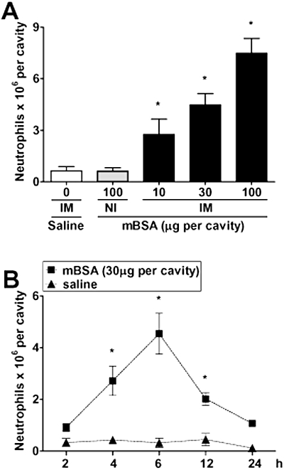
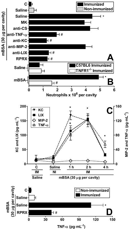
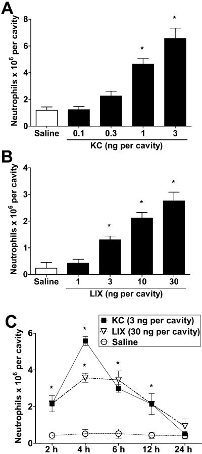
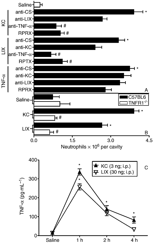
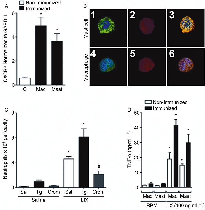
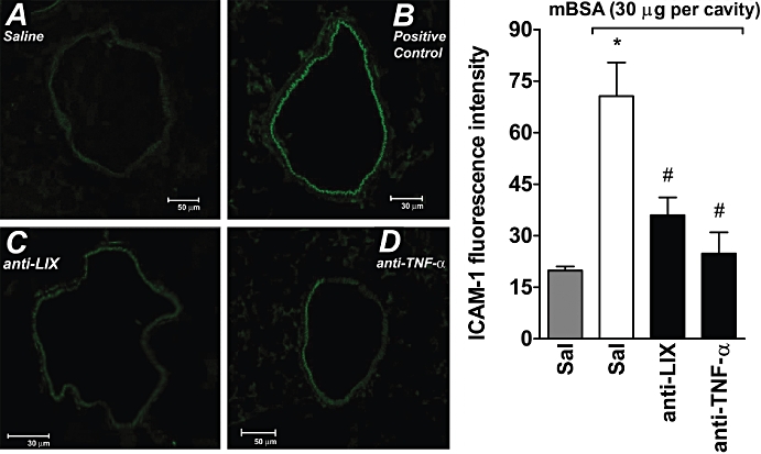
Key results: Antigen challenge induced dose- and time-dependent neutrophil recruitment and production of KC/CXCL1, LIX/CXCL5 and TNF-alpha, but not MIP-2/CXCL2, in peritoneal exudates. Neutrophil recruitment was inhibited by treatment with reparixin (CXCR1/2 antagonist), anti-KC/CXCL1, anti-LIX/CXCL5 or anti-TNF-alpha antibodies and in tumour necrosis factor receptor 1-deficient mice. Intraperitoneal injection of KC/CXCL1 and LIX/CXCL5 induced dose- and time-dependent neutrophil recruitment and TNF-alpha production, which were inhibited by reparixin or anti-TNF-alpha treatment. Macrophages and mast cells expressed CXCR2 receptors. Increased macrophage numbers enhanced, while cromolyn sodium (mast cell stabilizer) diminished, LIX/CXCL5-induced neutrophil recruitment. Macrophages and mast cells from immunized mice produced TNF-alpha upon LIX/CXCL5 stimulation. Methylated bovine serum albumin induced expression of ICAM-1 on mesenteric vascular endothelium, which was inhibited by anti-TNF-alpha or anti-LIX/CXCL5.

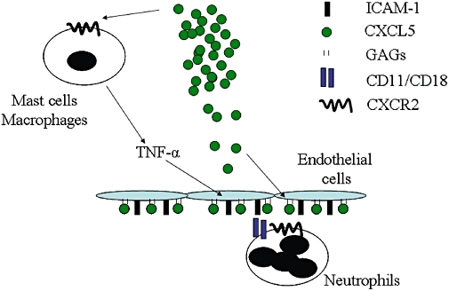
Conclusion and implications: Following antigen challenge, CXCR2 ligands are produced and act on macrophages and mast cells triggering the production of TNF-alpha, which synergistically contribute to neutrophil recruitment through induction of the expression of ICAM-1.


Figures

Figure 1
Figure 1
Antigen (methylated bovine serum albumin, mBSA) challenge in immunized (IM) mice induces dose- and time-dependent neutrophil recruitment to the peritoneal cavity. (A) mBSA was injected at the indicated doses into the peritoneal cavity of non-immunized (NI) or IM mice, and neutrophil recruitment was determined 6 h later. Neutrophil recruitment was also evaluated when saline was injected into IM mice (control). (B) Time dependence of the neutrophil recruitment in IM mice challenged with saline or mBSA. Data are means ± SEM (n= 5). *P <0.05 when compared with control mice (anova followed by the Bonferroni test).
Figure 2
Figure 2
Methylated bovine serum albumin (mBSA) challenge-induced neutrophil recruitment depends on tumour necrosis factor (TNF)-α, KC/CXCL1 and LIX/CXCL5, and time-dependent production of KC/CXCL1, LIX/CXCL5 and TNF-α in the peritoneal exudates. Reparixin reduced TNF-α levels in mBSA-challenged immunized (IM) mice. (A) IM mice were treated, 1 h before challenge, with MK 886 (inhibitor of 5-lipoxygenase activating protein; MK; 1 mg·kg−1, p.o.) or reparixin (CXCR1/2 antagonist; RPRX; 30 mg·kg−1, s.c.), or 30 min before with control antibody (anti-CS; 30 µL, i.p.), anti-TNF antiserum (anti-TNF-α; 30 µL, i.p.), chemokine antibodies, anti-KC/CXCL1 (anti-KC; 3 µg, i.p.), anti-MIP-2/CXCL2 (anti-MIP-2; 3 µg, i.p.) and anti-LIX/CXCL5 (anti-LIX; 3 µg, i.p.) and then challenged with mBSA (30 µg, i.p.). (B) Wild-type or tumour necrosis factor receptor (TNFR)1−/− mice were challenged with mBSA (30 µg, i.p.). Neutrophil recruitment was evaluated 6 h after mBSA or saline i.p. injection. (C) The concentrations of KC/CXCL1, MIP-2/CXCL2, LIX/CXCL5 and TNF-α were determined 1, 2 and 4 h after challenge with mBSA (30 µg, i.p.) in IM and in non-immunized (NI) mice or saline in IM mice (C; control). (D) Animals were treated 1 h before challenge with reparixin (RPRX; 30 mg·kg−1, s.c.) or saline, and the concentrations of TNF-α was determined 2 h after challenge with mBSA (30 µg, i.p.) in IM and NI mice or saline in IM mice (C; control). Data are means ± SEM (n= 5). *P <0.05 when compared with control group; #P <0.05 when compared with IM group after mBSA challenge. (anova followed by the Bonferroni test).
Figure 3
Figure 3
KC/CXCL1 and LIX/CXCL5 induce dose- and time-dependent neutrophil recruitment into peritoneal cavities of mice. (A,B) Saline (control), (A) KC/CXCL1 (KC; 0.1, 0.3, 1 or 3 ng) or (B) LIX/CXCL5 (LIX; 1, 3, 10 or 30 ng) were injected into the peritoneal cavity of mice and 4 h later, neutrophil recruitment was evaluated. (C) Time-dependent neutrophil recruitment after KC/CXCL1 (KC; 3 ng, i.p.) or LIX/CXCL5 (LIX; 30 ng, i.p.) injection. Data are means ± SEM (n= 5). *P <0.05 when compared with control mice (anova followed by the Bonferroni test).
Figure 4
Figure 4
KC/CXCL1 and LIX/CXCL5 induce neutrophil recruitment via tumour necrosis factor (TNF)-α. (A) Mice were treated, 1 h before challenge, with reparixin (RPRX; 30 mg·kg−1, s.c.), or 30 min before challenge, with control antibody (anti-CS; 30 µL, i.p.), anti-TNF antiserum (anti-TNF-α; 30 µL, i.p.), antibody anti-KC/CXCL1 (anti-KC; 3 µg, i.p.), antibody anti-LIX/CXCL5 (anti-LIX; 3 µg, i.p.) and then challenged with KC/CXCL1 (KC; 3 ng), LIX/CXCL5 (LIX; 30 ng) or TNF-α (40 ng) i.p. Neutrophil recruitment was evaluated 4 h after challenge in treated groups. (B) Wild-type or tumour necrosis factor receptor (TNFR)1−/−mice were challenged with KC/CXCL5 (KC; 3 ng, i.p.) or LIX/CXCL5 (LIX; 30 ng, i.p.), and 4 h later the neutrophil recruitment was estimated in mice. Neutrophil recruitment was also evaluated when saline was injected into mice (control). (C) The concentrations of TNF-α were determined 1, 2 and 4 h after challenge i.p. with saline, KC/CXCL1 (KC; 3 ng) or LIX/CXCL5 (LIX; 30 ng) in mice. Data are means ± SEM (n= 5). *P <0.05 when compared with control group; #P <0.05 when compared with group injected with cytokines or chemokines. (anova followed by the Bonferroni test).
Figure 5
Figure 5
LIX/CXCL5 induced neutrophil recruitment by acting on CXCR2 in macrophages and mast cells, stimulating production of tumour necrosis factor (TNF)-α. (A) Real-time PCR analysis of CXCR2 expression of purified macrophages (Mac) or mast cells (Mast) from peritoneal cavity of immunized and non-immunized mice 2 h after methylated bovine serum albumin (mBSA) (30 µg, i.p.) challenge. (B) Confocal analysis of mast cells and macrophages from immunized mice expressing CXCR2. (B1) 4,6-diamidino-2-phenylindole (DAPI) and anti-murine AA4 conjugated with Alexa488. (B2,B5) mAb anti-mouse CXCR2-phycoerythrin. (B4) DAPI and anti-murine F4/80 conjugated with Alexa488. (B3,B6) Double staining with Alexa488 and phycoerythrin was performed to discriminate the cell type and CXCR2 receptor respectively. Nuclei were counterstained with DAPI. (C) Thioglycollate 3% (Tg; 200 µL, i.p.) injected i.p. induces an increase in mononuclear cells (15.3 ± 1.8 × 10⁶; data not shown), and after 4 days the mice were further challenged with saline or LIX/CXCL5 (30 ng, i.p.). Mice were treated, 1 h before challenge, with cromolyn sodium (Crom; 10 mg·kg−1, i.p.) that induced stabilization of mast cells in the peritoneal cavity. Under these conditions, saline or LIX/CXCL5 (LIX; 30 ng) was injected i.p., and neutrophil recruitment was determined 4 h after stimulus injection. (D) Concentrations of TNF-α determined 6 h after saline (C; control) or LIX/CXCL5 (LIX; 100 ng·mL−1) in vitro stimulation of macrophages (Mac) or mast cells (Mast) from immunized mice. Data are means ± SEM (n= 5). *P <0.05 when compared with control group; #P <0.05 when compared with immunized group after mBSA challenge. (anova followed by the Bonferroni test).
Figure 6
Figure 6
Effects of anti-LIX/CXCL5 and anti-TNF-α on mBSA-induced ICAM-1 expression in the mesenteric microcirculation. ICAM-1 expression in the microcirculation in mesenteric tissues was determined by fluorescent immunohistochemical staining using anti-ICAM-1-specific mAb conjugated with FITC (A–D). (A) Immunofluorescence staining for ICAM-1 in PBS i.p. injected animals. (B) Mice treated with PBS (0.2 mL, s.c.) and then injected with mBSA (30 µg per cavity). (C) Mice treated with anti-LIX/CXCL5 (anti-LIX; 3 µg, i.p., 30 min before) and then injected with mBSA. (D) Mice treated with anti-TNF-α (4 µg, i.p., 30 min before) and then injected with mBSA. Panels show images representative of at least three independent experiments. In addition, the endothelial fluorescence intensity was quantified. Data represent mean ± SD from three independent experiments. *P <0.05 when compared with control group; #P <0.05 when compared with immunized group after mBSA challenge. (anova followed by the Bonferroni test). ICAM-1, intercellular adhesion molecule 1; mBSA, methylated bovine serum albumin; PBS, phosphate-buffered saline; TNF, tumour necrosis factor.
Figure 7
Figure 7
Schematic representation of the actions of LIX/CXCL5 and cooperation with tumour necrosis factor (TNF)-α-induced intercellular adhesion molecule 1 (ICAM-1) to drive neutrophil influx in vivo.

Similar articles

Cited by

References

    1. Anderson JA, Lentsch AB, Hadjiminas DJ, Miller FN, Martin AW, Nakagawa K, et al. The role of cytokines, adhesion molecules, and chemokines in interleukin-2-induced lymphocytic infiltration in C57BL/6 mice. J Clin Invest. 1996;97:1952–1959. - PMC - PubMed
    1. Bacon KB, Oppenheim JJ. Chemokines in disease models and pathogenesis. Cytokine Growth Factor Rev. 1998;9:167–173. - PubMed
    1. Baggiolini M, Walz A, Kunkel SL. Neutrophil-activating peptide-1/interleukin 8, a novel cytokine that activates neutrophils. J Clin Invest. 1989;84:1045–1049. - PMC - PubMed
    1. Begley LA, Kasina S, Mehra R, Adsule S, Admon AJ, Lonigro RJ, et al. CXCL5 promotes prostate cancer progression. Neoplasia. 2008;10:244–254. - PMC - PubMed
    1. Belanger C, Elimam H, Lefebvre J, Borgeat P, Marleau S. Involvement of endogenous leukotriene B4 and platelet-activating factor in polymorphonuclear leucocyte recruitment to dermal inflammatory sites in rats. Immunology. 2008;124:295–303. - PMC - PubMed

Publication types

MeSH terms