. 2009 Oct;158(3):738-48.
doi: 10.1111/j.1476-5381.2009.00361.x. Epub 2009 Aug 24.

Ginsenoside Rg1 protects dopaminergic neurons in a rat model of Parkinson's disease through the IGF-I receptor signalling pathway

Affiliations

Ginsenoside Rg1 protects dopaminergic neurons in a rat model of Parkinson's disease through the IGF-I receptor signalling pathway

Li Xu et al. Br J Pharmacol. 2009 Oct.

Abstract

Background and purpose: We have shown that ginsenoside Rg1 is a novel class of potent phytoestrogen and activates insulin-like growth factor-I receptor (IGF-IR) signalling pathway in human breast cancer MCF-7 cells. The present study tested the hypothesis that the neuroprotective actions of Rg1 involved activation of the IGF-IR signalling pathway in a rat model of Parkinson's disease, induced by 6-hydroxydopamine (6-OHDA).

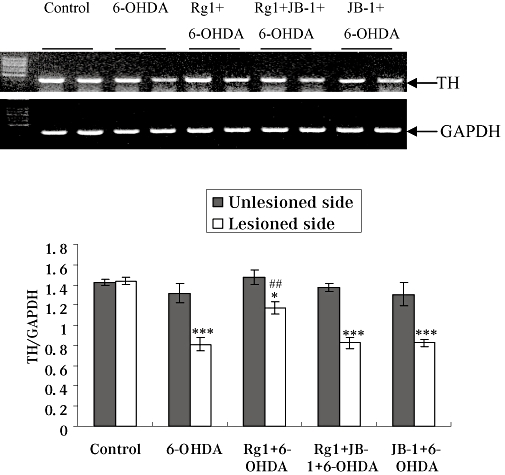
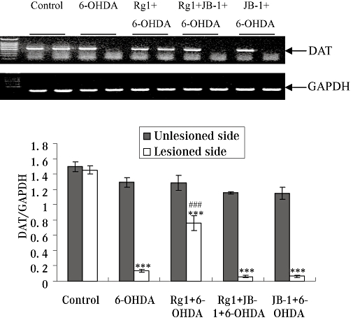
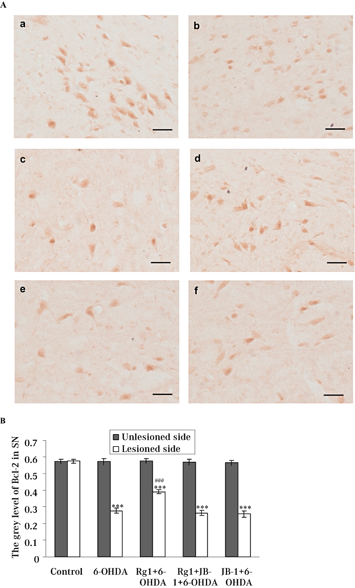
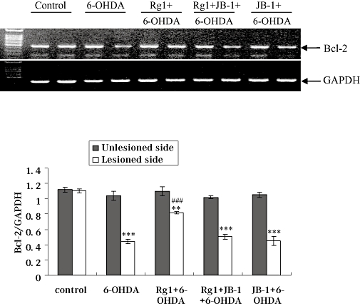
Experimental approach: Ovariectomized rats were infused unilaterally with 6-OHDA into the medial forebrain bundle to lesion the nigrostriatal dopamine pathway and treated with Rg1 (1.5 h after 6-OHDA injections) in the absence or presence of the IGF-IR antagonist JB-1 (1 h before Rg1 injections). The rotational behaviour induced by apomorphine and the dopamine content in the striatum were studied. Protein and gene expression of tyrosine hydroxylase, dopamine transporter and Bcl-2 in the substantia nigra were also determined.

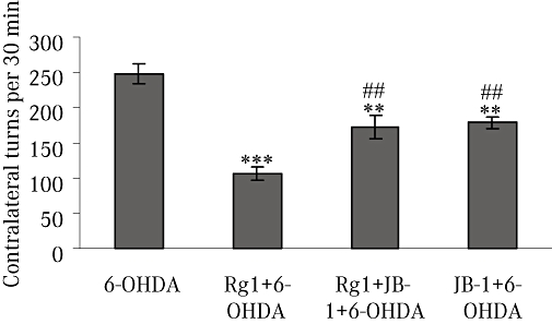
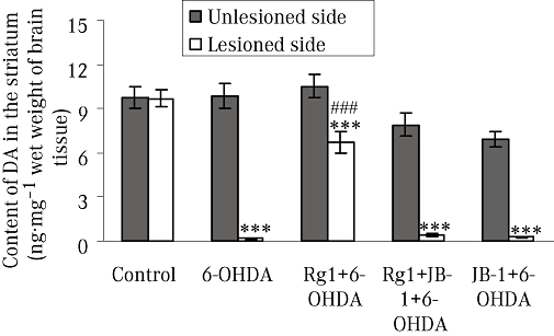
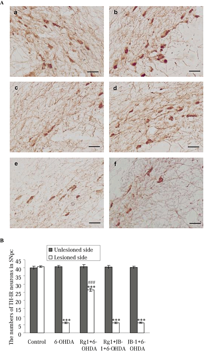
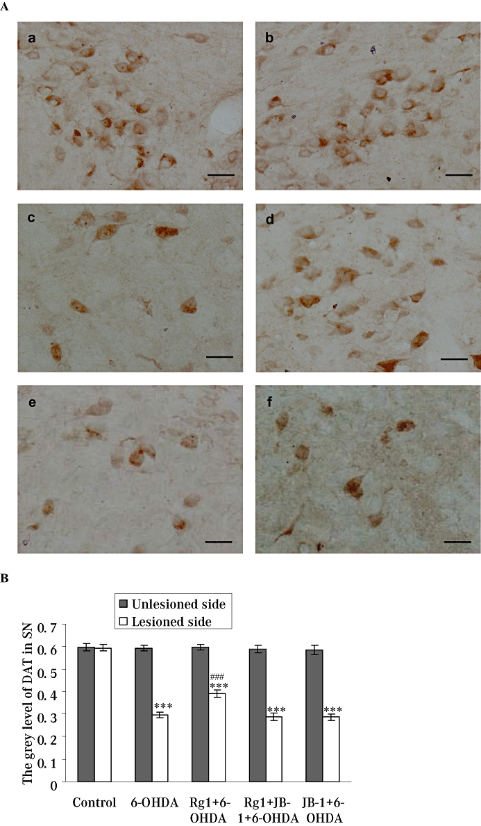
Key results: Rg1 treatment ameliorated the rotational behaviour induced by apomorphine in our model of nigrostriatal injury. This effect was partly blocked by JB-1. 6-OHDA significantly decreased the dopamine content of the striatum and treatment with Rg1 reversed this decrease. Treatment with Rg1 of 6-OHDA-lesioned rats reduced neurotoxicity, as measured by tyrosine hydroxylase, dopamine transporter and Bcl-2 protein and gene level in the substantia nigra. These effects were abolished by JB-1.

Conclusions and implications: These data provide the first evidence that Rg1 has neuroprotective effects on dopaminergic neurons in the 6-OHDA model of nigrostriatal injury and its actions might involve activation of the IGF-IR signalling pathway.


Figures

Figure 1
Figure 1
Apomorphine-induced rotation behaviour. Rotational behaviour was counted for 30 min after i.p. injection of 0.05 mg·kg−1 apomorphine in the 6-hydroxydopamine (6-OHDA) group, Rg1 + 6-OHDA group, Rg1 + JB-1 + 6-OHDA group and JB-1 + 6-OHDA group. All values are expressed as mean ± SEM. n= 12. **P < 0.01, ***P < 0.001 versus the 6-OHDA group, ##P < 0.01 versus the Rg1 + 6-OHDA group.
Figure 2
Figure 2
Effect of Rg1 on the dopamine (DA) content of the striatum of rats with 6-hydroxydopamine (6-OHDA) induced Parkinson's disease and the blocking effect of JB-1 (ng·mg−1 wet weight of brain tissue). The unlesioned and lesioned side of striatum were used to determine the content of dopamine by HPLC. All values are expressed as mean ± SEM. n= 6. ***P < 0.001 versus the control group, ###P < 0.001 versus the lesioned side of 6-OHDA group.
Figure 3
Figure 3
Effect of Rg1 on the tyrosine hydroxylase-immunoreactive (TH-IR) neurons in the substantia nigra pars compacta (SNpc) of the rat model of 6-hydroxydopamine (6-OHDA) induced Parkinson's disease and the blocking effect of JB-1. (A) Representative microphotographs of coronal sections immunostained for TH-IR neurons in the unlesioned side of the SNpc in the control (sham) group (a) and the lesioned side of the SNpc in the control (sham) group (b), 6-OHDA group (c), Rg1 + 6-OHDA group (d), Rg1 + JB-1 + 6-OHDA group (e) and JB-1 + 6-OHDA group (f). Bar = 30 µm. (B) Quantitative analysis of TH-IR neurons in the unlesioned and lesioned side of the SNpc. All values are expressed as mean ± SEM. n= 6. ***P < 0.001 versus the control group, ###P < 0.001 versus the lesioned side of 6-OHDA group.
Figure 4
Figure 4
Effect of Rg1 on the dopamine transporter-immunoreactivity (DAT-IR) neurons in the substantia nigra pars compacta (SNpc) of the rat model of Parkinson's disease induced by 6-hydroxydopamine (6-OHDA) and the blocking effect of JB-1. (A) Representative microphotographs of coronal sections immunostained for DAT-IR neurons the unlesioned side of the SNpc in the control (sham) group (a) and the lesioned side of the SNpc in the control (sham) group (b), 6-OHDA group (c), Rg1 + 6-OHDA group (d), Rg1 + JB-1 + 6-OHDA group (e) and JB-1 + 6-OHDA group (f). Bar = 30 µm. (B) Quantitative analysis of DAT-IR neurons in the unlesioned and lesioned side of the SNpc. All values are expressed as mean ± SEM. n= 6. ***P < 0.001 versus the control group, ###P < 0.001 versus the lesioned side of 6-OHDA group.
Figure 5
Figure 5
Effect of Rg1 on tyrosine hydroxylase (TH) gene expression in the substantia nigra (SN) of the rat model of Parkinson's disease induced by 6-hydroxydopamine (6-OHDA) and the blocking effect of JB-1. Total RNA was isolated from the unlesioned and lesioned side of SN and subjected to semi-quantitative RT-PCR analysis of TH and GAPDH mRNA expression. The TH mRNA expression level was expressed as a ratio of the expression of GAPDH. Summary results shown are expressed as mean ± SEM. n= 6. *P < 0.05, ***P < 0.001 versus the control group, ##P < 0.01 versus the lesioned side of 6-OHDA group.
Figure 6
Figure 6
Effect of Rg1 on dopamine transporter (DAT) gene expression in the substantia nigra (SN) of the rat model of Parkinson's disease induced by 6-hydroxydopamine (6-OHDA) and the blocking effect of JB-1. Total RNA was isolated from the unlesioned and lesioned side of the SN and subjected to semi-quantitative RT-PCR analysis of DAT and GAPDH mRNA expression. The DAT mRNA expression level was expressed as a ratio of the expression of GAPDH. Summary results shown are expressed as mean ± SEM. n= 6. ***P < 0.01 versus the control group, ###P < 0.01 versus the lesioned side of 6-OHDA group.
Figure 7
Figure 7
Effect of Rg1 on the Bcl-2 protein expression in the substantia nigra pars compacta (SNpc) of the rat model of Parkinson's disease induced by 6-hydroxydopamine (6-OHDA) and the blocking effect of JB-1. (A) Representative microphotographs of coronal sections immunostained for Bcl-2 protein expression in the unlesioned side of the SNpc in the control (sham) group (a) and the lesioned side of the SNpc in the control (sham) group (b), 6-OHDA group (c), Rg1 + 6-OHDA group (d), Rg1 + JB-1 + 6-OHDA group (e) and JB-1 + 6-OHDA group (f). Bar = 30µm. (B) Quantitative analysis of Bcl-2 protein expression in the unlesioned and lesioned side of the SNpc. All values are expressed as mean ± SEM. n= 6. ***P < 0.001 versus the control group, ###P < 0.001 versus the lesioned side of 6-OHDA group.
None
Effect of Rg1 on Bcl-2 gene expression in the substantia nigra (SN) of the rat model of Parkinson's disease induced by 6-hydroxydopamine (6-OHDA) and the blocking effect of JB-1. Total RNA was isolated from the unlesioned and lesioned side of SN and subjected to semi-quantitative RT-PCR analysis of Bcl-2 and GAPDH mRNA expression. The Bcl-2 mRNA expression level was expressed as a ratio of the expression of GAPDH. Summary results shown are expressed as mean ± SEM. n= 6. **P < 0.01, ***P < 0.001 versus the control group, ###P < 0.001 versus the lesioned side of 6-OHDA group.

References

    1. Abraham IM, Todman MG, Korach KS, Herbison AE. Critical in vivo roles for classical estrogen receptors in rapid estrogen actions on intracellular signaling in mouse brain. Endocrinology. 2004;145:3055–3061. - PubMed
    1. Bondy CA, Cheng CM. Signaling by insulin-like growth factor 1 in brain. Eur J Pharmacol. 2004;490:25–31. - PubMed
    1. Callier S, Morissette M, Grandbois M, Pelaprat D, Di Paolo T. Neuroprotective properties of 17beta-estradiol, progesterone, and raloxifene in MPTP C57Bl/6 mice. Synapse. 2001;41:131–138. - PubMed
    1. Cardona-Gomez GP, Mendez P, DonCarlos LL, Azcoitia I, Garcia-Segura LM. Interactions of estrogen and insulin-like growth factor-I in the brain: molecular mechanisms and functional implications. J Steroid Biochem Mol Biol. 2002;83:211–217. - PubMed
    1. Cerruti C, Pilotte NS, Uhl G, Kuhar MJ. Reduction in dopamine transporter mRNA after cessation of repeated cocaine administration. Brain Res Mol Brain Res. 1994;22:132–138. - PubMed

Publication types

MeSH terms