. 2009 Oct 15;587(Pt 20):4811-27.
doi: 10.1113/jphysiol.2009.173989. Epub 2009 Aug 24.

Tremor-related motor unit firing in Parkinson's disease: implications for tremor genesis

Affiliations

Tremor-related motor unit firing in Parkinson's disease: implications for tremor genesis

C N Christakos et al. J Physiol. .

Abstract

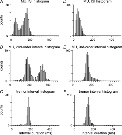
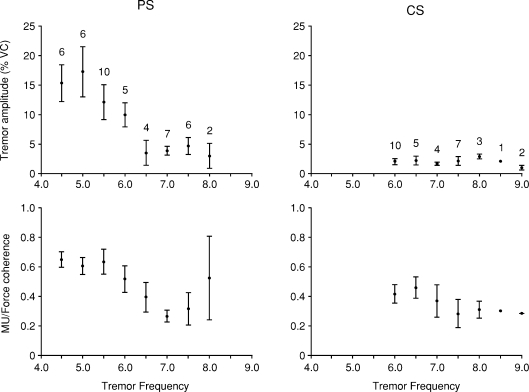
Muscle tremors reflect rhythmical motor unit (MU) activities. Therefore, the MU firing patterns and synchrony determine the properties of the parkinsonian force tremor (FT) and the neurogenic components of associated limb tremors. They may also be indicative of the neural mechanisms of tremor genesis which to date remain uncertain. We examined these MU behaviours during isometric contractions of a finger muscle in 19 parkinsonian subjects. Our results reveal that the parkinsonian FT is abnormally large. Like the physiological FT, it is accompanied by in-phase rhythms in all MU activities. However, there exist two important differences. Firstly, the synchrony during the parkinsonian FT is stronger than the normal one and therefore contributes to the FT enhancement. Secondly, the synchronous MU components partly represent rhythmical sequences of spike doublets and triplets whose incidences directly reflect the differences of the MU firing rates to the FT frequency. According to our analyses, the latter frequency coincides with the MU recruitment rate. Consequently, the numerous medium- and small-sized active MUs contribute rhythmical twitch doublets and triplets, i.e. large force pulses, to the parkinsonian FT. The impact of this effect on the FT amplitude is found to predominate over the impact of the augmented synchrony. Importantly, apart from the rule governing the occurrence of doublets/triplets, the mean interspike intervals within such spike events are fairly fixed around 50 ms. Such regularities in MU activities may reflect properties of the neural input underlying the FT, and thus represent a basis for more focused studies of the generator(s) of parkinsonian tremors.


Figures

Figure 7
Figure 7. Higher-order interval analyses for MUs belonging to categories II and III
A, ISI histogram; B, second-order interval histogram; and C, tremor interval histogram, for the case of the MU of Fig. 1. Note in the second-order histogram the high concentration of intervals around the tremor period, which indicates that two successive ISIs often combined so as to form one tremor interval. D, ISI histogram; E, third-order interval histogram; and F, tremor interval histogram, for the case of the MU of Fig. 2. Note in the third-order histogram the high concentration of intervals around the tremor period, which indicates that three successive ISIs often added up to one tremor interval.
Figure 1
Figure 1. Indices D and T as estimators of incidence of simulated doublets and triplets
Top: D plotted against the predetermined probability of doublet occurrence per tremor cycle. Bottom: T plotted against the predetermined probability of triplet occurrence per tremor cycle. Note in both cases the best-fit straight line with slope 1, which indicates that D and T represent appropriate measures of the incidence of doublets and triplets, respectively.
Figure 3
Figure 3. Firing pattern and synchrony for MUs of category III (parkinsonian subject No. 1)
All columns as in Fig. 1. Note: in the MU/force time record, the rhythmical spike triplets and doublets, all occurring near successive FT minima at 7.0 Hz (overall spike rate 19.5 Hz); in the ISI histogram, the main peak representing the ca 40 ms ISIs within triplets and doublets, and the small deflections near 60 ms and 90 ms, representing the longer ISIs that follow triplets and doublets; in the oscillatory MU/force cross-correlogram, the central trough at zero lag, which reflects the tendency for the MU spike events to occur near the FT minima; in the force auto-spectrum the dominant component at 7.0 Hz (vertical dashed line), which represents the tremor; in the MU auto-spectrum, the component at the FT frequency and harmonics (arrowheads), and the component at 26.0 Hz (arrow), which reflects the rhythm within the triplets; in the MU/force coherence spectrum, the very large component at 7.0 Hz which indicates the presence of MU correlations at the FT frequency.
Figure 2
Figure 2. Firing pattern and synchrony for MUs of category II (parkinsonian subject No. 4)
Left column (top to bottom): time record of force signal, intramuscular EMG and discriminated MU spikes; interspike interval (ISI) histogram; and normalized MU/force cross-correlogram. The MU/force time record shows rhythmical spike doublets and singlets near successive FT minima at 5.5 Hz (overall spike rate 9.0 Hz); the ISI histogram exhibits a dual peak representing the ca 50 ms ISIs within doublets and the longer ISIs that separate doublets and/or singlets; the oscillatory MU/force cross-correlogram has its central trough at zero lag, reflecting the tendency for the MU spike events to occur near the FT minima. Right column (top to bottom): auto-spectra and coherence of simultaneous MU spike activity and muscle force signal. The MU auto-spectrum shows a large component at 5.5 Hz (vertical dashed line), and harmonic components, manifesting the FT-related MU spike events. The force auto-spectrum exhibits a dominant component at 5.5 Hz, representing the tremor. The MU/force coherence spectrum shows a large component at 5.5 Hz, and harmonic components, indicative of MU spike event correlations at the FT frequency (the horizontal dashed line represents the threshold level of significance).
Figure 4
Figure 4. MU firing pattern and synchrony in normal subjects (control subject No. 2)
All columns as in Fig. 1. Note: in the MU/force time record, the rhythmical spikes at a rate of 9.0 Hz, a fraction of which occur near tremor minima at 6.5 Hz; in the ISI histogram, the main peak around the intrinsic MU period (110 ms), and the additional ISIs between 130 and 160 ms; in the oscillatory MU/force cross-correlogram, the central trough at zero lag, which reflects the tendency for some MU spikes to occur near the FT minima; in the force auto-spectrum, the large component at 6.5 Hz (vertical dashed line), which represents the tremor; in the MU auto-spectrum, the small deflection at the FT frequency, and the large component at 9.0 Hz (arrow), representing the intrinsic rhythm of the MU; in the MU/force coherence spectrum, the clear peak at 6.5 Hz, which indicates the presence of MU correlations at the FT frequency.
Figure 5
Figure 5. Dependence of the FT amplitude and the strength of the FT-related MU synchrony on the tremor frequency, for the parkinsonian (PS) and the control subjects (CS)
Top row: distribution for the tremor amplitude. Bottom row: distribution for the strength of the synchrony. Bars represent mean ±s.e.m. The values above bars indicate the number of contractions for each frequency. Note in the PS group the significant decrease of both amplitude and strength for tremor frequencies above 6 Hz. These relationships are not observed in the CS group.
Figure 6
Figure 6. Dependence of the FT amplitude on the strength of the tremor-related MU synchrony
Parkinsonian subjects: ▪, continuous line; control subjects: ○, dashed line. Note in the scatter diagrams for both groups the increase in tremor amplitude as the strength of the synchrony increases. Also note the much steeper slope of the best-fit straight line for the parkinsonian group.
Figure 8
Figure 8. Occurrence of spike doublets and triplets in relation to the MU firing rate
Index D (top) and T (bottom) plotted against the ratio of the MU firing rate (FR) to the tremor frequency (TF). Note the linear relationship between D, or T, and the difference of the MU firing rate to the tremor frequency, or to twice its value.

References

    1. Allum JH, Dietz V, Freund HJ. Neuronal mechanisms underlying physiological tremor. J Neurophysiol. 1978;41:557–571. - PubMed
    1. Amtage F, Henschel K, Schelter B, Vesper J, Timmer J, Lücking CH, Hellwig B. Tremor-correlated neuronal activity in the subthalamic nucleus of Parkinsonian patients. Neurosci Lett. 2008;442:195–199. - PubMed
    1. Bain PG. The management of tremor. J Neurol Neurosurg Psychiatry. 2002;72(Suppl. 1):I3–I9. - PMC - PubMed
    1. Baker JR, Davey NJ, Ellaway PH, Friedland CL. Short-term synchrony of motor unit discharge during weak isometric contraction in Parkinson's disease. Brain. 1992;115:137–154. - PubMed
    1. Barry BK, Pascoe MA, Jesunathadas M, Enoka RM. Rate coding is compressed but variability is unaltered for motor units in a hand muscle of old adults. J Neurophysiol. 2007;97:3206–3218. - PubMed

Publication types