. 2009 Oct 1;587(Pt 19):4681-94.
doi: 10.1113/jphysiol.2009.177105. Epub 2009 Aug 24.

Multifunctional rapidly adapting mechanosensitive enteric neurons (RAMEN) in the myenteric plexus of the guinea pig ileum

Affiliations

Multifunctional rapidly adapting mechanosensitive enteric neurons (RAMEN) in the myenteric plexus of the guinea pig ileum

Gemma Mazzuoli et al. J Physiol. .

Abstract

An important feature of the enteric nervous system (ENS) is its capability to respond to mechanical stimulation which, as currently suggested for the guinea-pig ileum, is encoded by specialized intrinsic primary afferent neurons (IPANs). We used von Frey hairs or intraganglionic volume injections to mimic ganglion deformation as observed in freely contracting preparations. Using fast voltage-sensitive dye imaging we identified rapidly adapting mechanosensitive enteric neurons (RAMEN, 25% of all neurons) in the myenteric plexus of the guinea pig ileum. RAMEN responded with phasic spike discharge to dynamic changes during ganglion deformation. This response was reproducible and increased with increasing forces. Deformation-evoked spike discharge was not changed by synaptic blockade with hexamethonium, omega-conotoxin or low Ca(2+)/high Mg(2+), defunctionalization of extrinsic afferents with capsaicin or muscle paralysis with nifedipine, suggesting direct activation of RAMEN. All RAMEN received hexamethonium-sensitive fast EPSPs, which were blocked by omega-conotoxin and low Ca(2+)/high Mg(2+). Seventy-two per cent of RAMEN were cholinergic, 22% nitrergic, and 44% were calbindin and NeuN negative, markers used to identify IPANs. Mechanosensitivity was observed in 31% and 47% of retrogradely traced interneurons and motor neurons, respectively. RAMEN belong to a new population of mechanosensitive neurons which differ from IPANs. We provided evidence for multifunctionality of RAMEN which may fulfil sensory, integrative and motor functions. In light of previously identified mechanosensitive neuron populations, mechanosensitivity appears to be a property of many more enteric neurons than generally assumed. The findings call for a revision of current concepts on sensory transmission within the ENS.


Figures
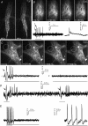
Figure 1
Figure 1
A, deformation of a Di-8-ANEPPS-stained ganglion during contractile activity of a flat sheet preparation from the ileum. The two frames were taken from a movie which can be accessed online as Supplemental material (Movie 1) and demonstrate that the lower part of the ganglion is distorted during muscle movements. B, neuronal deformation during intraganglionic volume injection. The four frames were taken from Movie 3 (online Supplemental material). Ganglion deformation is evident during volume injection. This is paralleled by deformation of individual neurons as illustrated for the neuron encircled by white lines. The response of this neuron to volume injection is shown in the left trace below the frames; signals have been filtered with a Butterworth filter (low pass 200 Hz, high pass 15 Hz). The bar below the trace indicates the onset and end of the volume injection. The very first deflections are mechanically induced artefacts (arrows) due to the volume injection. After that the neuron fires 4 action potentials. The trace on the right shows the response of the same neuron to electrical stimulation of interganglionic fibre tracts. The first action potential is a compound action potential reflecting axonal spike discharge (see Schemann et al. 2002). This is then followed by a fast EPSP. C, deformation during von Frey hair stimulation. The four frames were taken from Movie 2 (online Supplemental material). The white dotted circle indicates the stimulation site where the von Frey hair touched the ganglion. The von Frey hair deformed a relatively small area of the ganglion while distant areas of the ganglion showed no noticeable deformation. The white arrows mark two neurons: neuron 1 is located within the deformed area whereas neuron 2 is located in the non-deformed area. Below are the responses of the two neurons to von Frey hair stimulation; signals have been filtered with a Butterworth filter (low pass 150 Hz, high pass 25 Hz). The duration of the stimulation is indicated by the bar below the trace; the von Frey hair stimulation remained for the entire recording period. Neuron 1 fired 4 action potentials; the first deflection is the mechanical artefact (marked by an arrow). In contrast neuron 2 did not show any artefact and no action potential discharge. D, response of a neuron to two deformation stimuli (intraganglionic volume injection) which were applied 2 s apart. The neuron responded with action potential discharge after both stimulations (6 spikes with the first and 4 spikes with the second stimulation); signals have been filtered with a Butterworth filter (low pass 250 Hz, high pass 13 Hz). The bars below the traces indicate onset and end of the volume injection. The first deflection after the mechanical stimulus is the mechanical artefact (marked by arrows). The neuron in E (left trace) responded with 5 spikes to mechanical stimulation with the von Frey hair; the duration of the stimulation is indicated by the bar below the trace. The part highlighted in grey is shown on an expanded time scale in the trace on the right side in order to illustrate the fast onset of spike discharge; the first spike occurred 6 ms after stimulus onset. Signals have been filtered with a Butterworth filter (high pass 9.3 Hz).
Figure 2
Figure 2
A, spike discharge in mechanosensitive neurons gradually increased with increasing forces of the von Frey hairs (from 0.7 to 2.7 mN). Asterisks mark significant increase of spike discharge compared to a 0.7 mN von Frey hair. Number of neurons are given in parentheses. Data were obtained from different ganglia probed with different von Frey hair forces. Recordings are from a mixed population of neurons. B, spike discharge increased with increased durations of intraganglionic volume injections; increasing the pulse duration from 200 to 400 ms significantly enhanced the spike discharge (significance marked by asterisks). Number of neurons are given in parentheses. Data were obtained from the same neurons which all responded to both 200 and 400 ms injections. C and D, reproducibility of the responses to mechanical stimulation by intraganglionic volume injections. Four consecutive stimulations (c1–c4) were applied every 15 min in 13 ganglia and responses from 85 neurons were analysed. The injection pipette remained in the same position. C shows that the number of responding neurons remained unchanged. D illustrates that the spike discharge of the 85 neurons remained stable along the four injection stimuli.
Figure 3
Figure 3
Responses of mechanosensitive neurons to intraganglionic volume injections are not changed by nifedipine, hexamethonium, capsaicin or ω-conotoxin and increased in low Ca2+/high Mg2+ Krebs solution. Spike discharge during control stimulations (Ctrl) are shown as white whisker plots, spike discharge after treatment as grey whisker plots. Note that mechanical stimulation in low Ca2+/high Mg2+ evoked a significant increase in spike discharge which remained at a higher level after washout and reperfusion of normal Krebs solution; see text for further explanation. Number of neurons are given in parentheses. Significant increase is indicated by asterisks.
Figure 4
Figure 4
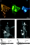
A, neurochemical code of mechanosensitive neurons. The image on the left illustrates a Di-8-ANEPPS-stained ganglion. The orange dots mark the neurons which responded to intraganglionic volume injection. Orange dots with white circles mark neurons which are neither Calb nor NeuN immunoreactive. Orange dot with green circle marks a neuron which is Calb and NeuN immunoreactive and orange dot with blue circle marks a neuron which is NeuN immunoreactive. Calb and NeuN staining of the ganglion is shown in the central and right images, respectively. B, responses in DiI-traced circular muscle motor neuron and interneuron. The arrows mark the locations of the DiI-traced neurons. Images were taken after recordings with Di-8-ANEPPS which explains the background staining. The traces below show the response of the circular muscle motor neuron (left trace) and the interneuron (right trace) to intraganglionic volume injection. In both traces strong mechanical artifacts are evident at the beginning of the injections (marked by arrows). These are followed by 3 spikes in the motor neuron and 5 spikes in the interneuron. The signals have been filtered with a Butterworth filter (low pass 300 Hz, high pass 20 Hz). Bars below traces indicate onset and end of intraganglionic volume injection.

References

    1. Blackshaw LA, Brookes SJH, Grundy D, Schemann M. Sensory trasmission in the gastrointestinal tract. Neurogastroenterol Motil. 2007;19(suppl. 1):1–19. - PubMed
    1. Brookes S. Retrograde tracing of enteric neuronal pathways. Neurogastroenterol Motil. 2001;13:1–18. - PubMed
    1. Christensen AP, Corey DP. TRP channels in mechanosensation: direct or indirect activation? Nat Rev Neurosci. 2007;8:510–521. - PubMed
    1. Furness JB, Kunze WAA, Bertrand PP, Clerc N, Bornstein JC. Intrinsic primary afferent neurons of the intestine. Prog Neurobiol. 1998;54:1–18. - PubMed
    1. Gershon MD. The enteric nervous system: a second brain. Hosp Pract (Minneap) 1999;34:31–32. 35–38, 41–42. passim. - PubMed

Substances

LinkOut - more resources