. 2009 Aug 31;206(9):1983-94.
doi: 10.1084/jem.20090480. Epub 2009 Aug 24.

Self-RNA-antimicrobial peptide complexes activate human dendritic cells through TLR7 and TLR8

Affiliations

Self-RNA-antimicrobial peptide complexes activate human dendritic cells through TLR7 and TLR8

Dipyaman Ganguly et al. J Exp Med. .

Abstract

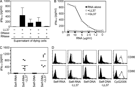
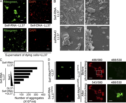
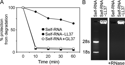
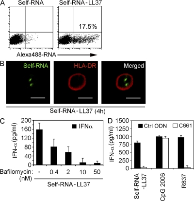
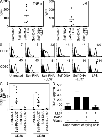
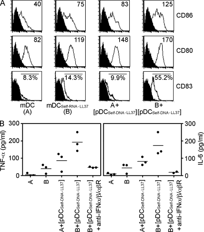
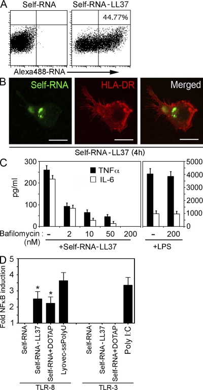
Dendritic cell (DC) responses to extracellular self-DNA and self-RNA are prevented by the endosomal seclusion of nucleic acid-recognizing Toll-like receptors (TLRs). In psoriasis, however, plasmacytoid DCs (pDCs) sense self-DNA that is transported to endosomal TLR9 upon forming a complex with the antimicrobial peptide LL37. Whether LL37 also interacts with extracellular self-RNA and how this may contribute to DC activation in psoriasis is not known. Here, we report that LL37 can bind self-RNA released by dying cells, protect it from extracellular degradation, and transport it into endosomal compartments of DCs. In pDC, self-RNA-LL37 complexes activate TLR7 and, like self-DNA-LL37 complexes, trigger the secretion of IFN-alpha without inducing maturation or the production of IL-6 and TNF-alpha. In contrast to self-DNA-LL37 complexes, self-RNA-LL37 complexes also trigger the activation of classical myeloid DCs (mDCs). This occurs through TLR8 and leads to the production of TNF-alpha and IL-6, and the differentiation of mDCs into mature DCs. We also found that self-RNA-LL37 complexes are present in psoriatic skin lesions and are associated with mature mDCs in vivo. Our results demonstrate that the cationic antimicrobial peptide LL37 converts self-RNA into a trigger of TLR7 and TLR8 in human DCs, and provide new insights into the mechanism that drives the auto-inflammatory responses in psoriasis.


Figures

Figure 1.
Figure 1.
LL37 converts self-RNA into a trigger of pDCs to produce IFN-α. (A) IFN-α produced by pDCs after stimulation with supernatants from dying (UV-irradiated) U937 cells, either alone or premixed with LL37. In some experiments, DNA and/or RNA was depleted from supernatants of dying cells by pretreatment with DNase I (2,000 U/ml) and/or RNase A (50 µg/ml). Data are the mean ± SEM of four independent experiments. (B) IFN-α produced by pDCs after stimulation with increasing concentrations of total human RNA purified from U937 cells (self-RNA), either alone (closed circles) or premixed with LL37 (self-RNA–LL37; open circles) or the scrambled peptide GL37 (self-RNA+GL37; open squares). (C) IFN-α produced by pDCs after stimulation with self-RNA or self-DNA (both at 5 µg/ml) alone or in complex with LL37 (self-RNA–LL37 and self-DNA–LL37). Each symbol represents an independent experiment; horizontal bars represent the mean. (D) Flow cytometric analysis of stimulated pDCs for CD80 and CD86 surface expression. CpG-2006 (1 µM) was used as a control to induce pDC maturation. Data are representative of at least three independent experiments.
Figure 2.
Figure 2.
LL37 binds self-RNA and forms aggregated particles. (A) Confocal microscopy images of self-RNA–LL37 and self-DNA–LL37 complexes generated in vitro (top panels) or formed by mixing supernatant of dying U937 cells with LL37 (bottom panels) and stained with Ribogreen (which stains both RNA and DNA) and DAPI (which stains DNA exclusively). RNA and DNA complexes were detected as Ribogreen+/DAPI and Ribogreen+/DAPI+ complexes, respectively. Bar, 20 µm. (B) Scanning electron microscopy of self-RNA–LL37 complexes formed by self-RNA fragments (top panels) or the short ssRNA sequence ssRNA40 (bottom panels). Bars: (left panels) 10 µm; (right panels) 1 µm. (C) Number of self-RNA–LL37 complexes counted as visible precipitates by phase-contrast microscopy. Increasing concentrations of NaCl (10, 50, 100, 200, 500, and 1,000 mM) were used to dissolve the complexes. Data are representative of three independent experiments. (D) Confocal microscopy of self-RNAAlexa488–LL37 (top left panel) and self-RNA–LL37Tamra (bottom left panel), or self-RNAAlexa488–LL37Tamra complexes (right panels). Self-RNAAlexa488–LL37Tamra complexes did not appear in the green channel after excitation at 488 nm (top right quadrant) but did appear in the red channel (top left quadrant), suggesting FRET. Photobleaching of the red fluorochrome TAMRA with the 543 laser (bottom left quadrant) resulted in recovery of green fluorescence of the Alexa Fluor 488 fluorochrome in response to excitation at 488 nm (bottom right quadrant). Numbers on the top of the right panels indicate excitation/emission wavelength. Bars: (left panels) 20 µm; (right panels) 10 µm.
Figure 3.
Figure 3.
Self-RNA–LL37 complexes are protected from enzymatic degradation. (A) Self-RNA alone, self-RNA–LL37 complexes, or self-RNA mixed with the control peptide GL37 were treated with RNase A and quantified over 60 min by a fluorometric assay. The percentage of protection was calculated as the ratio of remaining RNA over the initial RNA input. Data are representative of at least three experiments. (B) Agarose gel electrophoresis of self-RNA alone and self-RNA–LL37 complexes before and after RNase A treatment visualized by ethidium bromide staining.
Figure 4.
Figure 4.
LL37 transports self-RNA into pDCs to trigger endosomal TLR-7. (A) pDCs were stimulated for 4 h with self-RNAAlexa488 alone or self-RNAAlexa488–LL37 and analyzed by flow cytometry for self-RNAAlexa488–containing pDCs. (B) Confocal microscopy of pDCs stimulated for 4 h with self-RNAAlexa488–LL37 complexes and stained with Alexa 647–labeled anti–HLA-DR antibody to visualize the contour of the cell. (C) IFN-α produced by pDCs stimulated with self-RNA–LL37 complexes after pretreatment with increasing concentrations of bafilomycin. (D) IFN-α produced by pDCs after stimulation with self-RNA–LL37, CpG-2006 (1 µM), or R837 (10 µg/ml) after pretreatment with 1 µM TLR7 inhibitor C661 or a control oligonucleotide (ctrl-ODN). Data in A–D are representative of at least three independent experiments; error bars in C and D represent the SD of triplicate wells.
Figure 5.
Figure 5.
Self-RNA but not self-DNA in complex with LL37 activates mDCs to secrete TNF-α and IL-6 and undergo maturation. Immature monocyte-derived mDCs were stimulated with self-RNA, self-DNA, self-RNA–LL37, or self-DNA–LL37. (A) TNF-α and IL-6 secretion was measured after overnight culture. Each symbol represents an independent experiment, and horizontal bars represent the mean. (B) Flow cytometry for surface expression of CD86 and CD80 on stimulated mDCs. LPS was used as a control to induce mDC maturation. (C) Mean fluorescence intensity (MFI) changes for CD86 and CD80 expression on mDCs in response to stimulation with self-RNA–LL37 complexes. Each symbol represents an independent experiment, and horizontal bars represent the mean. *, P < 0.0005; paired Student's t test. (D) TNF-α production by mDCs stimulated with supernatants of dying U937 cells, either alone or premixed with LL37. In some experiments, DNA and/or RNA was depleted from supernatants of dying cells by pretreatment with DNase I and/or RNase A. Data indicate the mean ± SEM of three independent experiments.
Figure 6.
Figure 6.
IFN-α induced by concomitant pDC activation enhances maturation and cytokine production by mDCs. (A) Flow cytometry for CD86, CD80, and CD83 surface expression. Data are representative of at least three experiments. (B) TNF-α and IL-6 secretion measured by ELISA. Each symbol represents an independent experiment, and horizontal bars represent the mean.
Figure 7.
Figure 7.
LL37 transports self-RNA into mDCs and activates endosomal TLR-8. (A) mDCs were stimulated for 4 h with self-RNAAlexa488 alone or self-RNAAlexa488–LL37 and analyzed by flow cytometry for self-RNAAlexa488–containing mDCs. (B) Confocal microscopy of mDCs stimulated for 4 h with self-RNAAlexa488–LL37 complexes and stained with Alexa 647–labeled anti–HLA-DR antibody to visualize the contour of the cell. Data in A and B are representative of at least five independent experiments. (C) TNF-α and IL-6 secretion by mDCs stimulated with self-RNA–LL37 complexes after pretreatment with increasing concentrations of bafilomycin. Stimulation of the mDCs with LPS was used as negative control. Data are representative of three independent experiments. (D) NF-κB promoter activity of TLR8- or TLR3-transfected HEK293 cells measured by luciferase reporter assay after stimulation at the indicated conditions. Data in D are the mean ± SEM of five independent experiments. *, P < 0.05; paired Student's t test.
Figure 8.
Figure 8.
Extracellular RNA complexes are present in psoriatic skin and associated with mDC activation. (A) Confocal microscopy image of a representative psoriatic skin lesion stained with DAPI (red) and Ribogreen (green). DNA in cell nuclei appears yellow (Ribogreen+/DAPI+), and arrowheads indicate extracellular RNA aggregates appearing green (Ribogreen+/DAPI). Bar, 50 µm. (B) Numbers of visible extracellular RNA aggregates in the dermal compartment of healthy skin (n = 9), psoriatic skin (n = 11), and atopic dermatitis (n = 10). **, P = 0.0295; *, P = 0.042; unpaired Student's t test (both two-tailed). (C–F) High magnification images of extracellular RNA aggregates in the dermal compartment of psoriatic skin lesions. Bar, 10 µm. (E and F) SFP projection of panel D. (G-I) Representative confocal microscopy images of in vitro–generated self-RNA–LL37 complexes (G), and tissue self-RNA–LL37 aggregates stained with Ribogreen (green) and an anti-LL37 antibody (red) (H-I). Bars, (G) 10 µm; (H and I) 2 µm. (J) SFP projection of panel I. (K-N) Confocal microscopy images of DC-LAMP+ mature mDCs (red) in the dermal compartment of a representative psoriatic skin lesion co-stained with DAPI (blue) and Ribogreen (green). The images depict DC-LAMP (K), DAPI (L), and the merged image of DAPI and Ribogreen (M). (N) Merged image showing the colocalization of RNA complexes (Ribogreen+/DAPI; green) with DC-LAMP–positive organelles (red) appearing yellow (N). Bar, 10 µm. (O) Immunohistochemical results for DC-LAMP in a representative psoriatic skin lesion. Bar, 50 µm. (P) Correlation between the numbers of DC-LAMP+ mDCs and the numbers of visible extracellular RNA aggregates in multiple psoriatic skin lesions (n = 11). Pearson R = 0.615, two-tailed P = 0.044. SFP, simulated fluorescence projection.

References

    1. Agerberth B., Grunewald J., Castaños-Velez E., Olsson B., Jörnvall H., Wigzell H., Eklund A., Gudmundsson G.H. 1999. Antibacterial components in bronchoalveolar lavage fluid from healthy individuals and sarcoidosis patients.Am. J. Respir. Crit. Care Med. 160:283–290 - PubMed
    1. Akira S., Uematsu S., Takeuchi O. 2006. Pathogen recognition and innate immunity.Cell. 124:783–801 - PubMed
    1. Albanesi C., Scarponi C., Pallotta S., Daniele R., Bosisio D., Madonna S., Fortugno P., Gonzalvo-Feo S., Franssen J.D., Parmentier M., et al. 2009. Chemerin expression marks early psoriatic skin lesions and correlates with plasmacytoid dendritic cell recruitment.J. Exp. Med. 206:249–258 - PMC - PubMed
    1. Alexopoulou L., Holt A.C., Medzhitov R., Flavell R.A. 2001. Recognition of double-stranded RNA and activation of NF-kappaB by Toll-like receptor 3.Nature. 413:732–738 - PubMed
    1. Barrat F.J., Meeker T., Gregorio J., Chan J.H., Uematsu S., Akira S., Chang B., Duramad O., Coffman R.L. 2005. Nucleic acids of mammalian origin can act as endogenous ligands for Toll-like receptors and may promote systemic lupus erythematosus.J. Exp. Med. 202:1131–1139 - PMC - PubMed

Publication types

MeSH terms