. 2009 Aug 31;206(9):2027-35.
doi: 10.1084/jem.20090560. Epub 2009 Aug 24.

Aryl hydrocarbon receptor in combination with Stat1 regulates LPS-induced inflammatory responses

Affiliations

Aryl hydrocarbon receptor in combination with Stat1 regulates LPS-induced inflammatory responses

Akihiro Kimura et al. J Exp Med. .

Abstract

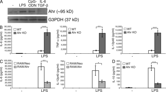
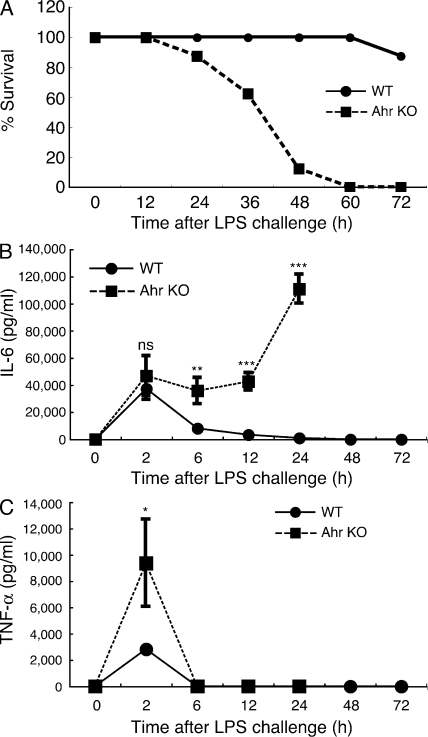
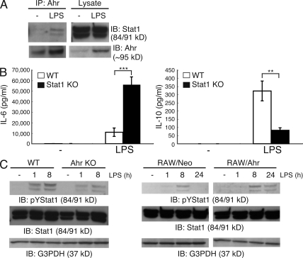
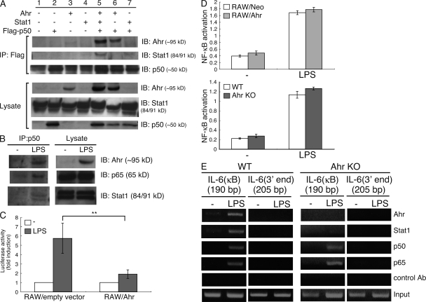
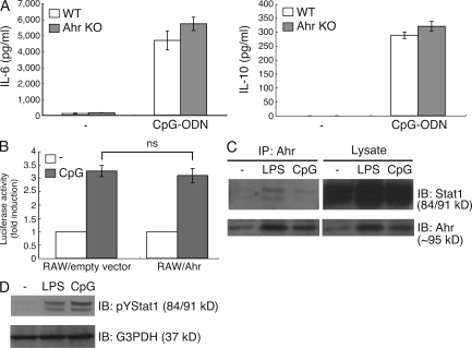
Toll-like receptor (TLR) signals perform a crucial role in innate immune responses to pathogens. In this study, we found that the aryl hydrocarbon receptor (Ahr) negatively regulates inflammatory responses mediated by lipopolysaccharide (LPS) in macrophages. Ahr was induced in macrophages stimulated by LPS, but not by transforming growth factor (TGF)-beta plus interleukin (IL)-6, which can induce Ahr in naive T cells. The production of IL-6 and tumor necrosis factor (TNF)-alpha by LPS was significantly elevated in Ahr-deficient macrophages compared with that in wild-type (WT) cells. Ahr-deficient mice were more highly sensitive to LPS-induced lethal shock than WT mice. Signal transducer and activator of transcription 1 (Stat1) deficiency, as well as Ahr deficiency, augmented LPS-induced IL-6 production. We found that Ahr forms a complex with Stat1 and nuclear factor-kappa B (NF-kappaB) in macrophages stimulated by LPS, which leads to inhibition of the promoter activity of IL-6. Ahr thus plays an essential role in the negative regulation of the LPS signaling pathway through interaction with Stat1.


Figures

Figure 1.
Figure 1.
Ahr deficiency augments LPS-induced proinflammatory responses in macrophages. (A) Peritoneal macrophages were stimulated with LPS, CpG-ODN, and TGF-β plus IL-6 for 24 h. The cells were lysed and subjected to immunoblotting (IB) analysis for the expression of Ahr and G3PDH. Data are from one representative of three independent experiments. (B–D) WT and Ahr KO peritoneal macrophages or RAW/Neo and RAW/Ahr cells were stimulated with LPS. Supernatants were collected 24 h after stimulation, and the production of IL-6, TNF-α, IL-12p40, and IL-10 were measured by means of ELISA. Data show means ± SEM of three independent experiments (*, P < 0.05; **, P < 0.005; ***, P < 0.001).
Figure 2.
Figure 2.
Hypersensitivity of Ahr KO mice to LPS in vivo. 6-wk-old Ahr KO mice and littermate WT mice (n = 10 for each) were i.p. injected with 7.5 mg/kg of LPS. (A) Lethality was observed over 60 h after LPS challenge. Data are representative of two independent experiments. (B and C) Serum levels of IL-6 and TNF-α between WT and Ahr KO mice were measured by ELISA at indicated time points after LPS challenge. Data show means ± SEM of three independent experiments (*, P < 0.05; **, P < 0.01; ***, P < 0.001).
Figure 3.
Figure 3.
Association between Ahr and Stat1 in macrophages. (A) Peritoneal macrophages were isolated from BALB/c mice and stimulated by LPS for 24 h. Interaction between Ahr and Stat1 was examined by means of immunoprecipitation (IP) and Western blotting. Data are from one representative of three independent experiments. IB, immunoblot. (B) WT and Stat1 KO peritoneal macrophages were stimulated with LPS. Supernatants were collected 24 h after stimulation, and the production of IL-6 and IL-10 were measured by means of ELISA. Data show means ± SEM of three independent experiments (**, P < 0.005; ***, P < 0.001). (C) WT and Ahr KO peritoneal macrophages or RAW/Neo and RAW/Ahr cells were incubated with LPS at the indicated time points. Whole-cell lysates were used for immunoblotting analysis with anti–phospho-tyrosine Stat1, Stat1, and G3PDH antibodies (Ab). Data are from one representative of three independent experiments.
Figure 4.
Figure 4.
Ahr inhibits LPS-induced NF-κB transcriptional activity together with Stat1. (A) COS7 cells were cotransfected with Ahr, Stat1, and p50-Flag. After 24 h, the cells were lysed and immunoprecipitated with anti-Flag Ab, followed by detection of Ahr, Stat1, and p50 by means of Western blotting. Data are from one representative of three independent experiments. IP, immunoprecipitation; IB, immunoblot. (B) Peritoneal macrophages were stimulated with LPS for 24 h. Whole-cell lysates were immunoprecipitated with anti-p50 antibody, after which Ahr, p65, and Stat1 were detected with Western blotting. Data are from one representative of three independent experiments. (C) RAW cells were transiently cotransfected with luciferase reporter gene construct of the murine IL-6 promoter and an expression vector for Ahr (RAW/Ahr) or empty control expression vector (RAW/empty vector). 6 h after transfection, cells were stimulated with LPS for 12 h. Luciferase assay and quantitation were performed as described in Materials and methods. Data show means ± SEM of three independent experiments (**, P < 0.02). (D) RAW/Neo and RAW/Ahr or WT and Ahr KO peritoneal macrophages were stimulated with LPS for 24 h NF-κB binding activity was examined using TransAM assay. Data show means ± SEM of three independent experiments. (E) Peritoneal macrophages from WT and Ahr KO mice were stimulated with LPS for 4 h, and the ChIP assay was performed using anti-p50, anti-p65, anti-Stat1, and anti-Ahr antibodies. Purified DNA fragments were amplified using primers specific for the IL-6 promoter. Data are from one representative of three independent experiments.
Figure 5.
Figure 5.
Ahr has no influence in CpG-ODN signaling pathway. (A) WT and Ahr KO peritoneal macrophages were stimulated with CpG-ODN for 24 h. The production of IL-6 and IL-10 were measured by means of ELISA. Data show means ± SE of three independent experiments. (B) RAW cells were transiently cotransfected with luciferase reporter gene construct of the murine IL-6 promoter and an expression vector for Ahr (RAW/Ahr) or empty control expression vector (RAW/empty vector). 6 h after transfection, cells were stimulated with CpG-ODN for 12 h. Luciferase assay and quantitation were performed as described in Materials and methods. Data show means ± SEM of three independent experiments. Peritoneal macrophages were stimulated with LPS and CpG-ODN. (C) Cells were lysed, immunoprecipitated by Ahr, and analyzed by Western blotting with anti-Stat1 Ab. IP, immunoprecipitation; IB, immunoblot. (D) Whole-cell lysates subjected to Western analysis with anti-pYStat1 antibodies. Data are from one representative of three independent experiments.

References

    1. Akira S., Takeda K. 2004. Toll-like receptor signalling.Nat. Rev. Immunol. 4:499–511 - PubMed
    1. Basu S., Dunn A.R., Marino M.W., Savoia H., Hodgson G., Lieschke G.J., Cebon J. 1997. Increased tolerance to endotoxin by granulocyte-macrophage colony-stimulating factor-deficient mice.J. Immunol. 159:1412–1417 - PubMed
    1. Bettelli E., Carrier Y., Gao W., Korn T., Strom T.B., Oukka M., Weiner H.L., Kuchroo V.K. 2006. Reciprocal developmental pathways for the generation of pathogenic effector TH17 and regulatory T cells.Nature. 441:235–238 - PubMed
    1. Beutler B., Rietschel E.T. 2003. Innate immune sensing and its roots: the story of endotoxin.Nat. Rev. Immunol. 3:169–176 - PubMed
    1. Bonnesen C., Eggleston I.M., Hayes J.D. 2001. Dietary indoles and isothiocyanates that are generated from cruciferous vegetables can both stimulate apoptosis and confer protection against DNA damage in human colon cell lines.Cancer Res. 61:6120–6130 - PubMed

Publication types

MeSH terms