. 2009 Nov;29(22):6074-85.
doi: 10.1128/MCB.00924-09. Epub 2009 Aug 24.

Global analysis of H3K4 methylation defines MLL family member targets and points to a role for MLL1-mediated H3K4 methylation in the regulation of transcriptional initiation by RNA polymerase II

Affiliations

Global analysis of H3K4 methylation defines MLL family member targets and points to a role for MLL1-mediated H3K4 methylation in the regulation of transcriptional initiation by RNA polymerase II

Pengfei Wang et al. Mol Cell Biol. 2009 Nov.

Abstract

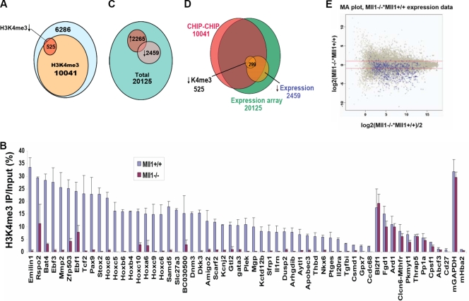
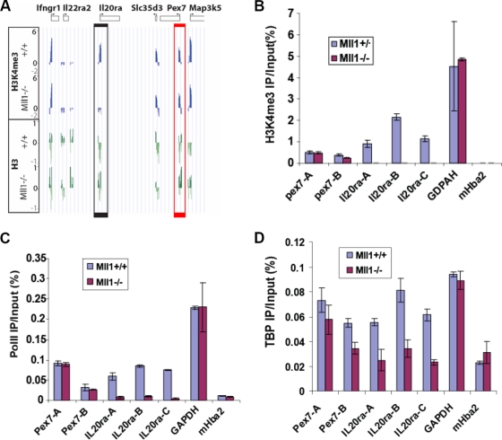
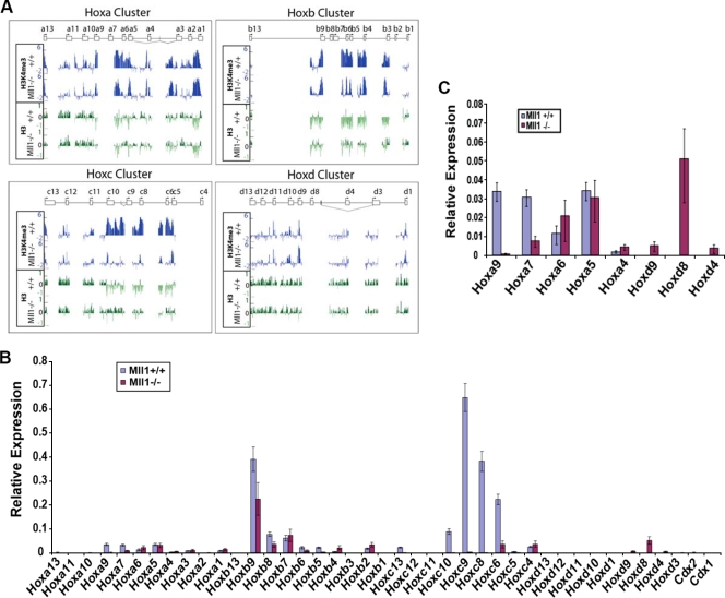
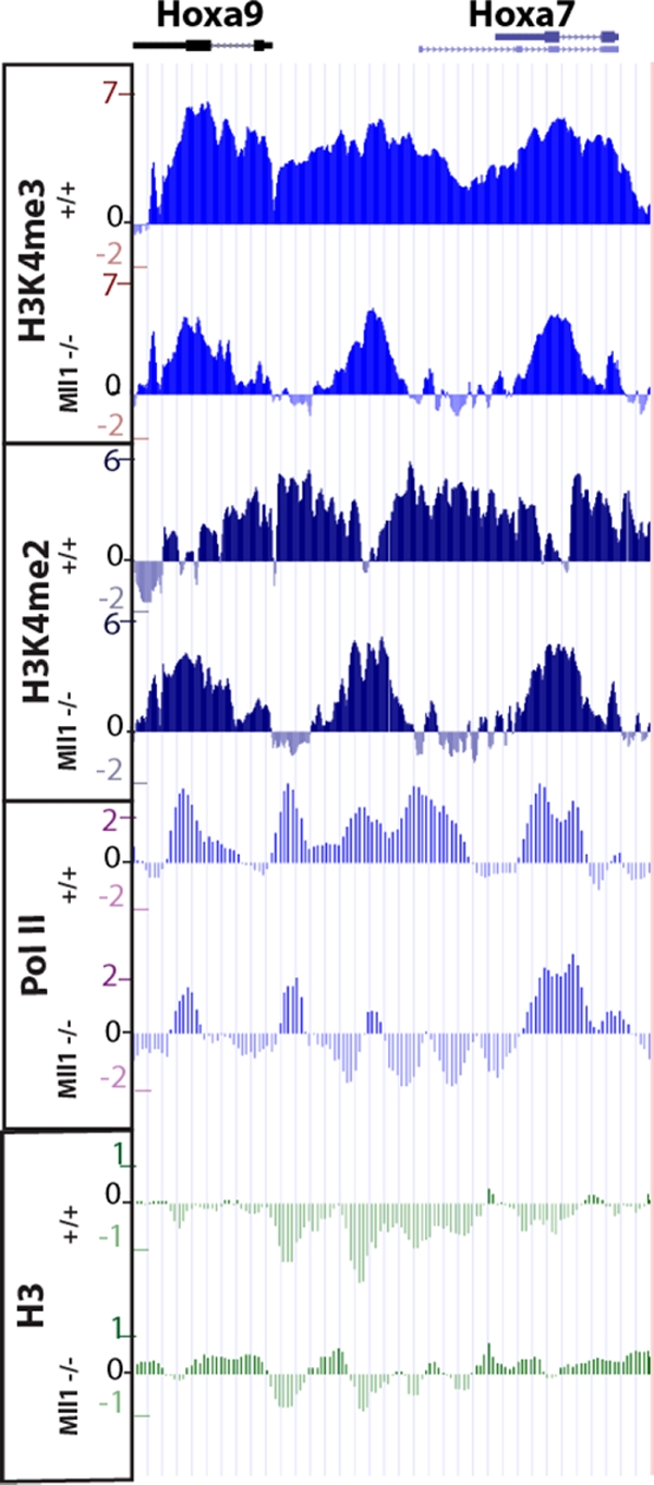
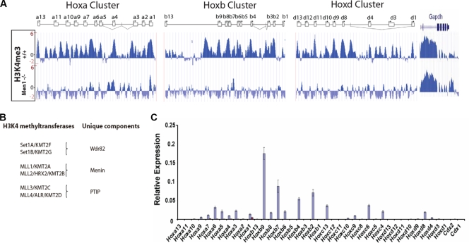
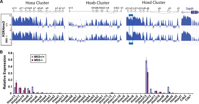
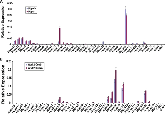
A common landmark of activated genes is the presence of trimethylation on lysine 4 of histone H3 (H3K4) at promoter regions. Set1/COMPASS was the founding member and is the only H3K4 methylase in Saccharomyces cerevisiae; however, in mammals, at least six H3K4 methylases, Set1A and Set1B and MLL1 to MLL4, are found in COMPASS-like complexes capable of methylating H3K4. To gain further insight into the different roles and functional targets for the H3K4 methylases, we have undertaken a genome-wide analysis of H3K4 methylation patterns in wild-type Mll1(+/+) and Mll1(-)(/)(-) mouse embryonic fibroblasts (MEFs). We found that Mll1 is required for the H3K4 trimethylation of less than 5% of promoters carrying this modification. Many of these genes, which include developmental regulators such as Hox genes, show decreased levels of RNA polymerase II recruitment and expression concomitant with the loss of H3K4 methylation. Although Mll1 is only required for the methylation of a subset of Hox genes, menin, a component of the Mll1 and Mll2 complexes, is required for the overwhelming majority of H3K4 methylation at Hox loci. However, the loss of MLL3/MLL4 and/or the Set1 complexes has little to no effect on the H3K4 methylation of Hox loci or their expression levels in these MEFs. Together these data provide insight into the redundancy and specialization of COMPASS-like complexes in mammals and provide evidence for a possible role for Mll1-mediated H3K4 methylation in the regulation of transcriptional initiation.


Figures

FIG. 1.
FIG. 1.
Identification of genes requiring Mll1 for H3K4 methylation and gene expression. (A) Numbers of promoters with changes in H3K4me3 levels as assessed by ChIP-chip with H3K4me3 antibodies in Mll1+/+ and Mll1/ MEFs. Ten thousand forty-one out of 16,852 gene promoters had detectable peaks of H3K4me3 in wild-type MEFs. Only 525 (5%) of these genes showed significant loss of H3K4me3 in the absence of Mll1. (B) ChIP-qPCR analysis confirmed the H3K4me3 changes on 45 of the genes found by ChIP-chip analysis to be most changed for H3K4me3, as well as the ChIP-chip results for 10 unchanged genes. The Hemoglobin gene, inactive in fibroblasts (Hba2), was used as a negative control for H3K4me3, and the housekeeping gene Gapdh was used as a positive control for the presence of H3K4me3. Error bars show standard deviations. (C) Gene expression analysis of 20,125 genes was performed with RNA isolated from Mll1+/+ and Mll1/ fibroblasts. The expression of 2,265 genes is upregulated and that of 2,459 genes is downregulated in the absence of Mll1. (D) Overlap of H3K4me3 and gene expression. About 299 of the 525 genes with loss of H3K4me3 also have decreased expression in the absence of Mll1. (E) Microarray (MA) plot of the gene expression data, with the 525 genes with significantly reduced H3K4 methylation highlighted in blue. The y axis represents the intensity ratio of mutant over wild type, and the x axis represents the average intensity of each spot. The red lines indicate a twofold up- or downregulation of expression. Most of the genes dependent on Mll1 for H3K4me3 show reduced expression.
FIG. 2.
FIG. 2.
Mll1 is required for the recruitment of the basal transcription machinery to chromatin. (A) The H3K4me3 level in the absence of Mll1 as determined by ChIP-chip analysis is dramatically decreased at the Il20ra promoter region (black box) but not at the neighboring Pex7 gene (red box). Enrichment profiles for H3K4me3 and total H3 expressed as log2 ChIP/input. (B) ChIP-qPCR with H3K4me3 antibodies was used to validate the results shown in panel A, using three primer sets spanning the transcription start site. (C and D) The results of ChIP-qPCR with Pol II antibodies (C) or TBP antibodies (D) show that RNA polymerase and TBP recruitment to Il20ra are significantly decreased in the absence of Mll1. Pol II and TBP recruitment on the Pex7 and Gapdh genes, however, did not change in the absence of Mll1. The murine Hba2 gene (mHba2), which is not transcribed in these cells, is used as a negative control. Error bars show standard deviations.
FIG. 3.
FIG. 3.
Mll1 is required for the H3K4 methylation and transcription of a subset of Hox genes. (A) Distribution of H3K4me3 and total H3 across 8-kb regions of each Hox gene promoter (5.5 kb before the transcription start site and 2.5 kb after the transcription start site) on the Agilent promoter array as determined by ChIP. Each individual Hox gene is represented as a box with an arrow indicating the major transcription start site and the direction of transcription. Vertical lines represent small alternative exons, and diagonal lines represent alternative splicing events. Reductions in H3K4me3 can be seen at several locations across the Hoxa, Hoxb, and Hoxc clusters. In contrast, Hoxd genes show little loss of H3K4me3 in Mll1/ cells. (B) qRT-PCR analysis of the expression of Hox genes in Mll1+/+ and Mll1/ fibroblasts normalized to the average value of reference genes Actb and Tbp. (C) Rescaling of the RT-PCR data presented in panel B. Shown are selected Hoxa and Hoxd genes, both of which have relatively low levels of expression in these fibroblasts. Some Hoxd genes that are not expressed in wild-type MEFs are expressed in the Mll1/ MEFs. Error bars show standard deviations.
FIG. 4.
FIG. 4.
H3K4 methylation is broadly lost at some coding and intergenic regions of Hox genes in the absence of Mll1. H3K4me3, H3K4me2, RNA Pol II, and total H3 profiles were determined across the Hoxa, Hoxb, and Hoxd loci by using a custom Agilent tiling array in the presence and absence of Mll1. Blue boxes indicate regions showing large reductions in both H3K4me3 and Pol II occupancy in the absence of Mll1. Green boxes indicate regions with unchanged H3K4 methylation and unchanged RNA Pol II levels. The orange box indicates a region in the Hoxd locus which shows little change in the H3K4me3 level but notable increases in H3K4me2 and Pol II occupancy in the Mll1/ cells. Red boxes indicate three regions of interest that correspond to the location of putative alternative promoters and enhancers regulated by Mll1. Also shown is the Gapdh gene, which is a control region on the Hox tiling array. Schematics for the Hox clusters are described in the Fig. 3A legend.
FIG. 5.
FIG. 5.
Enlargement of the results for the Hoxa9-Hoxa7 region shown in Fig. 4. Some regions showing loss of H3K4me3 in the absence of Mll1 still have residual peaks of this modification. In the region in between the H3K4me3 peaks, the H3K4me2 pattern “inverts” in the mutant, with valleys of H3K4me2 in the wild-type now showing peaks of this modification. Accordingly, peaks of H3K4me2 in the wild type decrease in the mutant. Together, these results suggest some redundancy or interplay among different H3K4 methyltransferases at the Hox loci.
FIG. 6.
FIG. 6.
Menin, a common component of Mll1 and Mll2 complexes, is required for the expression of nearly all Hox genes and for H3K4 trimethylation at Hox loci. (A) H3K4me3 ChIP profiles from Men1+/+ and Men1/ fibroblasts show that the vast majority of H3K4 methylation at Hox loci depends on menin. Schematics for the Hox clusters are described in the Fig. 3A legend. (B) Schematic of the association of subunits unique to Mll1/Mll2, Mll3/Mll4, and Set1A/Set1B complexes. Menin is found in Mll1/Mll2 but not other H3K4 methyltransferase complexes, PTIP is unique to Mll3/Mll4 but not other H3K4 methyltransferase complexes, and WDR82 is unique to the Set1A/Set1B but not the Mll1-to-Mll4 complexes. Alternate names for MLL2 and MLL4 are included to avoid confusion over alternative naming systems. (C) qRT-PCR analysis of Hox gene expression shows near total loss of Hox gene expression in Men1/ fibroblasts. Error bars show standard deviations.
FIG. 7.
FIG. 7.
Mll3 is not a major H3K4 histone methyltransferase at Hox loci in fibroblasts. (A) H3K4me3 ChIP profiles from Mll3+/+ and Mll3/ fibroblasts show relatively minor effects on the Hox gene H3K4me3 pattern by Mll3. A blue box outlines the results for the Hoxd12 gene, where a significant reduction in H3K4me3 can be observed in Mll3−/− fibroblasts. Schematics for the Hox clusters are described in the Fig. 3A legend. (B) qRT-PCR analysis of Hox gene expression reveals minor effects of Mll3. Hoxd12 shows reductions in both H3K4me3 and gene expression in Mll3/ fibroblasts. Error bars show standard deviations.
FIG. 8.
FIG. 8.
qRT-PCR analysis of Hox gene expression in the presence and absence of H3K4 methylation regulators. (A) Results for RNA from PTIP+/+ and PTIP/ fibroblasts. (B) Results for RNA from wild-type fibroblasts treated with an siRNA to Wdr82 or a nontargeting control siRNA. Error bars show standard deviations.

References

    1. Allis, C. D., S. L. Berger, J. Cote, S. Dent, T. Jenuwien, T. Kouzarides, L. Pillus, D. Reinberg, Y. Shi, R. Shiekhattar, A. Shilatifard, J. Workman, and Y. Zhang. 2007. New nomenclature for chromatin-modifying enzymes. Cell 131:633-636. - PubMed
    1. Ayton, P. M., and M. L. Cleary. 2001. Molecular mechanisms of leukemogenesis mediated by MLL fusion proteins. Oncogene 20:5695-5707. - PubMed
    1. Bernstein, B. E., M. Kamal, K. Lindblad-Toh, S. Bekiranov, D. K. Bailey, D. J. Huebert, S. McMahon, E. K. Karlsson, E. J. Kulbokas III, T. R. Gingeras, S. L. Schreiber, and E. S. Lander. 2005. Genomic maps and comparative analysis of histone modifications in human and mouse. Cell 120:169-181. - PubMed
    1. Charité, J., W. de Graaff, D. Consten, M. J. Reijnen, J. Korving, and J. Deschamps. 1998. Transducing positional information to the Hox genes: critical interaction of cdx gene products with position-sensitive regulatory elements. Development 125:4349-4358. - PubMed
    1. Cho, Y. W., T. Hong, S. Hong, H. Guo, H. Yu, D. Kim, T. Guszczynski, G. R. Dressler, T. D. Copeland, M. Kalkum, and K. Ge. 2007. PTIP associates with MLL3- and MLL4-containing histone H3 lysine 4 methyltransferase complex. J. Biol. Chem. 282:20395-20406. - PMC - PubMed

Publication types