. 2009 Nov;29(21):5645-56.
doi: 10.1128/MCB.00711-09. Epub 2009 Aug 24.

Regulation of protein synthesis by ionizing radiation

Affiliations

Regulation of protein synthesis by ionizing radiation

Steve Braunstein et al. Mol Cell Biol. 2009 Nov.

Abstract

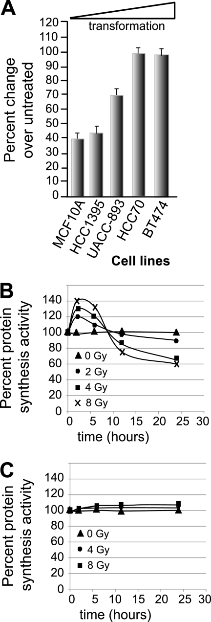
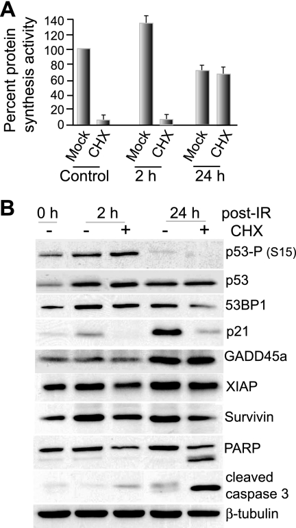
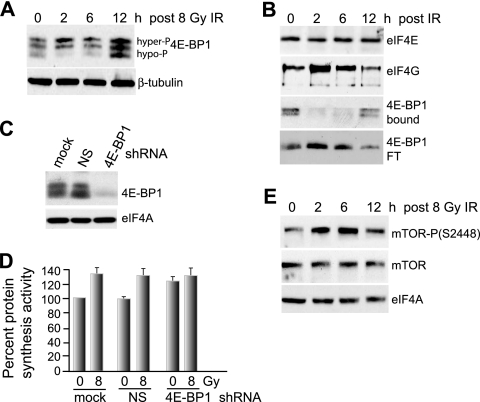
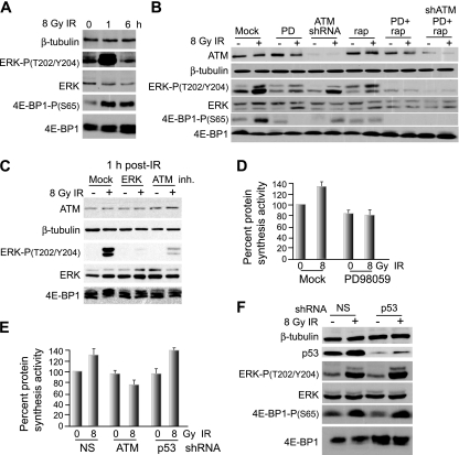
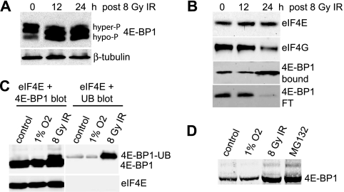
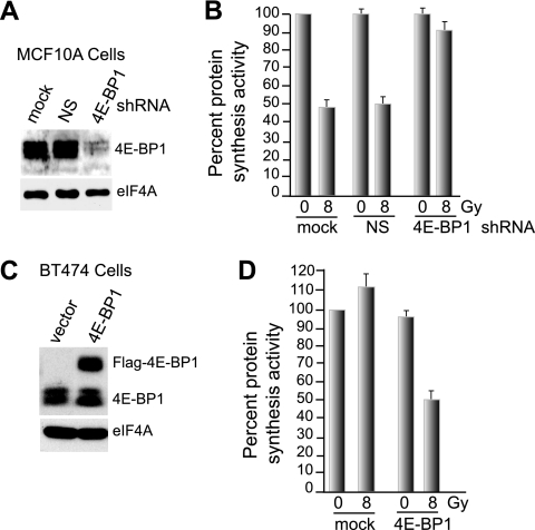
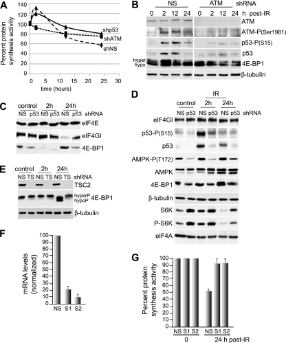
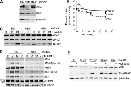
Ionizing radiation (IR) is a physiologically important stress to which cells respond by the activation of multiple signaling pathways. Using a panel of immortalized and transformed breast epithelial cell lines, we demonstrate that IR regulation of protein synthesis occurs in nontransformed cells and is lost with transformation. In nontransformed cells, IR rapidly activates the MAP kinases ERK1/2, resulting in an early transient increase in cap-dependent mRNA translation that involves mTOR and is radioprotective, enhancing the translation of a subset of mRNAs encoding proteins involved in DNA repair and cell survival. Following a transient increase in translation, IR-sensitive (nontransformed) cells inhibit cap-dependent protein synthesis through a mechanism that involves activation of p53, induction of Sestrin 1 and 2 genes, and stimulation of AMP kinase, inhibiting mTOR and hypophosphorylating 4E-BP1. IR is shown to block proteasome-mediated decay of 4E-BP1, increasing its abundance and the sequestration of eIF4E. The IR signal that impairs mTOR-dependent protein synthesis at late times is assembly of the DNA damage response machinery, consisting of Mre11, Rad50, and NBS1 (MRN); activation of the MRN complex kinase ATM; and p53. These results link genotoxic signaling from the DNA damage response complex to the control of protein synthesis.


Figures

FIG. 1.
FIG. 1.
IR differentially regulates protein synthesis in transformed and nontransformed cells. (A) Established cell lines with increasing transformation, from immortalized breast epithelial cells (MCF10A) to highly transformed breast cancer cells (BT474), were propagated under identical conditions and either left untreated or treated with 8 Gy IR (the highest tolerated dose for MCF10A cells), and protein-synthetic activity was measured 24 h later in the surviving fraction by [35S]methionine/cysteine incorporation and determination of protein specific activity. The data are presented as the ratio of treated to untreated specific activities of protein labeling plus standard errors of the mean. (B and C) Protein synthesis rates of MCF10A cells (B) and BT474 cells (C) with increasing IR doses. The rates were measured by [35S]methionine/cysteine incorporation and determination of protein specific activity. The data presented were derived from the mean of at least three independent experiments.
FIG. 2.
FIG. 2.
Effect of early-phase protein-synthetic activity on gene expression and viability. (A) MCF10A cells were left untreated (0 h) or treated with 8 Gy IR and then, at 2 or 24 h, treated with 10 μg/ml cycloheximide (CHX) or mock treated, and the protein-synthetic rates were determined by [35S]methionine incorporation for 1 h. (B) Immunoblot analysis of cellular lysates from cells treated in parallel with those in panel A. Equal amounts of total protein were resolved by 10% SDS-PAGE and identified by specific antibodies as indicated. The immunoblots are representative of typical results from at least three studies. All data presented were derived from the mean of at least three independent experiments, with standard errors of the mean shown.
FIG. 3.
FIG. 3.
Increased hyperphosphorylation of 4E-BP1 early after IR promotes eIF4F complex formation and protein synthesis. (A) 4E-BP1 was resolved by high-resolution SDS-15% PAGE using equal amounts of protein extracts obtained from MCF10A cells irradiated with a single fraction of 8 Gy IR and harvested at the indicated time points. Hyper- and hypophosphorylated forms of 4E-BP1 are indicated. (B) Cells irradiated at 8 Gy were harvested at the times shown, equal amounts of protein extracts were subjected to m7GTP-Sepharose cap chromatography, and retentates were recovered by elution with SDS, resolved by SDS-PAGE, and detected by immunoblot analysis with antibodies as indicated. The flowthrough fraction (FT) of 4E-BP1 that did not bind to m7GTP-Sepharose is shown. The data were quantified by densitometry of autoradiograms from at least three independent experiments. Representative results are shown. (C) MCF10A cells were stably transformed with lentivirus vectors expressing NS or 4E-BP1-silencing shRNAs, and equal amounts of protein lysates were subjected to immunoblot analysis with antibodies as shown. (D) MCF10A cells silenced as for panel C were subjected to 8 Gy irradiation, and protein synthesis rates were determined 2 h later by metabolic labeling with [35S]methionine/cysteine. (E) mTOR abundance and activating phosphorylation at Ser2448 were determined in MCF10A cells irradiated at 8 Gy at the times shown by immunoblot analysis using specific antibodies as shown. All results are representative of at least three independent experiments, with standard errors of the mean determined from all studies.
FIG. 4.
FIG. 4.
4E-BP1 is transiently phosphorylated at early times following IR treatment in a p53-independent, ATM-, ERK-, and mTOR-dependent manner. (A) Immunoblot analysis of total cell lysate from MCF10A cells treated with 8 Gy IR and harvested at the times shown. Equal amounts of total protein were resolved by SDS-12% PAGE and subjected to immunoblot analysis with protein- and phosphoprotein-specific antibodies as shown. (B) Immunoblot analysis of MCF10A cells at 1 h following 8 Gy IR, treated simultaneously with vehicle alone or the ERK inhibitor PD98059 (PD) at 20 μM or subjected to shRNA gene silencing for ATM alone or in combination with PD98059. Equal amounts of total protein (25 μg) were loaded for analysis on SDS-10% PAGE. Immunoblot analysis used protein- and phosphoprotein-specific antibodies as shown. rap, rapamycin. (C) Study conducted as for panel B but including high-resolution SDS-15% PAGE analysis of 4E-BP1 and use of the ATM inhibitor (inh.) KU55933 at 10 μM added 30 min prior to IR treatment and washed out 20 min following treatment. (D) Protein-synthetic rates in MCF10A cells treated with vehicle or the MEK inhibitor PD98059 at 20 μM for 2 h immediately following treatment with 8 Gy IR. Protein synthesis activity was determined by [35S]methionine/cysteine metabolic labeling of MCF10A cells, either untreated or treated with 8 Gy IR, with or without inhibition of ERK with PD98059. (E) Protein synthesis activity was determined as for panel D for MCF10A cells silenced for ATM or p53 or expressing NS shRNA from lentivirus vectors at 2 h following treatment with 8 Gy IR. (F) Immunoblot analysis of total cell lysate from MCF10A cells stably transformed with lentiviruses expressing NS or p53 shRNA. The lysates were reduced by SDS-10% PAGE and probed with protein- and phosphoprotein-specific antibodies as shown. Typical results of at least three independent experiments are shown for all studies. Standard errors of the mean (D and E) were calculated from the means of three independent experiments.
FIG. 5.
FIG. 5.
IR increases the stability of 4E-BP1 by preventing its ubiquitination and proteasome-mediated rapid decay. (A) 4E-BP1 was resolved by high-resolution SDS-15% PAGE using equal amounts of protein extract from MCF10A cells irradiated at 8 Gy and harvested at the times shown post-IR treatment. Proteins were detected by immunoblot analysis. hyper-P, hyperphosphorylated; hypo-P, hypophosphorylated. (B) MCF10A cells subjected to 8 Gy IR were harvested at the times shown, equal amounts of protein extract were subjected to m7GTP-Sepharose cap chromatography, and retentates were recovered by elution with SDS, resolved by SDS-10% PAGE, and detected by immunoblot analysis with antibodies as indicated. The flowthrough fraction (FT) of 4E-BP1 that did not bind to m7GTP-Sepharose is shown. Representative results are shown for three independent experiments. (C) MCF10A cells were transfected with a His-Myc-tagged pentameric ubiquitin (UB) expression vector. The cells were either untreated (control), hypoxia treated (24 h; 1% O2), or treated with 8 Gy IR, and equal amounts of protein extracts were resolved by SDS-PAGE and detected by immunoblotting for 4E-BP1 or ubiquitin and eIF4E. 4E-BP1 blots were overexposed to reveal the ubiquitinated 4E-BP1 protein. (D) MCF10A cells were treated as described for panel C, resolved by low-resolution SDS-8% PAGE, and detected by immunoblot analysis within the linear range of radiological imaging.
FIG. 6.
FIG. 6.
Increased abundance of 4E-BP1 is the primary means of protein synthesis inhibition in IR-treated cells. (A) MCF10A cells were stably transformed with lentivirus vectors expressing NS or 4E-BP1-silencing shRNAs, and equal amounts of protein lysates were subjected to immunoblot analysis with antibodies as shown. (B) MCF10A cells stably transformed with NS and 4E-BP1-silencing lentivirus vectors were subjected to 8 Gy IR, and protein synthesis activity in the cells was determined by [35S]methionine/cysteine labeling. Cells were harvested 24 h post-IR treatment. The results are the average of three independent experiments with standard errors of the mean shown. (C) A Flag-tagged 4E-BP1 protein was overexpressed in radioresistant BT474 cells transformed with a cDNA-expressing lentvirus vector. Proteins were detected by immunoblotting with antibodies to 4E-BP1. The decreased electrophoretic mobility of Flag-4E-BP1 is due to the additional Flag motif. (D) Vector control and 4E-BP1-overexpressing BT474 cells were subjected to 8 Gy IR, and protein synthesis rates were determined 24 h later as described for panel B. The data are the averages of three independent experiments, with standard errors of the mean shown.
FIG. 7.
FIG. 7.
Late-phase inhibition of protein synthesis by IR is dependent upon p53 and p53-induced Sestrin 1 and 2 proteins. (A) MCF10A cells were transformed with shRNA lentiviruses to ATM, p53, or NS as described in Materials and Methods and treated with 8 Gy IR, and protein-synthetic rates were measured over a 24-h period. (B) MCF10A cells were transformed with lentivirus vectors expressing shRNAs against ATM or NS, treated with 8 Gy IR, and harvested at 2 h, 12 h, or 24 h post-IR treatment. Equal amounts of protein from total cell lysates were analyzed by SDS-10% PAGE and subjected to immunoblotting with protein- and phosphoprotein-specific antibodies as shown. hyper, hyperphosphorylated; hypo, hypophosphorylated. (C) m7GTP cap chromatography and immunoblot analysis of NS and p53-silenced MCF10A cells treated with 8 Gy IR and harvested at the indicated times. (D) MCF10A cells were transformed with lentivirus vectors, silenced for p53, treated with 8 Gy IR, and analyzed by immunoblot analysis at 2 h and 24 h post-IR treatment. (E) NS or TSC2-silenced (TS) MCF10A cells were treated with 8 Gy IR, harvested at the indicated times, and analyzed by immunoblot analysis using equal amounts of protein from total cell lysates. (F) Sestrin 1 (S1) and 2 (S2) were silenced in MCF10A cells using shRNA-expressing lentivirus vectors. mRNA levels were determined by real-time qRT-PCR. (G) Sestrin-silenced MCF10A cells and control NS cells were subjected to 8 Gy IR, and protein synthesis rates were determined by metabolic labeling with [35S]methionine/cysteine. The results represent the average of three independent experiments, with standard errors of the mean shown.
FIG. 8.
FIG. 8.
Late-phase inhibition of protein synthesis is signaled by the MRN DNA DSB complex. (A) MCF10A cells were transduced with lentiviruses expressing shRNAs to ATM, NBS1, or NS. Immunoblot analysis was conducted with antibodies as shown. (B) MCF10A cells were transduced with shRNA lentiviruses to ATM, NBS1, or NS and treated with 8 Gy IR, and protein-synthetic rates were measured over a 24-h period by [35S]methionine/cysteine incorporation. (C) MCF10A cells silenced with shRNA-expressing lentiviruses as shown were subjected to 8 Gy IR and harvested at the times shown; equal amounts of protein extract were subjected to m7GTP-Sepharose cap chromatography, and retentates were recovered by elution with loading dye at elevated temperature, resolved by SDS-10% PAGE, and detected by immunoblot analysis with antibodies as indicated. (D) MCF10A cells were silenced with control NS or NBS1 shRNA lentivirus vectors, treated with 8 Gy IR, and analyzed by immunoblot analysis at the times shown. hyper, hyperphosphorylated; hypo, hypophosphorylated. (E) MCF10A cells were treated with the ATM inhibitor KU55933 at 10 μM added 30 min prior to treatment with 8 Gy IR and washed out 20 min following treatment and were analyzed by immunoblot analysis at the times shown post-IR treatment.

References

    1. Abedin, M. J., D. Wang, M. A. McDonnell, U. Lehmann, and A. Kelekar. 2007. Autophagy delays apoptotic death in breast cancer cells following DNA damage. Cell Death Differ. 14:500-510. - PubMed
    1. Albert, J. M., K. W. Kim, C. Cao, and B. Lu. 2006. Targeting the Akt/mammalian target of rapamycin pathway for radiosensitization of breast cancer. Mol. Cancer Ther. 5:1183-1189. - PubMed
    1. Bhandari, B. K., D. Feliers, S. Duraisamy, J. L. Stewart, A. C. Gingras, H. E. Abboud, G. G. Choudhury, N. Sonenberg, and B. S. Kasinath. 2001. Insulin regulation of protein translation repressor 4E-BP1, an eIF4E-binding protein, in renal epithelial cells. Kidney Int. 59:866-875. - PubMed
    1. Budanov, A. V., and M. Karin. 2008. p53 target genes sestrin1 and sestrin2 connect genotoxic stress and mTOR signaling. Cell 134:451-460. - PMC - PubMed
    1. Buttgereit, F., and M. D. Brand. 1995. A hierarchy of ATP-consuming processes in mammalian cells. Biochem. J. 312:163-167. - PMC - PubMed

Publication types

MeSH terms

Substances