. 2009 Aug 25:9:298.
doi: 10.1186/1471-2407-9-298.

Expression of the glioma-associated oncogene homolog (GLI) 1 in human breast cancer is associated with unfavourable overall survival

Affiliations

Expression of the glioma-associated oncogene homolog (GLI) 1 in human breast cancer is associated with unfavourable overall survival

Anette ten Haaf et al. BMC Cancer. .

Abstract

Background: The transcription factor GLI1, a member of the GLI subfamily of Krüppel-like zinc finger proteins is involved in signal transduction within the hedgehog pathway. Aberrant hedgehog signalling has been implicated in the development of different human tumour entities such as colon and lung cancer and increased GLI1 expression has been found in these tumour entities as well. In this study we questioned whether GLI1 expression might also be important in human breast cancer development. Furthermore we correlated GLI1 expression with histopathological and clinical data to evaluate whether GLI1 could represent a new prognostic marker in breast cancer treatment.

Methods: Applying semiquantitative realtime PCR analysis and immunohistochemistry (IHC) GLI1 expression was analysed in human invasive breast carcinomas (n = 229) in comparison to normal human breast tissues (n = 58). GLI1 mRNA expression was furthermore analysed in a set of normal (n = 3) and tumourous breast cell lines (n = 8). IHC data were statistically interpreted using SPSS version 14.0.

Results: Initial analysis of GLI1 mRNA expression in a small cohort of (n = 5) human matched normal and tumourous breast tissues showed first tendency towards GLI1 overexpression in human breast cancers. However only a small sample number was included into these analyses and values for GLI1 overexpression were statistically not significant (P = 0.251, two-tailed Mann-Whitney U-test). On protein level, nuclear GLI1 expression in breast cancer cells was clearly more abundant than in normal breast epithelial cells (P = 0.008, two-tailed Mann-Whitney U-test) and increased expression of GLI1 protein in breast tumours significantly correlated with unfavourable overall survival (P = 0.019), but also with higher tumour stage (P < 0.001) and an increased number of tumour-positive axillar lymph nodes (P = 0.027). Interestingly, a highly significant correlation was found between GLI1 expression and the expression of SHH, a central upstream molecule of the hedgehog pathway that was previously analysed on the same tissue microarray.

Conclusion: Our study presents a systematic expression analysis of GLI1 in human breast cancer. Elevated levels of GLI1 protein in human breast cancer are associated with unfavourable prognosis and progressive stages of disease. Thus GLI1 protein expression measured e.g. by an IHC based scoring system might have an implication in future multi-marker panels for human breast cancer prognosis or molecular sub typing. The highly significant correlation between SHH and GLI1 expression characterises GLI1 as a potential functional downstream target of the hedgehog signalling pathway in human breast cancer as well. Furthermore, our study indicates that altered hedgehog signalling may represent a key disease pathway in the progression of human breast cancer.


Figures

Figure 1
Figure 1
Elevated expression of GLI1 mRNA in breast cancer cell lines. Semiquantitative realtime PCR (Light Cycler) analysis for GLI1 expression was performed on reverse transcribed RNA extracted from non-malignant (grey bars) and malignant breast cell lines (black bars). Expression data were related to GLI1 mRNA expression in the human mammary epithelial cell line HMEC (set to 1). In five of eight analysed malignant mammary cell lines (SKBR3, MDA-MB468, BT20, MDA-MB231 and MDA-MB435s) increased levels of GLI1 mRNA expression were found. GLI1 mRNA expression in normal human epithelial mammary cell lines HMEC and MCF12A cells is relatively low, except for cell line MCF10A which also presents abundant GLI1 expression.
Figure 2
Figure 2
GLI1 expression in primary breast cancer. Realtime PCR analysis of GLI1 mRNA expression in a set of five matched pairs (N = normal vs. T = tumour) revealed increased levels of GLI1 expression in tumourous tissue for four of the five analysed matched pairs. Expression level of each sample is normalised to its GAPDH expression and related to normal breast tissue (N1) (set to 1).
Figure 3
Figure 3
Immunohistochemical expression analysis of GLI1 protein in normal breast tissue, non-invasive and invasive breast tumours using a TMA. (A, B) In normal breast tissue low levels of GLI1 expression are detectable in the cytoplasm (median IRS = 4) and in the nucleus (median IRS = 6). In ductal carcinomas in situ of high grade type (C, D) as well as in invasive breast carcinomas (E, F) nuclear GLI1 expression is more intense than in normal breast tissues (median IRS = 8), whereas the cytoplasmic GLI1 expression is weak (median IRS = 3). (G, H) Representative tissue section from human basal cell carcinoma which served as a positive control for GLI1 staining. (I, J) Negative control – benign breast tissue. Magnifications: A, C, E, G, I: 100×; B, D, F, H, J: 400×.
Figure 4
Figure 4
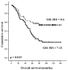
Breast cancer patients expressing nuclear GLI1 protein show unfavourable prognosis. Univariate Kaplan-Meier analysis was performed on the basis of GLI1 expression results derived from the TMA. Patients with low GLI1 expression (IRS = 0–6) displayed improved overall survival estimation (upper graph) compared to patients with abundant nuclear GLI1 expression (IRS 6–12; lower graph) (P = 0.019; univariate log-rank analysis).

References

    1. Bian YH, Huang SH, Yang L, Ma XL, Xie JW, Zhang HW. Sonic hedgehog-Gli1 pathway in colorectal adenocarcinomas. World J Gastroenterol. 2007;13:1659–1665. - PMC - PubMed
    1. Berman DM, Karhadkar SS, Maitra A, Montes De OR, Gerstenblith MR, Briggs K, Parker AR, Shimada Y, Eshleman JR, Watkins DN, Beachy PA. Widespread requirement for Hedgehog ligand stimulation in growth of digestive tract tumours. Nature. 2003;425:846–851. doi: 10.1038/nature01972. - DOI - PubMed
    1. Cowan R, Hoban P, Kelsey A, Birch JM, Gattamaneni R, Evans DG. The gene for the naevoid basal cell carcinoma syndrome acts as a tumour-suppressor gene in medulloblastoma. Br J Cancer. 1997;76:141–145. - PMC - PubMed
    1. Johnson RL, Rothman AL, Xie J, Goodrich LV, Bare JW, Bonifas JM, Quinn AG, Myers RM, Cox DR, Epstein EH Jr, Scott MP. Human homolog of patched, a candidate gene for the basal cell nevus syndrome. Science. 1996;272:1668–1671. doi: 10.1126/science.272.5268.1668. - DOI - PubMed
    1. Karhadkar SS, Bova GS, Abdallah N, Dhara S, Gardner D, Maitra A, Isaacs JT, Berman DM, Beachy PA. Hedgehog signalling in prostate regeneration, neoplasia and metastasis. Nature. 2004;431:707–712. doi: 10.1038/nature02962. - DOI - PubMed

Publication types