. 2009 Sep 1;106(35):14990-5.
doi: 10.1073/pnas.0812242106. Epub 2009 Aug 24.

Prevascularization of cardiac patch on the omentum improves its therapeutic outcome

Affiliations

Prevascularization of cardiac patch on the omentum improves its therapeutic outcome

Tal Dvir et al. Proc Natl Acad Sci U S A. .

Abstract

The recent progress made in the bioengineering of cardiac patches offers a new therapeutic modality for regenerating the myocardium after myocardial infarction (MI). We present here a strategy for the engineering of a cardiac patch with mature vasculature by heterotopic transplantation onto the omentum. The patch was constructed by seeding neonatal cardiac cells with a mixture of prosurvival and angiogenic factors into an alginate scaffold capable of factor binding and sustained release. After 48 h in culture, the patch was vascularized for 7 days on the omentum, then explanted and transplanted onto infarcted rat hearts, 7 days after MI induction. When evaluated 28 days later, the vascularized cardiac patch showed structural and electrical integration into host myocardium. Moreover, the vascularized patch induced thicker scars, prevented further dilatation of the chamber and ventricular dysfunction. Thus, our study provides evidence that grafting prevascularized cardiac patch into infarct can improve cardiac function after MI.


Conflict of interest statement

Conflict of interest statement: Y.E. and Mor Research Applications Ltd. have applied for a patent on the miniature bipolar hook electrode (International Patent Application No. PCT/IL2008/000161).

Figures

Fig. 1.
Fig. 1.
Construction of a cardiac patch in an alginate-sulfate/alginate scaffold capable of binding and releasing mixture factors. (A and B) The scaffold features before cell seeding; macroscopic view (A) and internal porosity by scanning electron microscopy (B). (C and D) Cardiac patch, seeded with 2.5 × 10⁶ cardiac cells and supplemented with factor mixture, after 48 h of cultivation. (C) Light microscope view of the cardiac patch, showing uniform distribution of cells in the matrix pores. (D) Cardiac cell organization within the scaffold, as judged by anti-actinin immunostaining (green) and nuclear staining (red). Some of the cells reveal the typical striation of cardiac tissue. [Scale bar: 200 μm (C); 10 μm (D).]
Fig. 2.
Fig. 2.
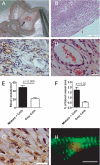
Vascularization of the 7 day omentum-transplanted, mixture-supplemented cardiac patch. (A) The cardiac patch (arrow) is stitched to the omentum. No contamination or inflammation were observed in any of the patches (n = 16). (B) H&E-stained cross-section from the omentum-generated cardiac patch supplemented with the prosurvival and angiogenic factors shows extensive tissue in-growth into the scaffold. Lower right is the patch edge. (C) Mature blood vessels populate the cardiac patch supplemented with mixture, as judged by anti-SMA immunostaining (brown). (D) The vessels are functional and anastomized with host vessels, as reflected in their red blood cell content. (E and F) Blood vessel density (E) and the area (F) (in %) occupied by the vessels in the omentum-implanted patches. The results represent mean values ± SEM. (n = 8 per group). Statistical evaluations were performed by unpaired Student's t tests, P < 0.05. (G) Anti-Tn-T immunostaining of thin section in the omentum-generated cardiac patch supplemented with mixture factors (brown). (H) Typical cardiac cell striation is revealed in an omentum-generated, mixture-supplemented cardiac patch, as revealed by anti-actinin immunostaining (green) and confocal microscopy. [Scale bar: 200 μm (B); 100 μm (C); 20 μm (D and G); 10 μm (H).]
Fig. 3.
Fig. 3.
Assessment of the scar zone, 28 days after patch grafting onto an infarcted heart. (A–C) Representative figures of Masson's trichrome-stained cross-sections in a stitched only scar (A), a scar implanted with an in vitro-grown patch (B), or a scar grafted with an omentum-generated cardiac patch (C). Collagen in the scar is stained blue and viable cardiac tissue is shown in red. (D) H&E staining of cross-sections in the interface (dashed black line) of the host myocardium (M) and grafted omentum-generated patch (P). (E) Typical cardiac cell striation could be observed by anti-Tn-T immunostaining (brown). (F) Cx-43 expression (brown) between adjacent cardiomyocytes in omentum-generated cardiac patch suggests mechanical coupling. (G and H) Morphometric analysis of scar area and calculation of relative scar thickness (G) and expansion index (H), [(LV cavity area/whole LV area)/relative scar thickness]. (I) Blood vessel density in the scar area was determined by counting anti-SMA-immunostained vessels. Results represent mean values ± SEM. (n = 4–6). Statistical evaluations were performed by unpaired Student's t tests, P < 0.05. [Scale bar: 500 μm (A–C); 200 μm (D); 20 μm (E and F).]
Fig. 4.
Fig. 4.
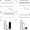
Electrical coupling of omentum-generated cardiac patch to host myocardium. Langendorff-perfused isolated hearts were blindly implanted with two bipolar epicardial electrodes located at the base of the healthy right ventricle myocardium (RV) and the scar zone as close as possible to the stitch (scar). (A and B) Nonpaced (spontaneous) electrical signals recorded from the healthy myocardium (RV) and scar zone (Scar) of hearts with an omentum-generated patch (A) or hearts with stitch only (B). (C and D) Pacing through the scar electrode using 2 ms square pulses (400 beats per min) in hearts treated with an omentum-generated patch (C) or stitched scar (D), at a stimulus intensity of 1.5 mA. This stimulus intensity could capture all hearts treated with omentum-generated patch, but none of the control hearts. (E) Comparison of signal amplitude in the scar zone after grafting an omentum-generated patch (black) or in stitched hearts (white). (F) Comparison of capture threshold intensity in hearts grafted with an omentum-generated patch (black) or only stitched (white).
Fig. 5.
Fig. 5.
Changes in left ventricle function after patch grafting. The FAC {[(LV end-diastolic area − LV end-systolic area)/LV end-diastolic area] ×100} of infarcted hearts treated with stitches only (A) (n = 7), hearts treated with in vitro-grown patches (B) (n = 6), treated with a-cellular omentum-grown scaffolds (C) (Om., n = 6), or treated with omentum-generated cardiac patches (D) (Om.+, n = 11), was determined by echocardiography. “Pre” indicates readings 6 days after MI induction by LAD ligation and 1 day before intervention. “Post” indicates readings taken 28 days after intervention. Comparison of FAC (E) change, LVEDD (F), and LVESD (G). Changes were calculated as follows: [(values obtained after 4 weeks – baseline values)/baseline values] × 100%. Statistical evaluations were performed by paired Student's t tests, P < 0.05 (A–D) or un-paired t test and one-way ANOVA, P < 0.05 (E–G).

References

    1. Pasumarthi KB, Field LJ. Cardiomyocyte cell cycle regulation. Circ Res. 2002;90:1044–1054. - PubMed
    1. Rubart M, Field LJ. Cardiac regeneration: Repopulating the heart. Ann Rev Physiol. 2006;68:29–49. - PubMed
    1. Assmus B, et al. Transplantation of progenitor cells and regeneration enhancement in acute myocardial infarction (TOPCARE-AMI) Circulation. 2002;106:3009–3017. - PubMed
    1. Schachinger V, et al. Transplantation of progenitor cells and regeneration enhancement in acute myocardial infarction: Final one-year results of the TOPCARE-AMI Trial. J Am Coll Cardiol. 2004;44:1690–1699. - PubMed
    1. Landa N, et al. Effect of injectable alginate implant on cardiac remodeling and function after recent and old infarcts in rat. Circulation. 2008;117(11):1388–1396. - PubMed

Publication types

MeSH terms