. 2009 Sep 8;106(36):15350-5.
doi: 10.1073/pnas.0907724106. Epub 2009 Aug 24.

Claudin-16 and claudin-19 interaction is required for their assembly into tight junctions and for renal reabsorption of magnesium

Affiliations

Claudin-16 and claudin-19 interaction is required for their assembly into tight junctions and for renal reabsorption of magnesium

Jianghui Hou et al. Proc Natl Acad Sci U S A. .

Abstract

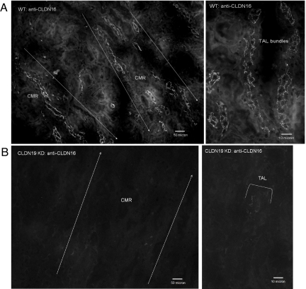
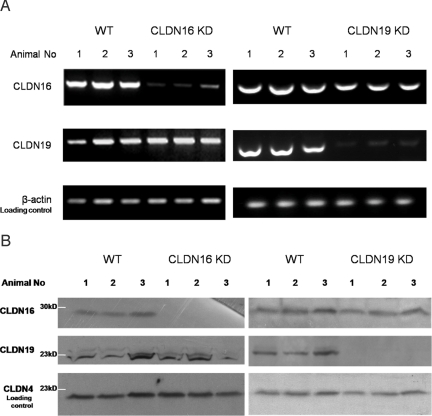
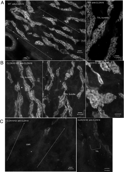
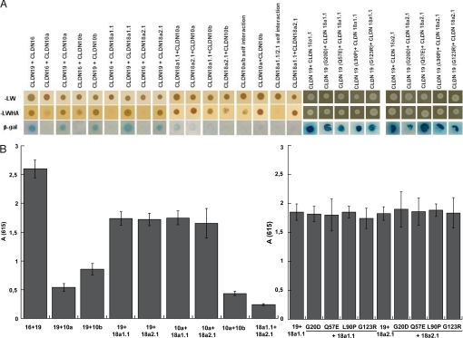
Claudins are tight junction integral membrane proteins that are key regulators of the paracellular pathway. Defects in claudin-16 (CLDN16) and CLDN19 function result in the inherited human renal disorder familial hypomagnesemia with hypercalciuria and nephrocalcinosis (FHHNC). Previous studies showed that siRNA knockdown of CLDN16 in mice results in a mouse model for FHHNC. Here, we show that CLDN19-siRNA mice also developed the FHHNC symptoms of chronic renal wasting of magnesium and calcium together with defective renal salt handling. siRNA knockdown of CLDN19 caused a loss of CLDN16 from tight junctions in the thick ascending limb (TAL) without a decrease in CLDN16 expression level, whereas siRNA knockdown of CLDN16 produced a similar effect on CLDN19. In both mouse lines, CLDN10, CLDN18, occludin, and ZO-1, normal constituents of TAL tight junctions, remained correctly localized. CLDN16- and CLDN19-depleted tight junctions had normal barrier function but defective ion selectivity. These data, together with yeast two-hybrid binding studies, indicate that a heteromeric CLDN16 and CLDN19 interaction was required for assembling them into the tight junction structure and generating cation-selective paracellular channels.


Conflict of interest statement

The authors declare no conflict of interest.

Figures

Fig. 1.
Fig. 1.
Immunofluorescence analysis of CLDN16 protein localization in the kidney. Cryostat sagittal sections (10 μm) from WT mouse kidneys (A) (Right, high magnification) show CLDN16 localization in the TJs of TAL tubules in the corticomedullary rays (CMR). In CLDN19 KD mouse kidneys, CLDN16 staining completely disappears from the TJs of the TAL (B).
Fig. 2.
Fig. 2.
Analysis of mRNA and protein levels of CLDN16 and CLDN19 in the kidney. (A) RT-PCR measurements showing CLDN16 and CLDN19 mRNA transcript levels in mouse kidneys of WT, CLDN16 KD, and CLDN19 KD (n = 3). (B) Western immunoblots showing CLDN16 and CLDN19 protein levels in the kidneys of WT, CLDN16 KD, and CLDN19 KD mice (n = 3).
Fig. 3.
Fig. 3.
Immunofluorescence analysis of CLDN19 protein localization in the kidney. Cryostat sagittal sections (10 μm) from WT mouse kidneys (A) (Right, high magnification) show CLDN19 localization in the TJs of TAL tubules in the corticomedullary rays (CMR). In CLDN16 KD mouse kidneys, CLDN19 staining is completely lost in TJs of the TAL (B). A strong intracellular background is seen in both WT and CLDN16 KD specimens. In CLDN19 KD mouse kidneys, CLDN19 staining disappears from both intracellular and junctional areas in the TAL (C).
Fig. 4.
Fig. 4.
Y2H assays of heteromeric claudin interactions. CLDN10 and CLDN18 are with weak binding affinity to CLDN16 and CLDN19, determined by using three reporter genes (HIS3, lacZ, and ADE2) in the yeast NMY51 strain. Shown are plates with selective medium lacking leucine and tryptophan (A) (SD-LW), indicating the transforming of both bait and prey vectors; with SD-LWHA (A), indicating the expression of reporter genes HIS3 and ADE2; and the β-galactosidase assay (B) [A(615) values] for quantification of interaction strength by using reporter gene LacZ.

References

    1. Tsukita S, Furuse M, Itoh M. Multifunctional strands in tight junctions. Nat Rev Mol Cell Biol. 2001;2:285–293. - PubMed
    1. Morita K, Furuse M, Fujimoto K, Tsukita S. Claudin multigene family encoding four transmembrane domain protein components of tight junction strands. Proc Natl Acad Sci USA. 1999;96:511–516. - PMC - PubMed
    1. Goodenough DA, Revel JP. A fine structural analysis of intercellular junctions in the mouse liver. J Cell Biol. 1970;45:272–290. - PMC - PubMed
    1. Staehelin LA. Structure and function of intercellular junctions. Int Rev Cytol. 1974;39:191–283. - PubMed
    1. Mitic LL, Unger VM, Anderson JM. Expression, solubilization, and biochemical characterization of the tight junction transmembrane protein claudin-4. Protein Sci. 2003;12:218–227. - PMC - PubMed

Publication types