. 2009 Sep 8;106(36):15472-7.
doi: 10.1073/pnas.0900141106. Epub 2009 Aug 24.

Reduction of seizures by transplantation of cortical GABAergic interneuron precursors into Kv1.1 mutant mice

Affiliations

Reduction of seizures by transplantation of cortical GABAergic interneuron precursors into Kv1.1 mutant mice

Scott C Baraban et al. Proc Natl Acad Sci U S A. .

Abstract

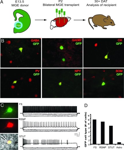
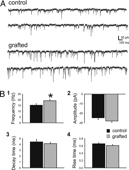
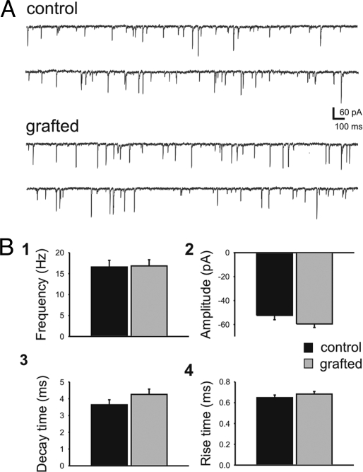
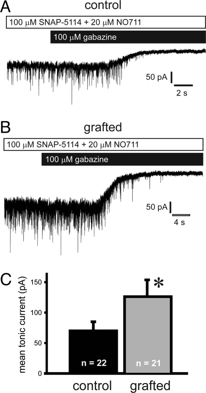
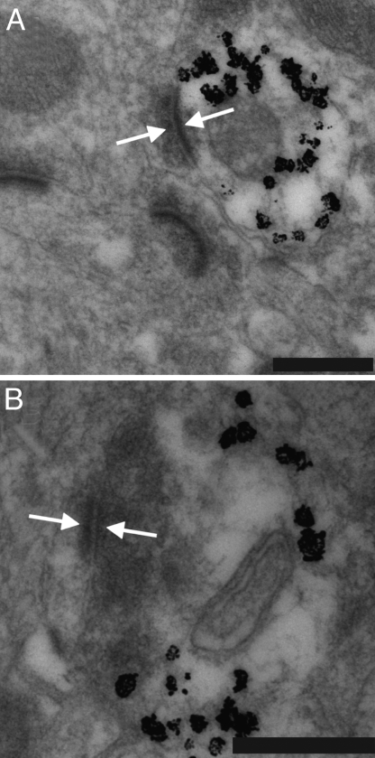
Epilepsy, a disease characterized by abnormal brain activity, is a disabling and potentially life-threatening condition for nearly 1% of the world population. Unfortunately, modulation of brain excitability using available antiepileptic drugs can have serious side effects, especially in the developing brain, and some patients can only be improved by surgical removal of brain regions containing the seizure focus. Here, we show that bilateral transplantation of precursor cells from the embryonic medial ganglionic eminence (MGE) into early postnatal neocortex generates mature GABAergic interneurons in the host brain. In mice receiving MGE cell grafts, GABA-mediated synaptic and extrasynaptic inhibition onto host brain pyramidal neurons is significantly increased. Bilateral MGE cell grafts in epileptic mice lacking a Shaker-like potassium channel (a gene mutated in one form of human epilepsy) resulted in significant reductions in the duration and frequency of spontaneous electrographic seizures. Our findings suggest that MGE-derived interneurons could be used to ameliorate abnormal excitability and possibly act as an effective strategy in the treatment of epilepsy.


Conflict of interest statement

Conflict of interest statement: S.C.B., J.L.R.R, and A.A.B are cofounders of, and have a financial interest in, Neurona Therapeutics.

Figures

Fig. 1.
Fig. 1.
Generation of interneurons from MGE precursors. (A) Schematic for MGE dissection (encircled region from GFP-expressing mouse embryos), bilateral transplantation into neonatal (P2) CD1 mice, and analysis at 30 DAT. (B) Immunohistochemical coexpression of GFP+ cells with GABA, GAD67, CR, PV, NPY, and SOM. Representative cortical neurons are shown at 30 DAT. (C) Representative in vitro recording from a GFP+ neuron in somatosensory cortex. (Top) Visualized patch-clamp recording from a GFP+ cell identified under epifluorescence. (Middle) GFP+ cell was filled with Alexa red via the patch pipette. (Bottom) Fluorescent images merged with IR-DIC image. Shown are sample current-clamp traces during depolarizing and hyperpolarizing steps to classify cells as fast-spiking (FS), regular-spiking nonpyramidal (RSNP), and stuttering (STUT) interneurons. (D) Summary plot for all GFP+ cells recorded in current-clamp at 30–40 DAT. Note: one cell classified as a putative astrocyte exhibited a hyperpolarized resting membrane potential and failed to generate action potentials upon depolarization.
Fig. 2.
Fig. 2.
MGE cell grafting increased inhibitory input onto host pyramidal cells. Spontaneous IPSCs were recorded from endogenous pyramidal cells near GFP+ cells in neocortex. (A) Representative voltage-clamp recordings from endogenous pyramidal cells in vehicle-treated control (Upper) and MGE-grafted (Lower) neocortical slices. (B) Summary bar plots for sIPSC frequency (Bi), amplitude (Bii), decay time (Biii), and rise time (Biv). sIPSC frequency recorded from pyramidal cells increased relative to controls by 25% from 15 ± 1 Hz (control) to 19 ± 1 Hz (grafted). There was no statistically significant difference in the amplitude, decay time, or rise times between control and grafted mice. Data are shown as mean ± SEM. *, P < 0.05; unpaired t test.
Fig. 3.
Fig. 3.
MGE cell grafting did not increase inhibitory input onto host interneurons. Spontaneous IPSCs were recorded from endogenous interneurons near GFP+ cells in neocortex. (A) Representative voltage-clamp recordings from endogenous interneurons in vehicle-treated control (Upper) and MGE-grafted (Lower) neocortical slices. Note that the electrical noise appears smaller in Fig. 3 (compared with Fig. 2) because the trace has been scaled down by a factor of 2 to show the larger-amplitude events. (B) Summary bar plots for sIPSC frequency (Bi), amplitude (Bii), decay time (Biii), and rise time (Biv). sIPSC frequency recorded from host interneurons in control (17 ± 1 Hz) and grafted (17 ± 2 Hz) mice was unchanged. There was no statistically significant difference in the amplitude, decay time, or rise times between control and grafted mice. Data are shown as mean ± SEM.
Fig. 4.
Fig. 4.
MGE cell grafting increased tonic current onto host pyramidal cells. Spontaneous IPSCs were recorded from endogenous pyramidal cells near GFP+ cells in neocortex. (A and B) Representative voltage-clamp recordings from pyramidal cells in neocortical slices from a vehicle-treated control (A) or an MGE-grafted mouse (B). Slices were first bath-perfused with 20 μM DNQX, 25 μM APV, 20 μM NO711, and 100 μM SNAP-5114. To measure the tonic GABA current, the GABAA receptor antagonist gabazine (100 μM) was bath-applied. Application of gabazine abolished all synaptic events. (C) MGE cell grafting increased the mean tonic by 80%, from 70 ± 15 pA in control to 126 ± 28 pA in grafted mice. Data are shown as mean ± SEM. *, P < 0.05; unpaired t test.
Fig. 5.
Fig. 5.
Integration of MGE-derived interneurons in the host brain. (A) Transmission electron micrographs of transplanted neurons labeled with immunogold for GFP (silver-enhanced black granules). Grafted cells had medium-sized soma characteristic of GABAergic interneurons, with invaginated nuclei and sparse endoplasmic reticulum. GFP-labeled neurons receive synaptic contacts from unlabeled host neurons. (B) GFP-labeled neurons make synaptic contacts onto unlabeled host neurons. Arrows indicate synaptic densities in the labeled transplanted cell and unlabeled host cell. (Scale bars: 200 μm.)
Fig. 6.
Fig. 6.
Seizure suppression in the Kv1.1 mouse model of epilepsy. (A) Sample EEG from a control vehicle-injected Kv1.1−/− mouse during a typical grade IV electrographic seizure. (B) The same seizure but with high resolution at four different stages of seizure progression (as noted by i–iv). (C and D) Sample EEG from Kv1.1−/− mouse grafted with MGE cells at P2. Note the seizure progression (Di–Div) is shorter. Summary bar plots show grade IV seizure duration (E) and the total number of seizures recorded (F). Data in E are shown as mean ± SEM. (G) Histogram showing durations for all electrographic seizures recorded in Kv1.1−/− (untreated, n = 4; vehicle-injected, n = 4; black bars) and Kv1.1−/− mice grafted with MGE cells (n = 8; gray bars). Total monitoring days and minutes are shown in the key. Given that control groups were monitored for a longer period (40 vs. 37 days; 16,695 vs. 11,875 min), it is possible that these results, which show a clear suppression of seizure activity, slightly overestimate the number of seizures occurring in grafted animals.

References

    1. Shetty AK, Zaman V, Hattiangady B. Repair of the injured adult hippocampus through graft-mediated modulation of the plasticity of the dentate gyrus in a rat model of temporal lobe epilepsy. J Neurosci. 2005;25:8391–8401. - PMC - PubMed
    1. Shetty AK, Hattiangady B. Restoration of calbindin after fetal hippocampal CA3 cell grafting into the injured hippocampus in a rat model of temporal lobe epilepsy. Hippocampus. 2007;17:943–956. - PMC - PubMed
    1. Huber K, et al. Grafts of adenosine-releasing cells suppress seizures in kindling epilepsy. Proc Natl Acad Sci USA. 2001;98:7611–7616. - PMC - PubMed
    1. Li T, et al. Suppression of kindling epileptogenesis by adenosine releasing stem cell-derived brain implants. Brain. 2007;130:1276–1288. - PubMed
    1. de Lanerolle NC, Kim JH, Robbins RJ, Spencer DD. Hippocampal interneuron loss and plasticity in human temporal lobe epilepsy. Brain Res. 1989;495:387–395. - PubMed

Publication types

Substances