. 2009 Sep 8;106(36):15332-7.
doi: 10.1073/pnas.0907864106. Epub 2009 Aug 20.

Metabolic labeling and direct imaging of choline phospholipids in vivo

Affiliations

Metabolic labeling and direct imaging of choline phospholipids in vivo

Cindy Y Jao et al. Proc Natl Acad Sci U S A. .

Abstract

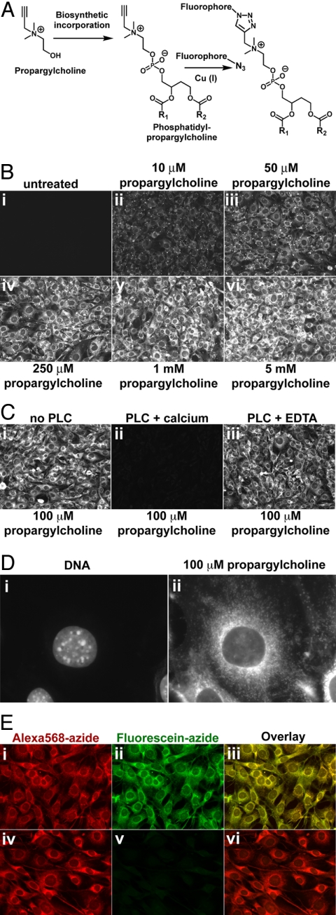
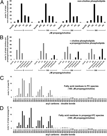
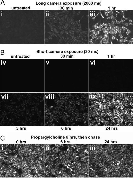
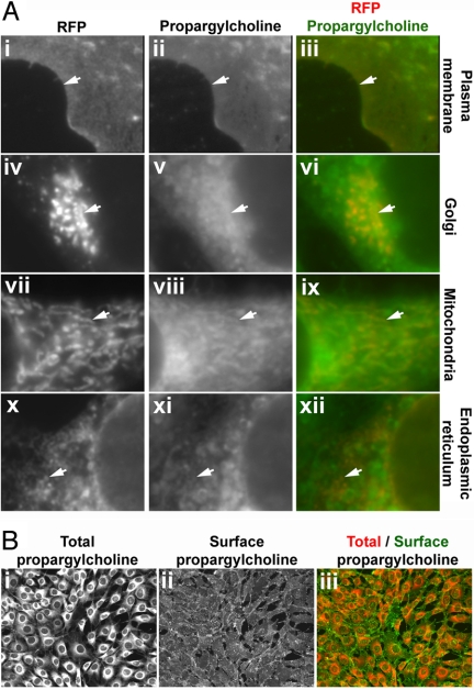
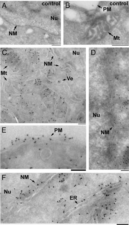
Choline (Cho)-containing phospholipids are the most abundant phospholipids in cellular membranes and play fundamental structural as well as regulatory roles in cell metabolism and signaling. Although much is known about the biochemistry and metabolism of Cho phospholipids, their cell biology has remained obscure, due to the lack of methods for their direct microscopic visualization in cells. We developed a simple and robust method to label Cho phospholipids in vivo, based on the metabolic incorporation of the Cho analog propargylcholine (propargyl-Cho) into phospholipids. The resulting propargyl-labeled phospholipid molecules can be visualized with high sensitivity and spatial resolution in cells via a Cu(I)-catalyzed cycloaddition reaction between the terminal alkyne group of propargyl-Cho and a labeled azide. Total lipid analysis of labeled cells shows strong incorporation of propargyl-Cho into all classes of Cho phospholipids; furthermore, the fatty acid composition of propargyl-Cho-labeled phospholipids is very similar to that of normal Cho phospholipids. We demonstrate the use of propargyl-Cho in cultured cells, by imaging phospholipid synthesis, turnover, and subcellular localization by both fluorescence and electron microscopy. Finally, we use propargyl-Cho to assay microscopically phospholipid synthesis in vivo in mouse tissues.


Conflict of interest statement

The authors declare no conflict of interest.

Figures

Fig. 1.
Fig. 1.
Imaging choline-containing phospholipids in cells with propargylcholine. (A) Propargyl-Cho, a biosynthetic label for Cho phospholipids. Phospholipid molecules bearing a terminal alkyne group can be detected by “click” chemistry using a fluorescent azide. (B) Propargyl-Cho incorporation into NIH 3T3 cells. Cells labeled overnight with varying concentrations of propargyl-Cho were fixed and stained with Alexa568-azide. Note the low background in the absence of propargyl-Cho (i) and the increase in staining intensity with increasing propargyl-Cho concentration (ii–vi). (C) Treatment of fixed cells with phospholipase C, which removes Cho head groups of phospholipids, strongly decreases propargyl-Cho staining by Alexa568-azide (compare ii to i). Phospholipase C requires calcium for activity; in the absence of calcium, propargyl-Cho staining is not affected by phospholipase C (iii). (D) Higher magnification fluorescence micrograph showing propargyl-Cho labeling of cellular organelles. (E) Reproducibility of propargyl-Cho staining. Cells labeled with 100 μM propargyl-Cho overnight were stained with 10 μM Alexa568-azide (i) and then with 20 μM fluorescein-azide (ii). Strong red and green signals indicate that the first staining reaction does not consume the incorporated propargyl-Cho. The red and green staining patterns are identical (iii), indicating that the two successive reactions detect the same phospholipid populations. The propargyl-Cho label can be consumed if, after Alexa568-azide (iv), cells are reacted with 5 mM non-fluorescent azide; this abolishes staining with fluorescein-azide (v and vi).
Fig. 2.
Fig. 2.
Total lipid analysis of propargylcholine-labeled cells. Total lipids isolated from NIH 3T3 cells labeled for 24 h with 0, 100, 250, and 500 μM propargyl-Cho were quantified by electrospray ionization-tandem mass spectrometry. Amounts of various phospholipids are shown as mole percentages of total non-Cho phospholipids (A), total Cho and propargyl-Cho phospholipids (B), total PC species (C), and total propargyl-PC species (D). (A) Distribution of non-Cho phospholipids (lyso-phosphatidylethanolamine, lyso-PE; ether-linked phosphatidylethanolamine, ePE; phosphatidylethanolamine, PE; phosphatidylinositol, PI; phosphatidylserine, PS; phosphatidic acid, PA) in cells labeled with propargyl-Cho. Propargyl-Cho labeling does not significantly affect the relative amount of non-Cho phospholipid classes. (B) Propargyl-Cho incorporates into all phosphatidylcholine (PC) classes (lysoPC; ether-linked PC, ePC; and PC) and into sphingomyelin (SM), proportional to the concentration of propargyl-Cho added to cells. (C and D) In NIH 3T3 cells labeled with 100 μM propargyl-Cho overnight, the sum fatty acid composition of PC species (C) is very similar to that of propargyl-PC species (D). Each PC and propargyl-PC species is identified by two numbers: the first is the sum of acyl carbons, and the second is the sum of double bonds present in the two fatty acid residues of the respective phospholipid species.
Fig. 3.
Fig. 3.
Rapid synthesis and slow turnover of propargylcholine-labeled phospholipids. (A) Time course of propargyl-Cho uptake and incorporation into phospholipids. NIH 3T3 cells were labeled with 1 mM propargyl-Cho in complete media for varying amounts of time and then stained with Alexa568-azide. The propargyl-Cho stain was photographed with a long exposure time (2 s, i–iii) or a short exposure time (30 ms, iv–ix). Note that propargyl-Cho incorporation is visible 30 min after addition to cells (ii) and continues to increase with time (ii–viii), saturating after about 24 h (ix). (B) Stability of propargyl-Cho-labeled phospholipids in cells. NIH 3T3 cells were labeled with 1 mM propargyl-Cho for 6 h and then chased with normal media for different amounts of time (i–iii). The intensity of the Alexa568-azide stain does not appreciably decrease after 24 h in culture, indicating the low turnover of propargyl-Cho phospholipids.
Fig. 4.
Fig. 4.
Subcellular distribution of propargylcholine-labeled phospholipids. (A) Co-localization of the propargyl-Cho stain with subcellular markers. Cultured NIH 3T3 cells were transfected with plasmids encoding red fluorescent protein fusions that mark various organelles. The cells were labeled with 100 μM propargyl-Cho overnight and stained with fluorescein-azide. Propargyl-Cho co-localizes with markers for the plasma membrane (i–iii), the Golgi (iv–vi), mitochondria (vii–ix), and endoplasmic reticulum (x–xii). The white arrows point to subcellular structures that stain for both propargyl-Cho and a given red fluorescent marker. (B) Staining propargyl-Cho phospholipids localized to the outer leaflet of the plasma membrane. Cells labeled with 100 μM propargyl-Cho were reacted with Alexa568-azide and biotin-azide. The latter is visualized specifically on the cell surface by staining with Alexa488-conjugated streptavidin (ii), which due to its size does not cross the plasma membrane. The right panel (iii) shows the overlay of the total (i) and surface (ii) propargyl-Cho stain.
Fig. 5.
Fig. 5.
Visualizing propargylcholine-labeled phospholipids by immuno-electron microscopy of cells. Cultured 293T cells were incubated overnight in the absence (A and B) or presence (C–F) of 100 μM propargyl-Cho, fixed, sectioned on an ultramicrotome and stained with biotin-azide. The sections were then stained with anti-biotin antibodies and protein A-gold (10 nm), counterstained with uranyl acetate and imaged by transmission electron microscopy. Arrows point to various cellular structures: Mt, mitochondria; Nu, nucleus; ER, endoplasmic reticulum; PM, plasma membrane; NM, nuclear membrane; Ve, vesicle. The scale bar in all panels is 100 nm. A negligible number of gold particles are seen on micrographs of control, unlabeled cells (A and B). Numerous gold particles are seen associated with membranes in cells labeled with propargyl-Cho (C–F).
Fig. 6.
Fig. 6.
Imaging the synthesis of choline phospholipids in vivo. Cryostat sections of organs from a propargyl-Cho-injected mouse and from an uninjected control mouse were stained in parallel with TMR-azide. The TMR-azide stain is shown in grayscale (black-and-white images) or in red, overlayed with DNA (blue) and DIC micrographs (color images). Strong propargyl-Cho incorporation is seen in all tissues surveyed: small intestine (A), kidney (B), liver (C), and spleen (D).

References

    1. Vance JE, Vance DE. Phospholipid biosynthesis in mammalian cells. Biochem Cell Biol. 2004;82:113–128. - PubMed
    1. Nagan N, Zoeller RA. Plasmalogens: Biosynthesis and functions. Prog Lipid Res. 2001;40:199–229. - PubMed
    1. Weiss SB, Smith SW, Kennedy EP. The enzymatic formation of lecithin from cytidine diphosphate choline and D-1,2-diglyceride. J Biol Chem. 1958;231:53–64. - PubMed
    1. Rostovtsev VV, Green LG, Fokin VV, Sharpless KB. A stepwise huisgen cycloaddition process: Copper(I)-catalyzed regioselective “ligation” of azides and terminal alkynes. Angew Chem Int Ed Engl. 2002;41:2596–2599. - PubMed
    1. Tornoe CW, Christensen C, Meldal M. Peptidotriazoles on solid phase: [1,2,3]-triazoles by regiospecific copper (i) -catalyzed 1,3-dipolar cycloadditions of terminal alkynes to azides. J Org Chem. 2002;67:3057–3064. - PubMed

Publication types

LinkOut - more resources