. 2009 Sep 8;106(36):15291-6.
doi: 10.1073/pnas.0905516106. Epub 2009 Aug 19.

A 3D structure model of the melibiose permease of Escherichia coli represents a distinctive fold for Na+ symporters

Affiliations

A 3D structure model of the melibiose permease of Escherichia coli represents a distinctive fold for Na+ symporters

Mohammad S Yousef et al. Proc Natl Acad Sci U S A. .

Abstract

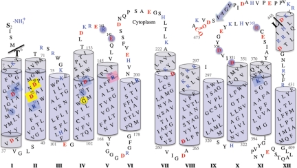
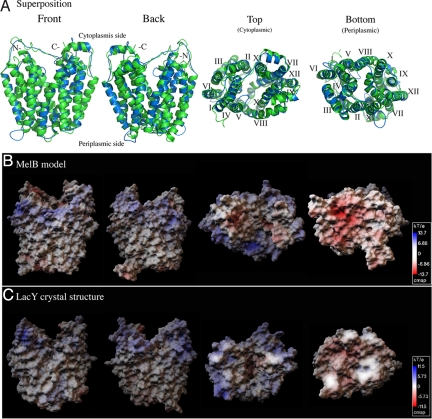
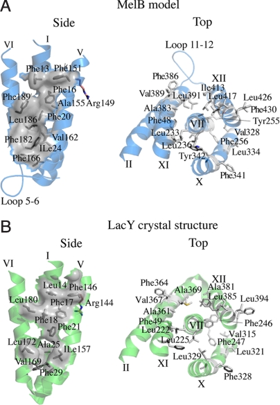
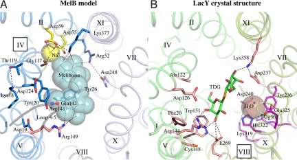
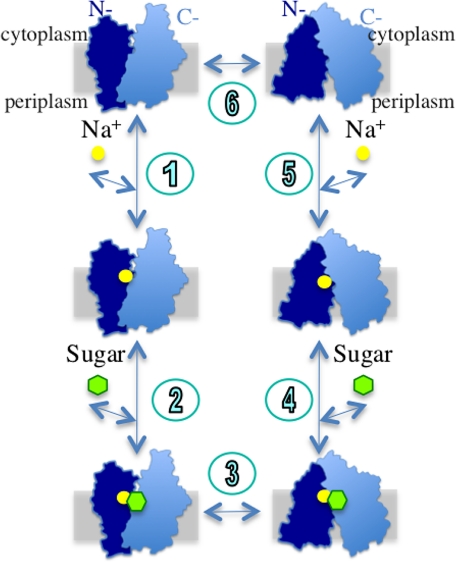
The melibiose permease of Escherichia coli (MelB) catalyzes the coupled stoichiometric symport of a galactoside with a cation (either Na(+), Li(+), or H(+)), using free energy from the downhill translocation of one cosubstrate to catalyze the accumulation of the other. Here, we present a 3D structure model of MelB threaded through a crystal structure of the lactose permease of E. coli (LacY), manually adjusted, and energetically minimized. The model contains 442 consecutive residues ( approximately 94% of the polypeptide), including all 12 transmembrane helices and connecting loops, with no steric clashes and superimposes well with the template structure. The electrostatic surface potential calculated from the model is typical for a membrane protein and exhibits a characteristic ring of positive charges around the periphery of the cytoplasmic side. The 3D model indicates that MelB consists of two pseudosymmetrical 6-helix bundles lining an internal hydrophilic cavity, which faces the cytoplasmic side of the membrane. Both sugar and cation binding sites are proposed to lie within the internal cavity. The model is consistent with numerous previous mutational, biochemical/biophysical characterizations as well as low-resolution structural data. Thus, an alternating access mechanism with sequential binding is discussed. The proposed overall fold of MelB is different from the available crystal structures of other Na(+)-coupled transporters, suggesting a distinctive fold for Na(+) symporters.


Conflict of interest statement

The authors declare no conflict of interest.

Figures

Fig. 1.
Fig. 1.
Membrane topology of MelB. The topology model of MelB (11) is matched to the threading 3D structure (see the text). Transmembrane helices are presented as blue cylinders; the light-blue color indicates extramembrane segments. Helices are numbered with Roman numerals. Charged residues are shown in red (negative) and blue (positive). Eight cavity-exposed charged residues from transmembrane regions are marked as filled square boxes. Residues known to participate in the sugar binding are highlighted with a pink background. Residues expected to be involved in cation binding are highlighted with a yellow background (see the text). Residues important for substrate(s) binding and/or coupling are highlighted with a blue background. The Met-1 residue is removed, as suggested by N-terminal sequencing (12). The threaded region is between residues Thr-6 and Leu-448, as indicated by black lines.
Fig. 2.
Fig. 2.
Comparison between the threading model of MelB and the crystal structure of LacY. (A) Superposition. The main chain coordinates of MelB (residues 6–448, blue), obtained from the LOOPP program, are superimposed on the LacY crystal structure (green, PDB ID 1pv6). The alignment was restricted to the corresponding transmembrane helices. (B and C) Surface electrostatic potential maps of the MelB threading model and the crystal structure of LacY were calculated using Adaptive Poisson-Boltzmann Solver (APBS) software (53). The scale indicates color-coded values of the electrostatic potentials (kT/e). A positive electrostatic potential is noticeable around the cytoplasmic opening of the central cavity in both permeases. Front and back, viewing parallel to the membrane; top, viewing from the cytoplasmic side; bottom, viewing from the periplasmic side.
Fig. 3.
Fig. 3.
Conserved helical packing. Hydrophobic patches (gray) between helices I, V, and VI in the N-terminal domain and around helix VII in the C-terminal domain of the MelB threading model (A, blue) and LacY crystal structure (B, green).
Fig. 4.
Fig. 4.
The central cavity in the MelB model containing putative binding sites for Na+ and sugar. Identical views from the cytoplasmic side down the central cavity of MelB (A, blue) and LacY (B, green). The N- and C-terminal helices of MelB and LacY are shown in blue/light-blue and green/light-green colors, respectively. Residues important for cosubstrate binding are shown as sticks. The large spheres reflect postulated positions of the ligands. Dotted lines show possible interactions. (A) In the threading model of MelB, a melibiose molecule and a Na+ ion are manually docked in putative binding sites. The modeling of melibiose (line, cyan) was guided by the X-ray coordinates of the sugar substrate in LacY, as shown in B. The Na+ and possible interacting residues (Asp-55, Asp-59, and Gly-117) are colored yellow. The functionally important loop 4–5 is shown in gray, with 2 important positions (Arg-141 and Glu-142) highlighted. Residues colored in blue are important for substrate(s) binding and/or coupling (see the text). The Arg-149 is important for sugar binding (26) and is colored pink. In LacY (B, PDB 1pv7), residues that are essential or important for the binding of sugar and H3O+ are shown in pink and magenta, respectively. A ß-D-galactopyranosyl 1-thio-ß-D-galactopyranoside (TDG) molecule is shown in green, and manually docked H3O+ is shown as an enlarged brown sphere.
Fig. 5.
Fig. 5.
A kinetic scheme of the efflux mode of galactoside/Na+ symport for MelB. A cross section of the membrane is shown as a gray rectangle.

References

    1. Yazyu H, et al. Nucleotide sequence of the melB gene and characteristics of deduced amino acid sequence of the melibiose carrier in Escherichia coli. J Biol Chem. 1984;259:4320–4326. - PubMed
    1. Saier MH., Jr Families of transmembrane sugar transport proteins. Mol Microbiol. 2000;35:699–710. - PubMed
    1. Tokuda H, Kaback HR. Sodium-dependent methyl 1-thio-β-d-galactopyranoside transport in membrane vesicles isolated from Salmonella typhimurium. Biochemistry. 1977;16:2130–2136. - PubMed
    1. Tsuchiya T, Raven J, Wilson TH. Co-transport of Na+ and methul-beta-d-thiogalactopyranoside mediated by the melibiose transport system of Escherichia coli. Biochem Biophys Res Commun. 1977;76:26–31. - PubMed
    1. Bassilana M, Pourcher T, Leblanc G. Facilitated diffusion properties of melibiose permease in Escherichia coli membrane vesicles. Release of co-substrates is rate limiting for permease cycling. J Biol Chem. 1987;262:16865–16870. - PubMed

Publication types

LinkOut - more resources