. 2009 Sep 1;106(35):14831-6.
doi: 10.1073/pnas.0900742106. Epub 2009 Aug 19.

A luminal flavoprotein in endoplasmic reticulum-associated degradation

Affiliations

A luminal flavoprotein in endoplasmic reticulum-associated degradation

Jan Riemer et al. Proc Natl Acad Sci U S A. .

Abstract

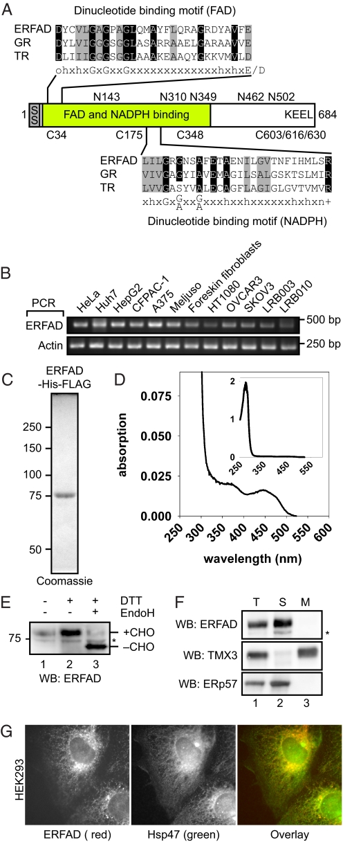
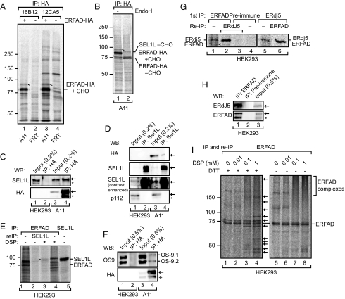
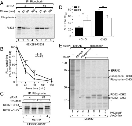
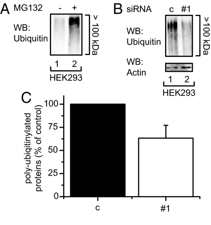
The quality control system of the endoplasmic reticulum (ER) discriminates between native and nonnative proteins. The latter are degraded by the ER-associated degradation (ERAD) pathway. Whereas many cytosolic and membrane components of this system are known, only few luminal players have been identified. In this study, we characterize ERFAD (ER flavoprotein associated with degradation), an ER luminal flavoprotein that functions in ERAD. Upon knockdown of ERFAD, the degradation of the ERAD model substrate ribophorin 332 is delayed, and the overall level of polyubiquitinated cellular proteins is decreased. We also identify the ERAD components SEL1L, OS-9 and ERdj5, a known reductase of ERAD substrates, as interaction partners of ERFAD. Our data show that ERFAD facilitates the dislocation of certain ERAD substrates to the cytosol, and we discuss the findings in relation to a potential redox function of the protein.


Conflict of interest statement

The authors declare no conflict of interest.

Figures

Fig. 1.
Fig. 1.
ERFAD is an ER flavoprotein. (A) Domain organization of the ERFAD protein. The two dinucleotide-binding motifs of the GXGXXG-type for FAD and NADPH binding are shown aligned with the corresponding motifs in GR, TR, and a consensus motif (amino acid residues: h = hydrophobic, o = polar/charged, + = positively charged, n = neutral). The sequence positions of the five N-glycosylation sites and the six cysteines in ERFAD are depicted. SS, signal sequence. (B) RT-PCR analysis of ERFAD. Total RNA was isolated from different human tissue culture cells, reverse transcribed and amplified with primers specific for ERFAD and actin. HeLa: cervical epithelial carcinoma; Huh7, HepG2: hepatocellular carcinoma; CF-PAC-1: pancreatic adeno carcinoma; A375, Meljuso: melanoma; HT1080: fibrosarcoma breast cancer; OVCAR3, SKOV3: ovarian epithelial carcinoma; LRB003, LRB010: embryonic stem cells (C) Purified recombinant ERFAD-His-FLAG visualized by Coomassie staining. (D) Absorption spectra of purified ERFAD-His-FLAG. The two peaks at 370 nm and 450 nm are indicative of the flavin cofactor. (Inset) Complete spectrum including the protein peak at 280 nm. (E) Glycosylation and oxidation state of human ERFAD. Lysates from HEK293 cells were treated as indicated, and analyzed by Western blotting against endogenous ERFAD. *, background band; CHO, N-glycans. (F) Subcellular fractionation of HEK293 cells. After isolation and sodium carbonate extraction of crude membranes, followed by ultracentrifugation through a sucrose cushion, the distribution of ERFAD, ERp57 (a soluble ER protein) and TMX3 (an ER membrane protein) was visualized by Western blot analysis. *, background band. (G) Immunofluorescence microscopy of ERFAD in HEK293 cells. Cells were fixed and stained with anti-ERFAD (Left, 1F6, red) and anti-Hsp47 (Center, green). A merged image is shown in the Right.
Fig. 2.
Fig. 2.
ERFAD interacts with the ERAD components SEL1L, OS-9, and ERdj5. (A) Immunoprecipitation of ERFAD-HA. Cells stably expressing ERFAD-HA (A11) and control cells (FRT) were [35S] pulse-labeled for 16 h, Triton X-100 lysates immunoprecipitated with anti-HA (16B12 and 12CA5), and samples separated by reducing SDS/PAGE. The position of a coimmunoprecipitating 90 kDa band is indicated by arrowheads. (B) Immunoprecipitation of ERFAD-HA with 16B12, undigested (lane 1) and digested (lane 2) with EndoH. The position of the protein identified by mass spectrometry—SEL1L—is indicated. CHO, N-glycans. (C) SEL1L coimmunoprecipitates with ERFAD-HA. Immunoprecipitations from lysates of A11 or HEK293 cells were performed with anti-HA, and subsequently analyzed by Western blotting as indicated. *, background band. (D) ERFAD-HA coimmunoprecipitates with SEL1L. Immunoprecipitations from lysates of A11 cells were performed with anti-SEL1L and subsequently analyzed by Western blotting with anti-HA, anti-SEL1L (contrast-enhanced blot included to better see the input), and anti-p112 as a specificity control. *, background band. (E) Endogenous ERFAD and SEL1L coimmunoprecipitate upon cross-linking with DSP. After incubation with DSP, ERFAD was immunoprecipitated from [35S] pulse-labeled HEK293 cells. The immunoprecipitate was either analyzed directly (lanes 1 and 4) or reimmunoprecipitated with antibodies against SEL1L (lane 2 and 3). For comparison an anti-SEL1L immunoprecipitate is loaded in lane 5. Samples were analyzed by reducing SDS/PAGE, which resolves the thiol-cleavable cross-link between ERFAD and SEL1L. (F) OS-9.1 and 9.2 coprecipitate with ERFAD-HA. Immunoprecipitations from A11 or HEK293 cell lysates were performed with anti-HA, and analyzed by Western blotting with antibodies against OS-9 and the HA tag. *, background band. (G) Endogenous ERFAD and ERdj5 coimmunoprecipitate. ERFAD was immunoprecipitated from [35S] pulse-labeled HEK293 cells with anti-ERFAD (SG2480). The immunoprecipitate was either analyzed directly (lane 1) or reimmunoprecipitated with anti-ERdj5 (lane 2). As a control, preimmune serum was used instead of anti-ERFAD (lanes 3 and 4). In another experiment, ERdj5 was immunoprecipitated from pulse-labeled HEK293 cells and either analyzed directly (lane 5) or immunoprecipitated with anti-ERFAD (lane 6). Samples were analyzed by reducing SDS/PAGE. Arrowhead, ERFAD. (H) Endogenous ERFAD and ERdj5 coimmunoprecipitate. Immunoprecipitations from lysates of HEK293 cells were performed with anti-ERFAD (1F6) or preimmune serum and subsequently analyzed by Western blotting with anti-ERdj5 and anti-ERFAD. *, background band. (I) Numerous proteins immunoprecipitate with endogenous ERFAD upon DSP cross-linking. Immunoprecipitates of ERFAD after treatment with increasing concentrations of DSP were either analyzed under reducing or nonreducing conditions. Arrows indicate proteins that coprecipitate with ERFAD upon cross-linking. For complete audiographs see Fig. S3.
Fig. 3.
Fig. 3.
ERFAD knockdown stabilizes the ERAD model substrate RI332. (A) Decay of RI332 upon ERFAD knockdown. HEK293 cells stably expressing RI332 were transfected with ERFAD siRNA#1 (#1) and nonsilencing control siRNA (c). Seventy-two hours after transfection cells were pulse labeled for 20 min and chased for the indicated times. SDS lysates were subjected to immunoprecipitation with anti-ribophorin. (B) Quantification of three independent RI332 decay experiments (RI332 signal normalized to full length RI). Mean ± SD; ***, P < 0.005; *, P < 0.5. (C) Ratio of the glycosylated ER form of RI332 versus the deglycosylated cytosolic form of RI332 upon ERFAD knockdown. ERFAD-silenced and control HEK-RI332 cells were pulse-labeled for 20 min and chased for 3 h in the presence of MG132. SDS lysates were subjected to immunoprecipitation with anti-ribophorin and half of the eluate was PNGaseF treated. The two glycosylation states of RI332 are indicated. (D) Quantification of three independent experiments as performed in C. **, P < 0.05. (E) Coimmunoprecipitation of ERFAD and RI332. HEK293-RI332 cells were pulse-labeled for 5 h in the presence of MG132. Anti-ERFAD immunoprecipitates were either analyzed directly (lane 1) or reimmunoprecipitated with anti-ERFAD (lane 2) or anti-ribophorin (lane 3). In lanes 4—7, SDS lysates from cells labeled in the presence or absence of zVAD-fmk (+MG132) were immunoprecipitated with anti-ribophorin with or without subsequent PNGaseF digest to allow the assignment of the three different forms of RI332 (RI332+CHO, RI332-CHO and *). The arrowhead indicates the minor fraction of deglycosylated RI332 (RI332-CHO) interacting with ERFAD. For a contrast-enhanced version, see Fig. S7A.
Fig. 4.
Fig. 4.
The knockdown of ERFAD decreases the cellular amount of polyubiquitinated proteins. (A) Accumulation of polyubiquitinated proteins upon MG132 treatment. HEK293 cells were either left untreated or treated with MG132, lysates were adjusted by a BCA assay to the same protein concentration and analyzed by Western blotting with anti-ubiquitin. (B) Decrease of polyubiquitinated proteins upon ERFAD knockdown. HEK293 cells were transfected with ERFAD siRNA#1 and nonsilencing control siRNA (c). Cells were lysed 72 h after transfection, lysates were adjusted to the same concentration and analyzed by Western blotting with anti-ubiquitin and anti-actin. (C) Quantification of three independent experiments performed as described in B and plotted as percentage of control, mean ± SD.

References

    1. Anelli T, Sitia R. Protein quality control in the early secretory pathway. EMBO J. 2008;27:315–327. - PMC - PubMed
    1. Kincaid MM, Cooper AA. ERADicate ER stress or die trying. Antioxid Redox Signal. 2007;9:2373–2387. - PubMed
    1. Nakatsukasa K, Brodsky JL. The recognition and retrotranslocation of misfolded proteins from the endoplasmic reticulum. Traffic. 2008;9:861–870. - PMC - PubMed
    1. Kikkert M, et al. Human HRD1 is an E3 ubiquitin ligase involved in degradation of proteins from the endoplasmic reticulum. J Biol Chem. 2004;279:3525–3534. - PubMed
    1. Mueller B, Lilley BN, Ploegh HL. SEL1L, the homologue of yeast Hrd3p, is involved in protein dislocation from the mammalian ER. J Cell Biol. 2006;175:261–270. - PMC - PubMed

Publication types