. 2009 Sep 1;106(35):14861-6.
doi: 10.1073/pnas.0907542106. Epub 2009 Aug 17.

Bacterial growth and motility in sub-micron constrictions

Affiliations

Bacterial growth and motility in sub-micron constrictions

Jaan Männik et al. Proc Natl Acad Sci U S A. .

Abstract

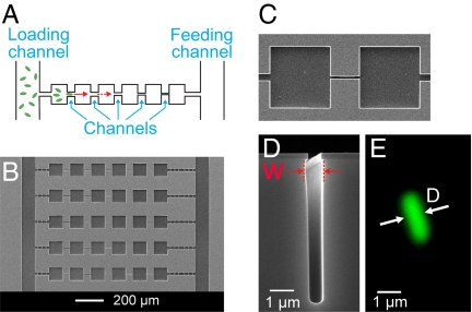
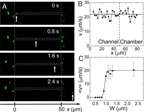
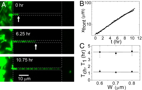
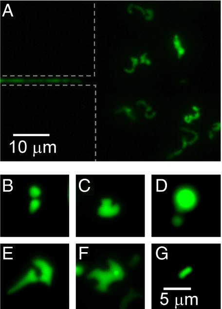
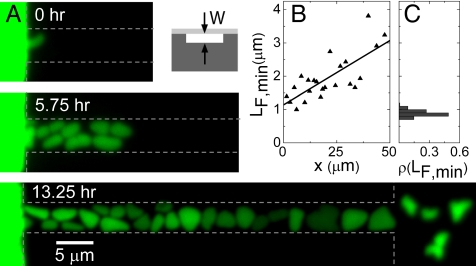
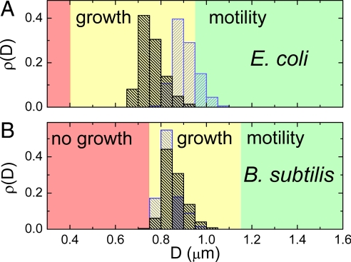
In many naturally occurring habitats, bacteria live in micrometer-size confined spaces. Although bacterial growth and motility in such constrictions is of great interest to fields as varied as soil microbiology, water purification, and biomedical research, quantitative studies of the effects of confinement on bacteria have been limited. Here, we establish how Gram-negative Escherichia coli and Gram-positive Bacillus subtilis bacteria can grow, move, and penetrate very narrow constrictions with a size comparable to or even smaller than their diameter. We show that peritrichously flagellated E. coli and B. subtilis are still motile in microfabricated channels where the width of the channel exceeds their diameters only marginally (approximately 30%). For smaller widths, the motility vanishes but bacteria can still pass through these channels by growth and division. We observe E. coli, but not B. subtilis, to penetrate channels with a width that is smaller than their diameter by a factor of approximately 2. Within these channels, bacteria are considerably squeezed but they still grow and divide. After exiting the channels, E. coli bacteria obtain a variety of anomalous cell shapes. Our results reveal that sub-micron size pores and cavities are unexpectedly prolific bacterial habitats where bacteria exhibit morphological adaptations.


Conflict of interest statement

The authors declare no conflict of interest.

Figures

Fig. 1.
Fig. 1.
Setup for studying bacterial movement in small constrictions. (A) Schematic of the experiment. Using time-lapse fluorescent microscopy, bacterial movement from left to right is observed in array structures consisting of multiple channels and chambers in series. (B) SEM image of a section of a microfluidic chip where five of these array structures can be seen. (C) Top view of a 3-μm wide and 50-μm long channel connecting two chambers. (D) SEM side view image of the cross-section of a 0.8-μm wide channel. The width of the channel (W) is measured at the widest cross-section. For small W, bacteria are confined to the slightly wider location near the top. (E) Fluorescent image of a typical E. coli bacterium shown at the same scale as the 0.8-μm wide channel on panel (D). The intensity profile across its cross-section is used to determine the bacterial diameter D, see SI Text.
Fig. 2.
Fig. 2.
Bacterial motility in channels. (A) Time-lapse images of an E. coli bacterium that swims through a 1.2-μm wide channel (three topmost images) and into the chamber area (Bottom image). The arrow points at the swimming bacterium. The vertical dashed lines mark the boundaries of the two chambers and the horizontal lines indicate the location of the channel. (B) Velocity of the bacterium v as a function of its coordinate along a 1.2-μm wide channel (left from the vertical dashed line) and in the chamber area (right from the vertical dashed line). The x-coordinate is measured along the channel with x = 0 at the channel entrance from the left chamber. (C) Average velocity <v> versus the width of the channel. The time-averaged velocity is first calculated from traces such as shown in panel B, excluding tumbling events which last >0.2 s. Subsequently, averaging over the population of bacteria in the same-size channel is carried out to yield <v>. The error bars correspond to the standard deviation of velocities among the population of bacteria in a given-size channel. Solid line represents a sigmoidial fit to the data, with a midpoint W = 0.95 μm.
Fig. 3.
Fig. 3.
Bacterial growth through narrow channels. (A) Time-lapse fluorescent images of bacterial growth in a 0.6-μm wide channel. Dashed gray lines show the approximate boundaries of the chambers and channels. The arrows point to the position of the bacterial front. (B) Position of the bacterial front vs. time for the growth process on panel A. The line presents a fit of the function xfront(t) = L0 + L1 2t/Tch. (C) Doubling time of the chain length Tch (squares) and division time of the first bacterium in the chain T1 (triangles) vs. channel width. Each Tch and T1 value in the Inset of B represents an average over several populations in channels of the given width on the same chip. Error bars represent standard deviations among different populations.
Fig. 4.
Fig. 4.
Aberrant bacteria exit narrow channels. (A) Fluorescence image of 0.6-μm wide channel and a chamber 5 h after the first bacterium appeared in this chamber. Variety of aberrantly shaped bacteria can be seen to populate the chamber. The image is sequence to the series shown in Fig. 4A. (B–F) Different aberrant bacterial shapes at higher magnification. (G) For comparison, fluorescence image of a regularly shaped and sized bacterium which has emerged from the same channel as the bacteria shown in panels E and F. The same scale bar applies for all of the panels from B to G.
Fig. 5.
Fig. 5.
Morphogenesis of E. coli in shallow horizontal channels. (A) Time lapse fluorescence images of bacteria in channel of 0.3 μm width (and other dimensions 5 and 50 μm). Dashed lines show approximate boundaries of chambers and channels. Inset: schematic side-view cross-section of such channel. Darker gray corresponds to silicon and lighter gray to PDMS. (B) Minimum Feret's diameter vs. bacterial position in the channel (right vertical axes). The position is measured from the channel entrance. The data points correspond to the bacteria on the Bottom image of panel A. (C) For comparison, distribution of minimum Feret's diameters in batch culture. Batch culture has grown to optical density OD600 = 2.3. The vertical axes of this plot is the same as in B.
Fig. 6.
Fig. 6.
Comparison of channel widths to bacterial diameters. (A) Distribution of bacterial diameters in log (OD600 = 0.6, blue) and in stationary phase (OD600 = 2.3, black) for E. coli as determined from fluorescence intensity profiles of individual bacteria from batch cultures. Details of the size determination are given in SI Text. The average diameters are = 0.91 ± 0.05 μm in log and = 0.76 ± 0.05 μm in stationary phase. The range of channel widths for which bacteria are motile or grow are indicated by the green or yellow colored background, respectively. The range of channel widths where no penetration is observed is shown in red. (B) The same for B. subtilis strain for the log (OD600 = 0.4, blue) and for stationary phase distributions (OD600 = 2.0, black) where the average diameters are = 0.84 ± 0.04 μm and = 0.86 ± 0.04 μm, respectively.

References

    1. Ranjard L, Richaume AS. Quantitative and qualitative microscale distribution of bacteria in soil. Res Microbiol. 2001;152:707–716. - PubMed
    1. Whitman WB, Coleman DC, Wiebe WJ. Prokaryotes: The unseen majority. Proc Natl Acad Sci USA. 1998;95:6578–6583. - PMC - PubMed
    1. Hahn MW. Broad diversity of viable bacteria in ‘sterile’ (0.2 μm) filtered water. Res Microbiol. 2004;155:688–691. - PubMed
    1. Wang Y, Hammes F, Boon N, Egli T. Quantification of the filterability of freshwater bacteria through 0.45, 0.22, and 0.1 μm pore size filters and shape-dependent enrichment of filterable bacterial communities. Environ Sci Technol. 2007;41:7080–7086. - PubMed
    1. do Nascimento C, et al. Bacterial leakage along the implant-abutment interface of premachined or cast components. Int J Oral Maxillofac Surg. 2008;37:177–180. - PubMed

Publication types

MeSH terms

LinkOut - more resources