. 2009 Sep 1;106(35):14814-9.
doi: 10.1073/pnas.0904553106. Epub 2009 Aug 19.

Human deoxyhypusine hydroxylase, an enzyme involved in regulating cell growth, activates O2 with a nonheme diiron center

Affiliations

Human deoxyhypusine hydroxylase, an enzyme involved in regulating cell growth, activates O2 with a nonheme diiron center

Van V Vu et al. Proc Natl Acad Sci U S A. .

Abstract

Deoxyhypusine hydroxylase is the key enzyme in the biosynthesis of hypusine containing eukaryotic translation initiation factor 5A (eIF5A), which plays an essential role in the regulation of cell proliferation. Recombinant human deoxyhypusine hydroxylase (hDOHH) has been reported to have oxygen- and iron-dependent activity, an estimated iron/holoprotein stoichiometry of 2, and a visible band at 630 nm responsible for the blue color of the as-isolated protein. EPR, Mössbauer, and XAS spectroscopic results presented herein provide direct spectroscopic evidence that hDOHH has an antiferromagnetically coupled diiron center with histidines and carboxylates as likely ligands, as suggested by mutagenesis experiments. Resonance Raman experiments show that its blue chromophore arises from a (mu-1,2-peroxo)diiron(III) center that forms in the reaction of the reduced enzyme with O2, so the peroxo form of hDOHH is unusually stable. Nevertheless we demonstrate that it can carry out the hydroxylation of the deoxyhypusine residue present in the elF5A substrate. Despite a lack of sequence similarity, hDOHH has a nonheme diiron active site that resembles both in structure and function those found in methane and toluene monooxygenases, bacterial and mammalian ribonucleotide reductases, and stearoyl acyl carrier protein Delta9-desaturase from plants, suggesting that the oxygen-activating diiron motif is a solution arrived at by convergent evolution. Notably, hDOHH is the only example thus far of a human hydroxylase with such a diiron active site.


Conflict of interest statement

The authors declare no conflict of interest.

Figures

Scheme 1.
Scheme 1.
Biosynthesis of hypusine on eIF5A.
Fig. 1.
Fig. 1.
The UV-Visible spectra of hDOHH at room temperature. Conversion of as-isolated hDOHH (—) by 2.2 equiv. dithionite to reduced hDOHH (···) and its regeneration upon exposure to air after 180 min of anaerobic incubation (−−−). Inset: Change of A630 with time.
Fig. 2.
Fig. 2.
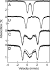
Mössbauer studies of hDOHH: 4.2 K zero field Mössbauer spectra of as-isolated hDOHH (A) and reduced with dithionite (B). The solid line in A is a simulation assuming 2 nested doublets as described in the text. (B) The solid line is a simulation of hDOHHred assuming 2 equally intense doublets representing ≈75% of Fe. Details of data reduction and analyses are given in SI. The 8.0 T Mössbauer spectra of as-isolated hDOHH recorded at 4.2 K (C) and 50 K (D). The solid lines are spectral simulations for an antiferromagnetically coupled diiron(III) center containing 2 high-spin (S = 5/2) Fe(III) sites. For the simulation of the 50-K spectrum we used J = 70 cm−1. At 50 K the central features of the 8.0 T spectrum contain contributions from the mononuclear Fe(III) contaminant (see Mössbauer spectroscopy section in SI Text).
Fig. 3.
Fig. 3.
Resonance Raman spectra of hDOHH samples. (A) as-isolated hDOHH. (BD) reduced hDOHH exposed to 16O2, 18O2, and mixed-labeled O2 (16O2, 18O2, and 16O18O). Background features because of the protein itself have been subtracted using the spectrum of fully reduced hDOHH collected under the same conditions. Experimental data are presented with thick lines and fits are presented with thin lines.
Fig. 4.
Fig. 4.
X-ray absorption spectroscopic analysis of hDOHH. (A) XANES spectra of hDOHHperoxo first scans (—), hDOHHphr (−−−), and hDOHHred (···). (B and C) Fe K-edge EXAFS data k3χ(k) of hDOHHphr and hDOHHperoxo and their Fourier transforms (thin lines) in k range = 2–14 Å−1 and 2–11.8 Å−1, respectively. Best fits are represented by the thick lines. Details of the fitting protocols are provided in Table S1.
Fig. 5.
Fig. 5.
Reaction of hDOHHperoxo with ≈1 equiv. of substrate, deoxyhypusine-containing eIF5A. The decay of hDOHHperoxo was monitored by following the change in A630 at different incubation times in the absence of eIF5A(Dhp) (◇) and in the presence of eIF5A(Dhp) (■). Yields of hypusine (●) are calculated on the basis of the observed amounts of hypusine (Hpu) and deoxyhypusine (Dhp); % Hpu = (Hpu × 100)/(Hpu + Dhp).
Fig. 6.
Fig. 6.
Proposed core structure of hDOHHperoxo

References

    1. Park MH. The post-translational synthesis of a polyamine-derived amino acid, hypusine, in the eukaryotic translation initiation factor 5A (eIF5A) J Biochem. 2006;139:161–169. - PMC - PubMed
    1. Wolff EC, Kang KR, Kim YS, Park MH. Posttranslational synthesis of hypusine: Evolutionary progression and specificity of the hypusine modification. Amino Acids. 2007;33:341–350. - PMC - PubMed
    1. Byers TL, Lakanen JR, Coward JK, Pegg AE. The role of hypusine depletion in cytostasis induced by S-adenosyl-L-methionine dercarboxylase inhibition: New evidence provided by 1-methylspermidine and 1,12-dimethylspermidine. Biochem J. 1994;303:363–368. - PMC - PubMed
    1. Park MH, Wolff EC, Leea YB, Folk JE. Antiproliferative effects of inhibitors of deoxyhypusine synthase: Inhibition of growth of Chinese hamster ovary cells by guanul diamines. J Biol Chem. 1994;269:27827–27832. - PubMed
    1. Hanauske-Abel HM, et al. Inhibition of the G1-S transition of the cell cycle by inhibitors of deoxyhypusine hydroxylation. Biochim Biophys Acta. 1994;1221:115–124. - PubMed

Publication types