. 2009 Sep 1;106(35):14960-5.
doi: 10.1073/pnas.0903805106. Epub 2009 Aug 17.

The human IgM pentamer is a mushroom-shaped molecule with a flexural bias

Affiliations

The human IgM pentamer is a mushroom-shaped molecule with a flexural bias

Daniel M Czajkowsky et al. Proc Natl Acad Sci U S A. .

Abstract

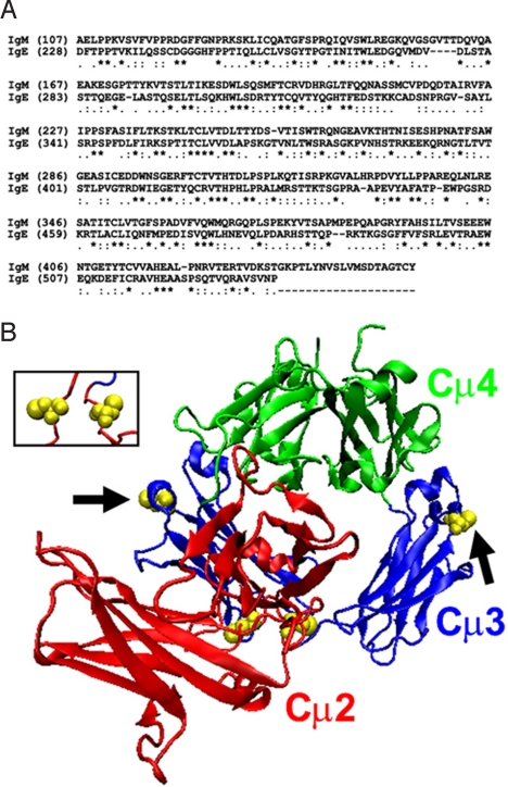
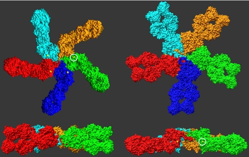
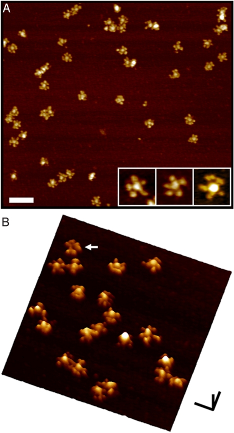
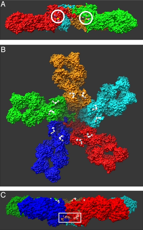
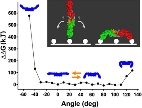
The textbook planar model of pentameric IgM, a potent activator of complement C1q, is based upon the crystallographic structure of IgG. Although widely accepted, key predictions of this model have not yet been directly confirmed, which is particularly important since IgG lacks a major Ig fold domain in its Fc region that is present in IgM. Here, we construct a homology-based structural model of the IgM pentamer using the recently obtained crystallographic structure of IgE Fc, which has this additional Ig domain, under the constraint that all of the cysteine residues known to form disulfide bridges both within each monomer and between monomers are bonded together. In contrast to the planar model, this model predicts a non-planar, mushroom-shaped complex, with the central portion formed by the C-terminal domains protruding out of the plane formed by the Fab domains. This unexpected conformation of IgM is, however, directly confirmed by cryo-atomic force microscopy of individual human IgM molecules. Further analysis of this model with free energy calculations of out-of-plane Fab domain rotations reveals a pronounced asymmetry favoring flexions toward the central protrusion. This bias, together with polyvalent attachment to cell surface antigen, would ensure that the IgM pentamer is oriented on the cell membrane with its C1q binding sites fully exposed to the solution, and thus provides a mechanistic explanation for the first steps of C1q activation by IgM.


Conflict of interest statement

The authors declare no conflict of interest.

Figures

Fig. 1.
Fig. 1.
Homology model of the human IgM Fc based on the structure of the human IgE Fc. (A) The sequence alignment used to generate the model is based on a BLAST search (22) for sequences homologous to IgM that identified IgE as being a highly similar match (E = 2 × 10−45). An asterisk “*” below the alignment denotes identical amino acids while “.” and “:” denote similar and highly similar residues, respectively, as determined by DeepView. (B) Initial model of Fcμ before energy minimization. Shown as yellow van der Waals spheres are the cysteine residues that are known to be involved in intra-monomer (Cys-214) and inter-monomer (Cys-291, arrows) disulfide bridges. Even at this initial stage of modeling, the Cys-214 residues are close to each other (see Inset), while both Cys-291 residues are on the outermost surface of the complex and so could possibly interact with similar residues in neighboring molecules. None of these residues are conserved in IgE, and so their disposition in this structure at locations consistent with expectations lends strong support to the proposed model.
Fig. 2.
Fig. 2.
Evaluation of the possible pentamer structures formed by the energy-minimized monomer, as judged by the relative disposition of the Cys-291 residues. The Left depicts a planar model, but the Cys-291 residues (circled) are not in a position to interact with a neighboring monomer. For this to occur, each must be rotated by 90 ° about its long axis. However, with this, as shown in the Right, the complex is not planar, but now exhibits a central protrusion, largely formed by the Cμ4 domains. In each of the Lower, the blue monomer was removed to facilitate visualization of the complex.
Fig. 3.
Fig. 3.
Cryo-AFM images of individual IgM complexes deposited on mica. (A) Individual, well-separated pentamers are well resolved as star-shaped complexes, with a central ≈20 nm circular region, from which approximately 11 nm radial arms emerge. Most of the complexes have five radial arms that emanate from the central protrusion (see Inset), which are likely the closely apposed pairs of Fab domains of each monomer. Some of the oligomers exhibit more than five radial arms, which is likely a result of a slight separation of Fab domains within some of the monomers. These images are the so-called top-view representations, as if looking down onto the sample along the axis perpendicular to the substrate. (B) The surface view of the complexes clearly shows that the central region in most of the complexes protrudes out from the plane defined by the Fab domains. This is a demonstration of the non-planar topography of these IgM pentamers, and is in agreement with the predictions of the modeling presented here. A few (2%–3%) of the complexes appear to have a lower central region (arrow), which might be a result of mushroom-shaped complexes adsorbed in the opposite orientation on the mica surface. In this surface view representation, the data are presented as if viewed from the side, at an angle, thereby enabling an easier appreciation of the relative differences in height in the sample. [Scale bars (A) 100 nm; (B) x,y: 50 nm, z: 10 nm.]
Fig. 4.
Fig. 4.
Locations of critical residues previously identified in mutational analyses. (A) Encircled are the residues (pink) that were concluded to be involved in monomer-monomer interactions (34). Their location at the monomer-monomer interface in this structure lends further support to the proposed model. (B) Colored in white are the residues that are believed to directly interact with C1q (–36). All of the residues are located on the surface of the complex, as they must be to interact with C1q, providing additional support for the proposed model. Also note that these residues are all located on the same side of the complex, which, as described in the text, places constraints on proposed mechanisms of C1q binding to cell-associated IgM. (c) The residues (light pink) in the white box were recently identified as likely directly interacting with a protein, PfEMP1 that is present in the membranes of erythrocytes infected with the bacteria that causes malaria, Plasmodium falcipaum (37). The location of these residues in this structure is consistent with the observed properties of the interaction with PfEMP1 (requiring oligomerization of IgM, not inhibited by binding by C1q) (37), further supporting the proposed structure.
Fig. 5.
Fig. 5.
Energies associated with out-of-plane Fab domain rotations. The polyvalent attachment of the Fab domains to cell surface antigen can be accomplished if, after a first Fab-antigen interaction, the complex rotates in one of two directions, which results in one of two sides of the structure facing the solution (as portrayed in the Inset). The energies associated with the out-of-plane rotations of the Fab domains in the proposed model were calculated by manually rotating the Fab domains in increments of 10 °, followed by energy minimization. These results identify a pronounced steric hindrance to Fab flexions away from the central protrusion, and an absence of any similar prohibition to flexions toward the protrusion by up to 110 °. This result suggests that the complex would likely be oriented with the flat side facing the solution, when the Fab domains from more than a single monomer are associated with cell-surface antigen. This is also the side in which the C1q-binding sites are located (see Fig. 4B).

References

    1. Boes M, Prodeus AP, Schmidt T, Carroll MC, Chen J. A critical role of natural immunoglobulin M in immediate defense against systemic bacterial infection. J Exp Med. 1998;188:2381–2386. - PMC - PubMed
    1. Ochsenbein AF, et al. Control of early viral and bacterial distribution and disease by natural antibodies. Science. 1999;286:2156–2159. - PubMed
    1. Vollmers HP, Brandlein S. Natural IgM antibodies: The orphaned molecules in immune surveillance. Adv Drug Deliv Rev. 2006;58:755–765. - PubMed
    1. Beutner U, et al. Neoadjuvant therapy of gastric cancer with the human monoclonal IgM antibody SC-1: Impact on the immune system. Oncol Rep. 2008;19:761–769. - PubMed
    1. Illert B, et al. Human antibody SC-1 reduces disseminated tumor cells in nude mice with human gastric cancer. Oncol Rep. 2005;13:765–770. - PubMed

Publication types

Substances

LinkOut - more resources