. 2009 Sep 1;106(35):15002-6.
doi: 10.1073/pnas.0907339106. Epub 2009 Aug 17.

Measuring prions by bioluminescence imaging

Affiliations

Measuring prions by bioluminescence imaging

Gültekin Tamgüney et al. Proc Natl Acad Sci U S A. .

Abstract

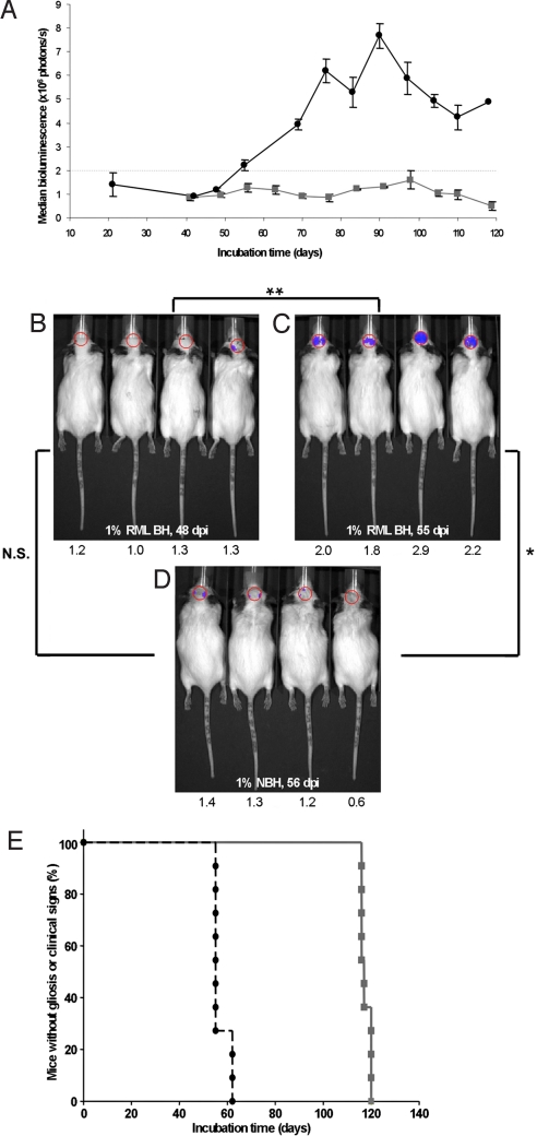
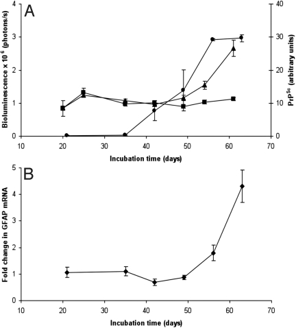
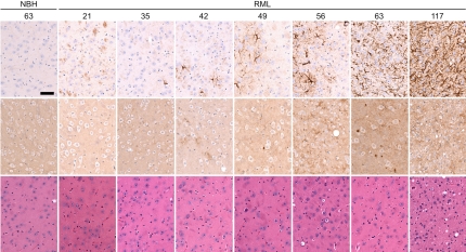
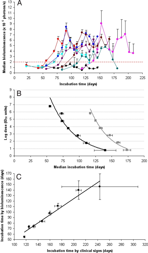
Prions are infectious proteins that cause fatal neurodegenerative diseases. Because astrocytic gliosis marked by the deposition of fibrils composed of GFAP is a prominent feature of prion disease, we asked whether GFAP might be used as a surrogate marker for prions. To interrogate this posit, we inoculated prions into transgenic (Tg) mice expressing luciferase (luc) under the GFAP gene (Gfap) promoter, denoted Tg(Gfap-luc) mice. Weekly noninvasive, bioluminescence imaging (BLI) detected an increase in light emitted from the brains of Tg(Gfap-luc) mice at approximately 55 d after inoculation and approximately 62 d before neurologic deficits appeared. To determine whether BLI could be used as a proxy bioassay for prion infectivity, we performed endpoint titrations of prions in Tg(Gfap-luc) mice. BLI bioassays were as or more sensitive than those determined by the onset of neurological dysfunction, and were completed in approximately half the time. Our studies argue that BLI is likely to be a suitable surrogate for measuring prion infectivity, and might be useful in the study of Tg mouse models for other neurodegenerative illnesses.


Conflict of interest statement

The authors declare no conflict of interest.

Figures

Fig. 1.
Fig. 1.
BL in Tg(Gfap-luc) mice i.c. inoculated with RML prions (n = 12) indicates reactive astrocytic gliosis. (A) BL measured from the brains of prion-inoculated mice (black circles) begins to increase at 55 dpi. BL in control Tg(Gfap-luc) mice inoculated with 1% normal brain homogenate (NBH) (n = 4, gray squares) remains low throughout the incubation period. (B–D) Photos of representative Tg(Gfap-luc) mice, with overlays of the circular area above the brain from which BL was quantified. The BL measured, ×106 photons/s, from each mouse brain is shown below each image. The BL measured from the brains of prion-infected mice significantly increased (**, P < 0.001, Bonferroni t test) from 48 dpi (B) to 55 dpi (C). Similarly, BL measured from infected mice at 55 dpi (C) was also significantly (*, P < 0.005) greater than in control mice inoculated with NBH and imaged at 56 dpi (D). No significant difference (N.S., P > 0.5) was measured between RML-inoculated mice at 48 dpi (B) and control mice at 56 dpi (D). Based on this result, we determined that astrocytic gliosis was detectable at BL values ≥2.0 × 10⁶ photons/s. (E) Kaplan–Meier plots for incubation times based on the appearance clinical signs (gray squares) or on the onset of reactive astrocytic gliosis (black circles). The median onset time for clinical signs was 117 ± 1.1 dpi (95% confidence interval), whereas that for gliosis was 55 ± 2.8 dpi.
Fig. 2.
Fig. 2.
During prion infection, increases in PrPSc preceded increases in BL (A) and GFAP mRNA (B). (A) Protease-resistant PrPSc (circles, right ordinate) in the brains of RML-infected Tg(Gfap-luc) mice began to accumulate at 42 dpi and increased steadily. PrPSc levels were quantified at each time point by densitometry of Western blottings. BL (triangles, left ordinate) in the brains of infected Tg(Gfap-luc) mice increased beginning at 56 dpi, ≈14 d after PrPSc accumulation. BL measured from the brains of control mice inoculated with 1% NBH did not increase (squares). (B) Correlating with an increase of BL, GFAP mRNA also increased at ≈56 dpi. Error bars indicate the SE, based on at least 9 mice for BL measurements and 3 mice for all other measurements.
Fig. 3.
Fig. 3.
Immunohistochemistry of thalamic brain sections from Tg(Gfap-luc) mice show time-dependent progression of reactive astrocytic gliosis and neuropathology after RML inoculation. Tg(Gfap-luc) mice inoculated with 1% NBH are shown as controls. Mice were killed at the time (dpi) indicated above each column. Sections were stained with peroxidase for GFAP (Top), anti-PrP antibodies for PrPSc deposition (Middle), and hematoxylin and eosin for vacuolation (Bottom). At 21 dpi, both RML-inoculated and control mice developed some mild gliosis due to inoculation-induced trauma that disappeared at 35 dpi. In contrast to control mice, gliosis increased in RML-inoculated mice at 42 dpi and continued until 117 dpi, when the mice developed clinical signs. PrPSc deposits were detected beginning at 42 dpi (Middle), and vacuolation was not evident until late in the disease process (Bottom). (Scale bar, 150 μm.)
Fig. 4.
Fig. 4.
The inoculated prion dose is related to BL in Tg(Gfap-luc) mice. (A) Median BL measured from the brains of Tg(Gfap-luc) mice inoculated with 10% RML brain homogenate at the following serial log dilutions: −1 (red), −2 (turquoise), −3 (blue), −4 (violet), −5 (brown), −6 (green), and −7 (pink). BL from Tg(Gfap-luc) mice inoculated with 1% NBH is shown as a control (black). (B) Using the method of Spearman–Kärber, log doses were calculated for the log dilutions shown in A, based on the median incubation times for astrocytic gliosis (BL exceeding 2 × 10⁶ photons/s; black curve) and median incubation times for clinical signs (gray curve). BLI detects prion infection from −1 log dilution higher than the incubation-time assay. (C) For each dilution, median incubation times for clinical signs plotted against the median incubation times for astrocytic gliosis show a linear relationship. Error bars indicate the SE.

References

    1. Chesebro B. Introduction to the transmissible spongiform encephalopathies or prion diseases. Br Med Bull. 2003;66:1–20. - PubMed
    1. Weissmann C. The state of the prion. Nat Rev Microbiol. 2004;2:861–871. - PubMed
    1. Caughey B, Baron GS. Prions and their partners in crime. Nature. 2006;443:803–810. - PubMed
    1. Prusiner SB. Prions. In: Knipe DM, et al., editors. Fields Virology. 5th Ed. Philadelphia: Lippincott Williams & Wilkins; 2007. pp. 3059–3092.
    1. Aguzzi A, Sigurdson C, Heikenwaelder M. Molecular mechanisms of prion pathogenesis. Annu Rev Pathol. 2008;3:11–40. - PubMed

Publication types

LinkOut - more resources