. 2009 Aug 25;106(34):14536-41.
doi: 10.1073/pnas.0907560106.

Antiestrogen-resistant subclones of MCF-7 human breast cancer cells are derived from a common monoclonal drug-resistant progenitor

Affiliations

Antiestrogen-resistant subclones of MCF-7 human breast cancer cells are derived from a common monoclonal drug-resistant progenitor

Kathryn R Coser et al. Proc Natl Acad Sci U S A. .

Abstract

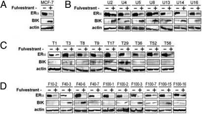
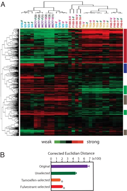
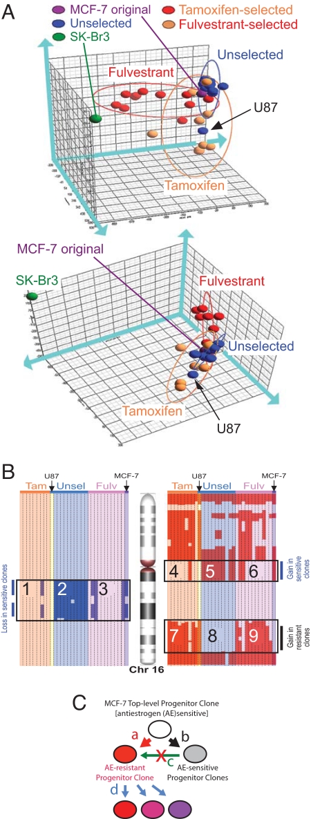
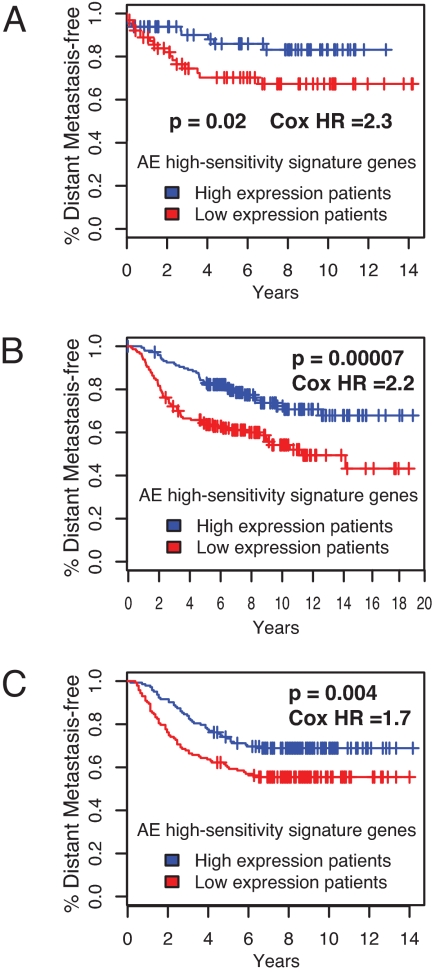
Emergence of antiestrogen-resistant cells in MCF-7 cells during suppression of estrogen signaling is a widely accepted model of acquired breast cancer resistance to endocrine therapy. To obtain insight into the genomic basis of endocrine therapy resistance, we characterized MCF-7 monoclonal sublines that survived 21-day exposure to tamoxifen (T-series sublines) or fulvestrant (F-series sublines) and sublines unselected by drugs (U-series). All T/F-sublines were resistant to the cytocidal effects of both tamoxifen and fulvestrant. However, their responses to the cytostatic effects of fulvestrant varied greatly, and their remarkably diversified morphology showed no correlation with drug resistance. mRNA expression profiles of the U-sublines differed significantly from those of the T/F-sublines, whose transcriptomal responsiveness to fulvestrant was largely lost. A set of genes strongly expressed in the U-sublines successfully predicted metastasis-free survival of breast cancer patients. Most T/F-sublines shared highly homogeneous genomic DNA aberration patterns that were distinct from those of the U-sublines. Genomic DNA of the U-sublines harbored many aberrations that were not found in the T/F-sublines. These results suggest that the T/F-sublines are derived from a common monoclonal progenitor that lost transcriptomal responsiveness to antiestrogens as a consequence of genetic abnormalities many population doublings ago, not from the antiestrogen-sensitive cells in the same culture during the exposure to antiestrogens. Thus, the apparent acquisition of antiestrogen resistance by MCF-7 cells reflects selection of preexisting drug-resistant subpopulations without involving changes in individual cells. Our results suggest the importance of clonal selection in endocrine therapy resistance of breast cancer.


Conflict of interest statement

The authors declare no conflict of interest.

Figures

Fig. 1.
Fig. 1.
AE sensitivities of MCF-7 monoclonal sublines. (A) Phase-contrast images of the original MCF-7 cell culture and its monoclonal sublines isolated in the presence of Fv: 40 nM (F40–3, -6, -7) or 100 nM (F100–1, -3, -7, -16). (B and C) Drug resistance of Tam- or Fv-selected sublines. Cells were exposed to Fv (0.1 μM), 4-hydroxyTam (1 μM), paclitaxel (1 μM), or vehicle (0.1% ethanol) for 10 days, followed by visualization (B) and quantitation (C) of survived cells by sulforhodamine-B staining. Survived cell numbers relative to vehicle controls (mean ± SEM) are shown for individual sublines (open symbols) and each group (closed symbols). (D) AE-induced activation of caspase-7. Relative numbers of DEVDase-positive cells (mean ± SEM, n ≥ 3) after 48-hour exposure to 0.1 μM Fv (open symbols) or vehicle (closed symbols) are plotted. Small symbols represent individual sublines. Large symbols represent groups. (E) Cell cycle arrest by Fv. Unselected, Tam-selected, and Fv-selected sublines were exposed to Fv (0.1 μM) or vehicle (0.1% ethanol) for 48 h. Symbols indicate the relative S-phase cell population in cultures exposed to vehicle (open symbols) or to Fv (closed symbols). All data indicate the mean of at least 3 independent assays whose SEs were not greater than 10% of the means.
Fig. 2.
Fig. 2.
Expression of ERα and BIK proteins in MCF-7 sublines. Cells were exposed to Fv (0.1 μM) or vehicle (0.1% ethanol) for 48 h and subjected to Western blotting for detection of ER↑ and BIK. (A) Original MCF-7 cells, (B) unselected, AE-sensitive sublines, (C) Tam-selected, and (D) Fv-selected AE-resistant sublines.
Fig. 3.
Fig. 3.
Transcriptomal profiles of MCF-7 monoclonal sublines. (A) Two-way hierarchical clustering of Tam-selected (T8, T17, T29, and T52), Fv-selected (F40–6, F40–7, F100–3, and F100–16), and unselected (U2, U4, U5, and U16) sublines and original MCF-7 culture (3 independent cultures) exposed to Fv (-F suffix; 0.1 μM) or vehicle (-V suffix; 0.1% ethanol) for 48 h. Columns represent cell culture samples; rows represent genes. Red and green vertical color bars indicate genes differentially expressed between AE-sensitive and -resistant cells; blue and gray vertical color bars represent genes that do not respond to Fv in AE-resistant cells. (B) Corrected Euclidian transcriptomal distances between the Fv- and vehicle-exposed cultures. Asterisks indicate significant difference (P < 0.001) from unselected sublines.
Fig. 4.
Fig. 4.
gDNA copy number profiling of MCF-7 sublines. (A) Principal component analysis (PCA). Whole-genome copy number profiles of the MCF-7 sublines, the original MCF-7 culture, and SK-Br3 cells are subjected to dimensionality reduction and projected in a 3D space that is shown from 2 different angles of view. Blue, orange, and red ellipsoids represent the PCA spaces for the U-, T-, and F-series sublines, respectively. The arrow indicates the U87 subline, which belongs to the U-series but has its gDNA aberration profile and located within the T-series ellipsoid. (B) Copy number profiling of chromosome 16. Columns represent individual sublines. gDNA segments with increased and decreased copy numbers are indicated by red and blue, respectively. Fulv, fulvestrant selected; Tam, tamoxifen selected; Unsel, unselected series of monoclonal sublines. The original MCF-7 cells and the U-series sublines were sensitive to the cytocidal fulvestrant actions; the T- and F-series sublines and U87 subline (which belongs to the U-series) were resistant. (C) Evolution of the AE-resistant subpopulations in MCF-7 cells. From the top-level progenitor clone, an AE-resistant progenitor clone and an AE-sensitive progenitor clone were generated through accumulation of different sets of gDNA aberrations (arrows a and b, respectively). The AE-sensitive progenitor clone propagated to form the major subpopulation in MCF-7 cells (arrow d); the progenies of the AE-resistant progenitor clone remained as a minor subpopulation. The AE-resistant progenitor clone was not a progeny of the AE-sensitive progenitor clone (arrow c). Progenies of the AE-resistant clone evolved diversified phenotypic features through genetic and nongenetic changes.
Fig. 5.
Fig. 5.
Prediction of a metastasis-free period of breast cancer patients by tumor expression of AE-sensitivity signature genes identified by MCF-7 monoclonal subline analysis. Patients were divided into 2 groups based on the expression of the AE-sensitivity signature genes. Kaplan-Meier curves for emergence of distant metastases in these 2 groups are based on large-scale clinical data described by (A) Chin et al. (16), (B) van de Vijver et al. (17), and (C) Wang et al. (18). One-sided p-values and Cox hazard ratios between the 2 groups are shown.

References

    1. Jordan VC, Brodie AM. Development and evolution of therapies targeted to the estrogen receptor for the treatment and prevention of breast cancer. Steroids. 2007;72:7–25. - PMC - PubMed
    1. Lacroix M, Leclercq G. Relevance of breast cancer cell lines as models for breast tumours: An update. Breast Cancer Res Treat. 2004;83:249–289. - PubMed
    1. Brodie A, et al. Therapeutic observations in MCF-7 aromatase xenografts. Clin Cancer Res. 2005;11:884s–888s. - PubMed
    1. Hur J, et al. Regulation of expression of BIK proapoptotic protein in human breast cancer cells: p53-dependent induction of BIK mRNA by fulvestrant and proteasomal degradation of BIK protein. Cancer Res. 2006;66:10153–10161. - PubMed
    1. Hur J, et al. The Bik BH3-only protein is induced in estrogen-starved and antiestrogen-exposed breast cancer cells and provokes apoptosis. Proc Natl Acad Sci USA. 2004;101:2351–2356. - PMC - PubMed

Publication types

MeSH terms

Associated data