. 2009 Oct 23;284(43):29514-25.
doi: 10.1074/jbc.M109.027896. Epub 2009 Aug 25.

MicroRNA expression signature and the role of microRNA-21 in the early phase of acute myocardial infarction

Affiliations

MicroRNA expression signature and the role of microRNA-21 in the early phase of acute myocardial infarction

Shimin Dong et al. J Biol Chem. .

Abstract

Several recent reports have suggested that microRNAs (miRNAs) might play critical roles in acute myocardial infarction (AMI). However, the miRNA expression signature in the early phase of AMI has not been identified. In this study, the miRNA expression signature was investigated in rat hearts 6 h after AMI. Compared with the expression signature in the noninfarcted areas, 38 miRNAs were differentially expressed in infarcted areas and 33 miRNAs were aberrantly expressed in the border areas. Remarkably, miR-21 expression was significantly down-regulated in infarcted areas, but was up-regulated in border areas. The down-regulation of miR-21 in the infarcted areas was inhibited by ischemic preconditioning, a known cardiac protective method. Overexpression of miR-21 via adenovirus expressing miR-21 (Ad-miR-21) decreased myocardial infarct size by 29% at 24 h and decreased the dimension of left ventricles at 2 weeks after AMI. Using both gain-of-function and loss-of-function approaches in cultured cardiac myocytes, we identified that miR-21 had a protective effect on ischemia-induced cell apoptosis that was associated with its target gene programmed cell death 4 and activator protein 1 pathway. The protective effect of miR-21 against ischemia-induced cardiac myocyte damage was further confirmed in vivo by decreased cell apoptosis in the border and infarcted areas of the infarcted rat hearts after treatment with Ad-miR-21. The results suggest that miRNAs such as miR-21 may play critical roles in the early phase of AMI.


Figures

FIGURE 1.
FIGURE 1.
Verification of some aberrantly expressed miRNAs in infarcted areas of the hearts by qRT-PCR. miRNAs were isolated in the noninfarcted area and the infarcted area of the infarcted hearts. The expression of some miRNAs that were aberrantly expressed in the infarcted area was verified by qRT-PCR. Note: Data presented as mean ± S.E. (error bars), n = 5, *, p < 0.05 compared with the noninfarcted control.
FIGURE 2.
FIGURE 2.
miR-21 expression in different areas of the infarcted hearts and the effect of IP on the expression in the early phase of AMI. miRNAs were isolated in different areas of the infarcted hearts as well as in sham-opened hearts at 6 (A) and 24 h (B) after AMI. The expression of miR-21 was determined by qRT-PCR. miR-21 expression was down-regulated in the infarcted area at both 6 and 24 h after AMI compared with that in other areas and in sham-opened hearts. Notably, compared with that in other areas and sham-opened hearts, miR-21 expression in the border area was significantly increased. C, the effect of IP on miR-21 expression at 6 h after AMI. The IP was performed 6 h before the AMI. Note: Data presented as mean ± S.E. (error bars), n = 6, *, p < 0.05 compared with the noninfarcted control in A and C, and with the IP group in C. #, p < 0.05 compared with that in the border area.
FIGURE 3.
FIGURE 3.
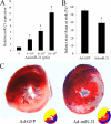
The effect of adenovirus-mediated miR-21 gene transfer on myocardial infarct size. A, Ad-miR-21 increased miR-21 expression in rat hearts in a dose-dependent manner. Ad-miR-21 was delivered into the rat hearts using the local delivery method at doses: 0 (Ad-GFP control), 4 × 10⁸, 8 × 10⁸, or 12 × 108 pfu/rat. Three days later, rat hearts were isolated for miR-21 expression assay by qRT-PCR. Note: Data presented as mean ± S.E. (error bars), n = 5, *, p < 0.05 compared with 0 group. B, Ad-miR-21 (8 × 10⁸ pfu) reduced infarct size in rat hearts at 24 h after AMI. Note: Data presented as mean ± S.E. (error bars), n = 10; *, p < 0.05 compared with Ad-GFP control. C, representative TTC-stained heart slices from rats treated with control adenovirus, Ad-GFP, or Ad-miR-21. The infarcted area in the Ad-GFP-treated image was 56% and the infarcted area in the Ad-21-treated image was 37%. Corresponding color drawings (right corner) show the risk area (red and blue), infracted area (blue), and the Evans blue-stained area (yellow).
FIGURE 4.
FIGURE 4.
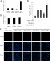
The effect of miR-21 on ischemia-related cell injury in vitro. Cultured cardiac cell injury was induced by hypoxia for 4 h in a serum- and glucose-free medium followed by reoxygenation (H/R) for 3 h in normal culture medium. Cell apoptosis was determined by TUNEL staining. A, miR-21 inhibitor, LNA-anti-miR-21 (30 nm) decreased, but pre-miR-21 (30 nm) increased miR-21 expression in cultured cardiac cells. Note: Data presented as mean ± S.E. (error bars), n = 5, *, p < 0.05 compared with the vehicle control. B, hypoxia/reoxygenation resulted in an increase in apoptosis. Pre-miR-21 decreased the cardiac myocyte apoptosis. In contrast, cardiac myocyte apoptosis was exacerbated after treatment with LNA-anti-miR-21. Note: n = 5, *, p < 0.05 compared with the noninjured (without H/R) control. #, p < 0.05 compared with H/R treated with the vehicle control. C, representative TUNEL-stained photomicrographs from uninjured cells, and cardiac myocytes treated with vehicle, control oligos, pre-miR-21, or LNA-anti-miR-21. Note: red is TUNEL staining representing apoptotic cells; blue is the cell nucleus stained by 4′,6-diamidino-2-phenylindole (DAPI). D, the effect of miR-382 on H/R-mediated cell apoptosis.
FIGURE 5.
FIGURE 5.
The effect of miR-21 overexpression on cardiac cell apoptosis in the border and infarcted areas of rat hearts. A, representative hematoxylin and eosin (HE), myosin heavy chain (MHC), and TUNEL-stained photomicrographs from cardiac myocytes in heart sections from rats treated with Ad-GFP or Ad-miR-21 (8 × 10⁸ per rat). Note: I, infarcted area; B, border area; and n, non-infarcted area. Red is TUNEL staining representing apoptotic cells; blue is the cell nucleus stained by 4′,6-diamidino-2-phenylindole (DAPI). B, quantitative analysis of the apoptotic cells in heart sections. Note: Data presented as mean ± S.E. (error bars), n = 6, *, p < 0.05 compared with the Ad-GFP control.
FIGURE 6.
FIGURE 6.
The effect of adenovirus-mediated miR-21 gene transfer on LV dimensions. A, representative heart sections from sham-opened, Ad-GFP, or Ad-miR-21-treated rats at 2 weeks after AMI. B, Ad-miR-21 (8 × 10⁸ pfu) reduced internal diastolic diameter of left ventricles in rat hearts at 2 weeks after AMI. Note: Data presented as mean ± S.E. (error bars), n = 8; *, p < 0.05 compared with Ad-GFP control.
FIGURE 7.
FIGURE 7.
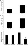
PDCD4 is a miR-21 target gene that is involved in miR-21-mediated anti-apoptotic effect on cardiac cells. A, overexpression of Ad-PDCD4 by Ad-PDCD4 (30 m.o.i.) in cultured cardiac myocytes. Note: n = 3, *, p < 0.05 compared with the Ad-GFP control. B, Ad-PDCD4 (30 m.o.i.) increased cardiac myocyte apoptosis induced by hypoxia/reoxygenation as determined by TUNEL staining. Note: n = 5, *, p < 0.05 compared with the Ad-GFP control. C, modulation of PDCD4 expression in cardiac myocytes by LNA-anti-miR-21 (30 nm) and pre-miR-21 (30 nm). Note: Data presented as mean ± S.E. (error bars), n = 6, *, p < 0.05 compared with the vehicle control.
FIGURE 8.
FIGURE 8.
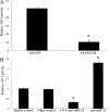
AP-1 is a downstream signaling molecule of PDCD4 that may be involved in miR-21-mediated effect on cardiac myocytes. A, overexpression of PDCD4 by Ad-PDCD4 (30 m.o.i.) inhibited AP-1 activity. Note: n = 5, *, p < 0.05 compared with the Ad-GFP control. B, modulation of AP-1 activity in cardiac myocytes by LNA-anti-miR-21 (30 nm) and pre-miR-21 (30 nm). Note: Data presented as mean ± S.E. (error bars), n = 5, *, p < 0.05 compared with the vehicle control.

References

    1. Ambros V. (2003) Cell 113, 673–676 - PubMed
    1. Farh K. K., Grimson A., Jan C., Lewis B. P., Johnston W. K., Lim L. P., Burge C. B., Bartel D. P. (2005) Science 310, 1817–1821 - PubMed
    1. Pasquinelli A. E., Hunter S., Bracht J. (2005) Curr. Opin. Genet. Dev. 15, 200–205 - PubMed
    1. Zaug A. J., Cech T. R. (1986) Science 231, 470–475 - PubMed
    1. Kong Y., Han J. H. (2005) Genomics Proteomics Bioinformatics 3, 62–72 - PMC - PubMed

Publication types