. 2009 Nov 20;284(47):32454-61.
doi: 10.1074/jbc.M109.023630. Epub 2009 Aug 25.

T cell receptor signaling co-regulates multiple Golgi genes to enhance N-glycan branching

Affiliations

T cell receptor signaling co-regulates multiple Golgi genes to enhance N-glycan branching

Hung-Lin Chen et al. J Biol Chem. .

Abstract

T cell receptor (TCR) signaling enhances beta1,6GlcNAc-branching in N-glycans, a phenotype that promotes growth arrest and inhibits autoimmunity by increasing surface retention of cytotoxic T lymphocyte antigen-4 (CTLA-4) via interactions with galectins. N-Acetylglucosaminyltransferase V (MGAT5) mediates beta1,6GlcNAc-branching by transferring N-acetylglucosamine (GlcNAc) from UDP-GlcNAc to N-glycan substrates produced by the sequential action of Golgi alpha1,2-mannosidase I (MIa,b,c), MGAT1, alpha1,2-mannosidase II (MII, IIx), and MGAT2. Here we report that TCR signaling enhances mRNA levels of MIa,b,c and MII,IIx in parallel with MGAT5, whereas limiting levels of MGAT1 and MGAT2. Blocking the increase in MI or MII enzyme activity induced by TCR signaling with deoxymannojirimycin or swainsonine, respectively, limits beta1,6GlcNAc-branching, suggesting that enhanced MI and MII activity are both required for this phenotype. MGAT1 and MGAT2 have an approximately 250- and approximately 20-fold higher affinity for UDP-GlcNAc than MGAT5, respectively, and increasing MGAT1 expression paradoxically inhibits beta1,6GlcNAc branching by limiting UDP-GlcNAc supply to MGAT5, suggesting that restricted changes in MGAT1 and MGAT2 mRNA levels in TCR-stimulated cells serves to enhance availability of UDP-GlcNAc to MGAT5. Together, these data suggest that TCR signaling differentially regulates multiple N-glycan-processing enzymes at the mRNA level to cooperatively promote beta1,6GlcNAc branching, and by extension, CTLA-4 surface expression, T cell growth arrest, and self-tolerance.


Figures

FIGURE 1.
FIGURE 1.
mRNA levels of N-glycan GlcNAc branching pathway genes in resting Jurkat T cells. A, N-glycosylation pathway in mammals, with enzyme and human gene names above and below the arrows, respectively. OT/DDOST, dolichyl-diphosphooligosaccharide-protein glycosyltransferase; GI/GCS1, glucosidase I; GII/GANAB, glucosidase II; MIa,b,c/MAN1A1, MAN1A2, MAN1C1, α-mannosidase I; MII,IIx/MAN2A1, MAN2A2, α-mannosidase II; TI, TII, TIV, TV/MGAT1, MGAT2, MGAT4a,b, and MGAT5, N-acetylglucosaminyltransferases I, II, IV, and V. B, relative mRNA expression of N-glycan pathway genes in resting Jurkat T cells by TaqMan quantitative real-time PCR, normalized to MAN2A1. Error bars represent mean ± S.E. of triplicate values. C, relative mRNA decay by TaqMan quantitative real-time PCR following the addition of the transcriptional inhibitor actinomycin D to unstimulated Jurkat T cells. mRNA level at 0 h was arbitrarily set at log 3 for all genes. The half-life of each gene was calculated by converting Ct values via a standard curve generated for each primer.
FIGURE 2.
FIGURE 2.
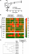
mRNA levels of N-glycan pathway genes in activated Jurkat T cells. A, flow cytometry analysis of Jurkat T cells stained with L-PHA-FITC, a marker of GlcNAc branching in N-glycans, following stimulation with soluble anti-CD3 (OKT3) and co-stimulatory soluble anti-CD28 for 72 h; MFI, mean fluorescence intensity. Error bars represent mean ± S.E. of triplicate values. B, quantitative real-time PCR of N-glycan pathway (MAN1A1, MAN1A2, MAN1C1, MAN2A1, MAN2A2, MGAT1, MAGT2, and MGAT5) and cytokine (IFNγ, IL-2, and IL-4) genes in Jurkat T cells stimulated with soluble anti-CD3 + soluble anti-CD28 (1 μg/ml). The -fold difference was calculated by the ΔΔCt method using glyceraldehyde-3-phosphate dehydrogenase as an endogenous control, with colorimetric results displaying comparisons with unstimulated mock control cells at each time point (i.e. 0, 3, 6, 9, 12, 24, 48, and 72 h). C, 10 kb of 5′-untranslated region nucleotide sequences of N-glycan pathway genes were retrieved from the Geneatlas data base and analyzed by the BioEdit software. The phylogenetic graph and distance were generated by the Neighbor-Joining/UPGMA method Version 3.62a2.1. N/A, not applicable.
FIGURE 3.
FIGURE 3.
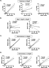
mRNA correlation of MGAT5 with N-glycan and cytokine genes following TCR activation. A–D, expression data from Fig. 2B (both 0.5 μg/ml and 1 μg/ml anti-CD3-stimulated cells) were plotted to compare MGAT5 with cytokines (IL-2, IL-4, and INF-γ) (A), MGAT1 and MGAT2 (B), mannosidase II (MAN2A1 and MAN2A2) (C), and mannosidase I (MAN1A1, MAN1A2, and MAN1C1) (D). The correlation coefficient R2, p value, and 95% confidence intervals (dotted line) were calculated by the Prism software.
FIGURE 4.
FIGURE 4.
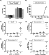
N-Glycan branching and mRNA levels of Golgi genes in TCR activated human T cells. A and C–F, PBMCs from five healthy human control subjects at rest or stimulated with either 1 μg/ml soluble anti-CD3 or 1 μg/ml soluble anti-CD28 were analyzed for L-PHA binding at 72 h by flow cytometry (A) and mRNA levels of the indicated genes at 3, 6, 9, and 12 h by TaqMan quantitative real-time PCR (C–F) as described in the legend for Fig. 2. Cells in A are gated on the CD4+ population. p values in C–E were determined by one-way analysis of variance and Neuman-Keuls multiple comparison test (only p values < 0.05 are shown). MFI, mean fluorescence intensity. B, PBMCs were stimulated for 48 h with increasing amounts of plate-bound anti-CD3 antibody (1, 2, 4, 8, 16, and 32 ng/ml) + 2 μg/ml soluble anti-CD28. L-PHA binding in CD4+ T cells and IL-2 levels in the supernatant were measured by FACS and ELISA, respectively. Error bars represent mean ± S.E. of triplicate values.
FIGURE 5.
FIGURE 5.
TCR signaling-mediated enhancement of mannosidase I and mannosidase II enzyme activity increases N-glycan GlcNAc branching in Jurkat T cells. A, splenocytes from a non-immunized control mouse and a mouse with EAE (clinical score = 2) induced by immunization with MBP + CFA were analyzed by flow cytometry for T cell activation markers (CD69 and CD25) and L-PHA binding (stained in triplicate). Cells are gated on the CD4+ population. MFI, mean fluorescence intensity. Error bars represent mean ± S.E. of triplicate values. p values were determined by Student's t test. B, flow cytometry analysis of concanavalin A-FITC (ConA)- and L-PHA-FITC-stained resting Jurkat T cells treated with increasing concentrations of a mannosidase I or mannosidase II inhibitor for 72 h. Concanavalin A-FITC binds high mannose N-glycans, whereas L-PHA binds β1,6GlcNAc-branched N-glycans. C, Jurkat T cells were stimulated with phorbol 12-myristate 13-acetate (PMA) and ionomycin (Ionom.) in the presence or absence of minimal concentrations of DMN or SW and analyzed by L-PHA-FITC flow cytometry. The low DMN and SW concentrations were chosen because they have little or no effect on L-PHA binding in unstimulated Jurkat T cells.

References

    1. Demetriou M., Granovsky M., Quaggin S., Dennis J. W. (2001) Nature 409, 733–739 - PubMed
    1. Morgan R., Gao G., Pawling J., Dennis J. W., Demetriou M., Li B. (2004) J. Immunol. 173, 7200–7208 - PubMed
    1. Comelli E. M., Sutton-Smith M., Yan Q., Amado M., Panico M., Gilmartin T., Whisenant T., Lanigan C. M., Head S. R., Goldberg D., Morris H. R., Dell A., Paulson J. C. (2006) J. Immunol. 177, 2431–2440 - PubMed
    1. Toscano M. A., Bianco G. A., Ilarregui J. M., Croci D. O., Correale J., Hernandez J. D., Zwirner N. W., Poirier F., Riley E. M., Baum L. G., Rabinovich G. A. (2007) Nat. Immunol. 8, 825–834 - PubMed
    1. Amano M., Galvan M., He J., Baum L. G. (2003) J. Biol. Chem. 278, 7469–7475 - PubMed

Publication types