. 2009 Oct 9;284(41):28476-28484.
doi: 10.1074/jbc.M109.050864. Epub 2009 Aug 25.

Nitric oxide regulates lung carcinoma cell anoikis through inhibition of ubiquitin-proteasomal degradation of caveolin-1

Affiliations

Nitric oxide regulates lung carcinoma cell anoikis through inhibition of ubiquitin-proteasomal degradation of caveolin-1

Pithi Chanvorachote et al. J Biol Chem. .

Abstract

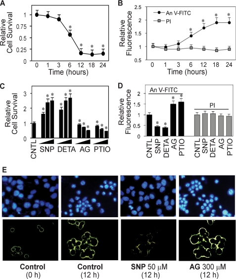
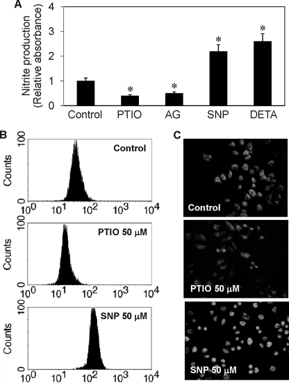
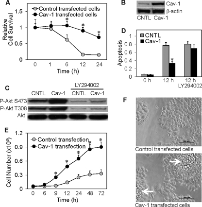
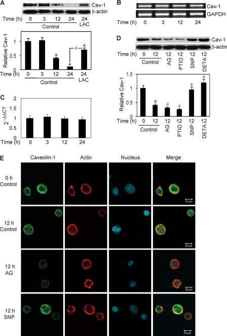
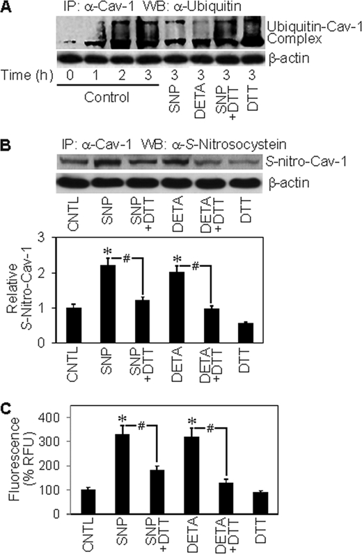
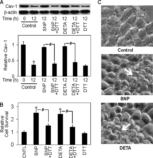
Anoikis, a detachment-induced apoptosis, is a principal mechanism of inhibition of tumor cell metastasis. Tumor cells can acquire anoikis resistance which is frequently observed in metastatic lung cancer. This phenomenon becomes an important obstacle of efficient cancer therapy. Recently, signaling mediators such as caveolin-1 (Cav-1) and nitric oxide (NO) have garnered attention in metastasis research; however, their role and the underlying mechanisms of metastasis regulation are largely unknown. Using human lung carcinoma H460 cells, we show that NO impairs the apoptotic function of the cells after detachment. The NO donors sodium nitroprusside and diethylenetriamine NONOate inhibit detachment-induced apoptosis, whereas the NO inhibitors aminoguanidine and 2-(4-carboxyphenyl) tetramethylimidazoline-1-oxyl-3-oxide promote this effect. Resistance to anoikis in H460 cells is mediated by Cav-1, which is significantly down-regulated after cell detachment through a non-transcriptional mechanism involving ubiquitin-proteasomal degradation. NO inhibits this down-regulation by interfering with Cav-1 ubiquitination through a process that involves protein S-nitrosylation, which prevents its proteasomal degradation and induction of anoikis by cell detachment. These findings indicate a novel pathway for NO regulation of Cav-1, which could be a key mechanism of anoikis resistance in tumor cells.


Figures

FIGURE 1.
FIGURE 1.
Detachment-induced apoptosis and its inhibition by NO. A, effect of cell detachment on cell survival determined by XTT assay. Lung epithelial H460 cells were detached as described under “Materials and Methods” and suspended in HEMA-coated plates for various times (0–24 h). B, effect of cell detachment on apoptosis and necrosis determined by flow cytometry using annexin V-FITC (An V-FITC) and PI assays. C, effect of NO modulators on detachment-induced cell death. Detached cells were treated with various concentrations of NO donor, SNP (10, 50, 100 μm), or DETA NONOate (10, 50, 100 μm) or with NO inhibitor, AG (100, 200, 300 μm), or PTIO (10, 50, 100 μm) for 12 h. Cell survival was then determined by XTT assay. CNTL, control. D, effects of NO modulators on detachment-induced apoptosis and necrosis. Detached cells were treated with SNP (50 μm), DETA NONOate (50 μm), AG (300 μm), or PTIO (50 μm) for 12 h, and cell apoptosis and necrosis were determined as described above. E, upper panel, effect of NO modulators on detachment-induced apoptosis determined by Hoechst 33342 nuclear fluorescence staining. Lower panel, effect of NO modulators on detachment-induced apoptosis determined by annexin V-FITC fluorescence microscopy. Data are the mean ± S.D. (n = 3). *, p < 0.05 versus non-treated control.
FIGURE 2.
FIGURE 2.
Effect of NO modulators on cellular NO and nitrite levels. H460 cells were detached and either left untreated or treated with SNP (50 μm), DETA NONOate (50 μm), AG (300 μm), or PTIO (50 μm) for 2 h. A, nitrite production determined by the Griess assay. B and C, NO production determined by flow cytometry and fluorescence microscopy using DAF-DA as a probe. Data are the mean ± S.D. (n = 3). *, p < 0.05 versus non-treated control.
FIGURE 3.
FIGURE 3.
Cav-1 overexpression increases cell death resistance and Akt phosphorylation, alters growth pattern, and increases growth rate. A, H460 cells were stably transfected with Cav-1 or control plasmid as described under “Materials and Methods.” Transfected cells were grown in culture medium and analyzed for cell survival at various times after detachment using XTT assay. Attached cells showed no significant apoptosis during the test period of 24 h (data not shown). B, Western blot analysis of Cav-1 expression in control and Cav-1-transfected cells. Cell extracts were prepared and separated on 10% polyacrylamide-SDS gels, transferred, and probed with Cav-1 antibody. β-Actin was used as a loading control. CNTL, control. C, effect of Cav-1 overexpression on Akt phosphorylation. Transfected cells were detached and incubated in HEMA-coated plates for 12 h in the presence or absence of LY294002 (10 μm). Cell lysates were prepared and analyzed for Akt phosphorylation by Western blotting. Blots were probed with antibodies specific to phospho-Akt (Ser-473 and Thr-308) and Akt. D, apoptosis of the treated cells was analyzed by Hoechst 33342 assay and expressed as the ratio of apoptotic nuclei to total nuclei. E, effect of Cav-1 overexpression on cell proliferation. Cav-1 and control-transfected cells were grown in normal tissue culture plates and analyzed for cell proliferation at various times by hemocytometry. F, morphology of control and Cav-1-transfected cells in culture. Data are the mean ± S.D. (n = 3). *, p < 0.05 versus vector-transfected control.
FIGURE 4.
FIGURE 4.
Effect of cell detachment on Cav-1 expression and its regulation by NO. A, H460 cells were detached and seeded in HEMA-coated plates for various times (0–24 h) in the presence or absence of lactacystin (LAC) (10 μm). Cells extracts were prepared and analyzed for Cav-1 protein expression by Western blotting. Blots were reprobed with β-actin antibody to confirm equal loading of samples. The immunoblot signals were quantified by densitometry, and mean data from independent experiments (one of which is shown here) were normalized to the results in control cells at 0 h. B, RT-PCR analysis of Cav-1 and glyceraldehyde-3-phosphate dehydrogenase (GAPDH) mRNA expression at various times (0–24 h) after cell detachment. C, relative Cav-1 mRNA expression determined by quantitative real-time PCR. The relative mRNA expression was determined by using the comparative CT method as described under “Materials and Methods.” D, detached cells were treated with NO inhibitor, AG (300 μm) or PTIO (50 μm), or with NO donor, SNP (50 μm), or DETA NONOate (50 μm) for 12 h, after which they were analyzed for Cav-1 expression by Western blotting. E, detached cells were either left untreated or treated with SNP (50 μm) or AG (300 μm) for 12 h and analyzed for Cav-1 by immunofluorescence confocal microscopy. Cells were also stained with Alexa Fluor 546-conjugated phalloidin and ToPro-3 to aid visualization of actin cytoskeleton and nucleus. Data are the mean ± S.D. (n = 3). *, p < 0.05 versus attached cell control; #, p < 0.05 versus the indicated control or 12-h detached cell control.
FIGURE 5.
FIGURE 5.
Effects of NO modulators on Cav-1 ubiquitination and S-nitrosylation. A, H460 cells were detached and either left untreated or treated with SNP (50 μm) or DETA NONOate (50 μm) in the presence or absence of DTT (1 mm) in HEMA-coated plates. Cells lysates were prepared and immunoprecipitated (IP) with anti-Cav-1 antibody. The resulting immune complexes were then analyzed for ubiquitin by Western blotting (WB) at various times. Maximum ubiquitination of Cav-1 was observed at ∼3 h after cell detachment. Lysate input was determined by probing β-actin. CNTL, control. B, detached cells were similarly treated with the test agents, and Cav-1 S-nitrosylation was determined by immunoprecipitation using anti-Cav-1 antibody followed by Western blot analysis of the immunoprecipitated protein using anti-S-nitrosocysteine antibody. Densitometry was performed to determine the relative S-nitrosocysteine/β-actin levels. C, Cav-1 S-nitrosylation was determined by fluorometry. Immunoprecipitates from above were incubated with 200 μm HgCl2 and 200 μm diaminonaphthalene in phosphate-buffered saline. NO released from S-nitrosylated Cav-1 was quantified at 375/450 nm. Plots are the mean ± S.D. (n = 3). *, p < 0.05 versus non-treated control; #, p < 0.05 versus the indicated treatment controls.
FIGURE 6.
FIGURE 6.
NO inhibits detachment-induced Cav-1 down-regulation and cell death. A, H460 cells were detached and either left untreated or treated with SNP (50 μm) or DETA NONOate (50 μm) in the presence or absence of DTT (1 mm) in HEMA-coated plates. A, cell lysates were prepared and analyzed for Cav-1 protein expression by Western blotting after 12 h. Densitometry was performed to determine the relative levels of Cav-1 after reprobing the membranes with β-actin antibody. B, cells survival was determined by XTT assay after 12 h. C, morphology of cells treated with SNP (50 μm) or DETA NONOate (50 μm) for 12 h. Data are the mean ± S.D. (n = 3). *, p < 0.05 versus non-treated control; #, p < 0.05 versus the indicated treatment controls.

References

    1. Galbiati F., Volonté D., Liu J., Capozza F., Frank P. G., Zhu L., Pestell R. G., Lisanti M. P. (2001) Mol. Biol. Cell 12, 2229–2244 - PMC - PubMed
    1. Glenney J. R., Jr., Zokas L. (1989) J. Cell Biol. 108, 2401–2408 - PMC - PubMed
    1. Rothberg K. G., Heuser J. E., Donzell W. C., Ying Y. S., Glenney J. R., Anderson R. G. (1992) Cell 68, 673–682 - PubMed
    1. Scherer P. E., Okamoto T., Chun M., Nishimoto I., Lodish H. F., Lisanti M. P. (1996) Proc. Natl. Acad. Sci. U.S.A. 93, 131–135 - PMC - PubMed
    1. Razani B., Schlegel A., Liu J., Lisanti M. P. (2001) Biochem. Soc. Trans. 29, 494–499 - PubMed

Publication types

MeSH terms