. 2009 Sep 1;69(17):7013-21.
doi: 10.1158/0008-5472.CAN-09-0523. Epub 2009 Aug 25.

MUC1-C oncoprotein functions as a direct activator of the nuclear factor-kappaB p65 transcription factor

Affiliations

MUC1-C oncoprotein functions as a direct activator of the nuclear factor-kappaB p65 transcription factor

Rehan Ahmad et al. Cancer Res. .

Abstract

Nuclear factor-kappaB (NF-kappaB) is constitutively activated in diverse human malignancies. The mucin 1 (MUC1) oncoprotein is overexpressed in human carcinomas and, like NF-kappaB, blocks cell death and induces transformation. The present studies show that MUC1 constitutively associates with NF-kappaB p65 in carcinoma cells. The MUC1 COOH-terminal subunit (MUC1-C) cytoplasmic domain binds directly to NF-kappaB p65 and, importantly, blocks the interaction between NF-kappaB p65 and its inhibitor IkappaBalpha. We show that NF-kappaB p65 and MUC1-C constitutively occupy the promoter of the Bcl-xL gene in carcinoma cells and that MUC1-C contributes to NF-kappaB-mediated transcriptional activation. Studies in nonmalignant epithelial cells show that MUC1-C interacts with NF-kappaB in the response to tumor necrosis factor-alpha stimulation. Moreover, tumor necrosis factor-alpha induces the recruitment of NF-kappaB p65-MUC1-C complexes to NF-kappaB target genes, including the promoter of the MUC1 gene itself. We also show that an inhibitor of MUC1-C oligomerization blocks the interaction with NF-kappaB p65 in vitro and in cells. The MUC1-C inhibitor decreases MUC1-C and NF-kappaB p65 promoter occupancy and expression of NF-kappaB target genes. These findings indicate that MUC1-C is a direct activator of NF-kappaB p65 and that an inhibitor of MUC1 function is effective in blocking activation of the NF-kappaB pathway.


Figures

Figure 1
Figure 1. MUC1-C associates with NF-κB p65
A. Lysates from ZR-75-1 cells were immunoprecipitated with anti-p65 or a control IgG. The precipitates were immunoblotted with anti-MUC1-C and anti-p65. B. Amino acid sequence of the MUC1 cytoplasmic domain is shown with the PKCδ, GSK3β, c-Src and c-Abl phosphorylation sites and the serine-rich (SRM) motif. GST and the indicated MUC1-CD fusion proteins bound to glutathione beads were incubated with purified p65. The adsorbates were immunoblotted with anti-p65. Input of the GST and GST-MUC1-CD fusion proteins was assessed by Coomassie blue staining. C. Schematic representation of the NF-κB p65 protein. GST and GST-MUC1-CD bound to glutathione beads were incubated with purified p65(1-306) or p65(354-551). The adsorbates and inputs were immunoblotted with anti-p65. D. Amino acid sequences of peptides derived from MUC1-CD(46-72). GST-p65(1-306) bound to glutathione beads was incubated with MUC1-CD in the absence (Control) and presence of the indicated A and B peptides at a 20-fold excess compared to MUC1-CD. The adsorbates were immunoblotted with anti-MUC1-C (left). GST, GST-MUC1-CD and GST-MUC1-CD(mSRM) bound to glutathione beads were incubated with p65(1-306). The adsorbates were immunoblotted with anti-p65 (right).
Figure 2
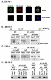
Figure 2. MUC1 attenuates binding of IκBα and NF-κB p65
A-C. Cytosolic lysates from the indicated ZR-75-1/vector, ZR-75-1/MUC1siRNA (A), HeLa/vector, HeLa/MUC1 (B), 3Y1/vector and 3Y1/MUC1-CD (C) cells were immunoprecipitates with anti-p65 or a control IgG. The precipitates were immunoblotted with antibodies against IκBα and p65. D. GST and GST-IκBα bound to glutathione beads were incubated with p65(186-306) in the absence and presence of increasing amounts of MUC1-CD. The adsorbates were immunoblotted with anti-p65 (upper). Input of the MUC1-CD was assessed by immunoblotting with anti-MUC1-C (middle). Input of the GST and GST-IκBα proteins was assessed by Coomassie blue staining (lower).
Figure 3
Figure 3. MUC1-C promotes occupancy of NF-κB p65 on the Bcl-xL gene promoter
A. ZR-75-1/vector and ZR-75-1/MUC1siRNA cells were fixed and double stained with anti-MUC1-C (green) and anti-NF-κB p65 (red). Nuclei were stained with TO-PRO-3. B and C. Soluble chromatin from ZR-75-1/vector, ZR-75-1/MUC1siRNA (B), HeLa/vector and HeLa/MUC1 (C) cells was immunoprecipitated with anti-p65 or a control IgG. The final DNA extractions were amplified by PCR with pairs of primers that cover the NF-κB-RE (-597 to -304) or control region (-1001 to -760) in the Bcl-xL promoter. D. Soluble chromatin from ZR-75-1 cells was immunoprecipitated with anti-MUC1-C or a control IgG and analyzed for Bcl-xL NF-κB-RE or control region sequences (left). In Re-ChIP experiments, the anti-MUC1-C precipitates were released, reimmunopreciptiated with anti-p65 and then analyzed for Bcl-xL promoter sequences (right).
Figure 4
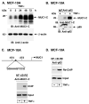
Figure 4. MUC1-C interacts with NF-κB p65 in the response of MCF-10A cells to TNFα
A. MCF-10A cells were stimulated with 20 ng/ml TNFα for the indicated times. Lysates were immunoblotted with anti-MUC1-C and anti-β-actin. B. Lysates from MCF-10A cells left untreated or stimulated with 20 ng/ml TNFα for 24 h were subjected to immunoprecipitation with anti-p65 or a control IgG. The precipitates were immunoblotted with the indicated antibodies. C. Soluble chromatin from MCF-10A cells left untreated and stimulated with 20 ng/ml TNFα for 24 h was immunoprecitated with anti-MUC1-C and then analyzed for MUC1 NF-κB binding motif promoter sequences. D. In Re-ChIP experiments, the anti-MUC1-C precipitates were released, reimmunoprecipitated with anti-p65 and then analyzed for MUC1 NF-κB binding motif promoter sequences.
Figure 5
Figure 5. MUC1-C promotes NF-κB p65-mediated activation of the MUC1 promoters
A and B. MCF-10A cells were transfected with control or p65 siRNA pools for 72 h. The transfected cells were left untreated or stimulated with TNFα for 24 h. Lysates were immunoblotted with the indicated antibodies (A). The cells were then transfected to express a NF-κB-Luc reporter or a MUC1 promoter-Luc reporter (pMUC1-Luc) and, as a control, the SV-40-Renilla-Luc plasmid (B). C and D. MCF-10A cells were transfected with control or MUC1 siRNA pools for 72 h. The transfected cells were left untreated or stimulated with TNFα for 24 h. Lysates were immunoblotted with the indicated antibodies (C). The cells were then transfected to express a NF-κB-Luc reporter or a MUC1 promoter-Luc reporter (pMUC1-Luc) and, as a control, the SV-40-Renilla-Luc plasmid (D). Luciferase activity was measured at 48 h after transfection. The results are expressed as the fold-activation (mean±SD from three separate experiments) compared to that obtained with cells transfected with the control siRNA and left untreated (assigned a value of 1).
Figure 6
Figure 6. GO-201 blocks the interaction between MUC1 and NF-κB p65
A. Sequence of GO-201 and CP-1 with the poly-dArg transduction domain. GST-MUC1-CD was incubated with purified NF-κB p65 in the presence of GO-201 or CP-1 for 1 h at room temperature. Adsorbates to glutathione beads were immunoblotted with anti-p65 (left). MCF-10A cells were left untreated or stimulated with TNFα in the presence of 5 μM GO-201 or CP-1 added each 24 h for 72 h. Anti-p65 precipitates were immunoblotted with the indicated antibodies (right). B and C. MCF-10A cells were left untreated or stimulated with TNFα in the presence of 5 μM GO-201 or CP-1 added each 24 h for 72 h. Soluble chromatin was precipitated with anti-MUC1-C (left) or anti-p65 (right) and then analyzed for MUC1 NF-κB binding motif promoter sequences (B). Lysates were immunoblotted with the indicated antibodies (C). D. Model for the proposed effects of MUC1-C on activation of the NF-κB pathway through interactions with IKKs and p65 in an auto-inductive regulatory loop. The available findings do not exclude the possibility that MUC1-C forms complexes that include both IKKβ and p65.

References

    1. Hayden MS, Ghosh S. Shared principles in NF-kappaB signaling. Cell. 2008;132:344–62. - PubMed
    1. Karin M, Lin A. NF-kappaB at the crossroads of life and death. Nat Immunol. 2002;3:221–7. - PubMed
    1. Hoffman A, Natoli G, Ghosh G. Transcriptional regulation via the NF-kappaB signaling module. Oncogene. 2006;25:6706–16. - PubMed
    1. Chen FE, Ghosh G. Regulation of DNA binding by Rel/NF-kappaB transcription factors: structural views. Oncogene. 1999;18:6845–52. - PubMed
    1. Li Y, Liu D, Chen D, Kharbanda S, Kufe D. Human DF3/MUC1 carcinoma-associated protein functions as an oncogene. Oncogene. 2003;22:6107–10. - PMC - PubMed

Publication types

MeSH terms