. 2009 Sep 1;69(17):7121-9.
doi: 10.1158/0008-5472.CAN-08-4151. Epub 2009 Aug 25.

Ovarian cancer cell detachment and multicellular aggregate formation are regulated by membrane type 1 matrix metalloproteinase: a potential role in I.p. metastatic dissemination

Affiliations

Ovarian cancer cell detachment and multicellular aggregate formation are regulated by membrane type 1 matrix metalloproteinase: a potential role in I.p. metastatic dissemination

Natalie M Moss et al. Cancer Res. .

Abstract

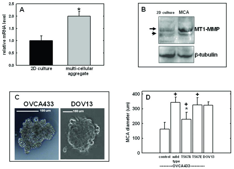
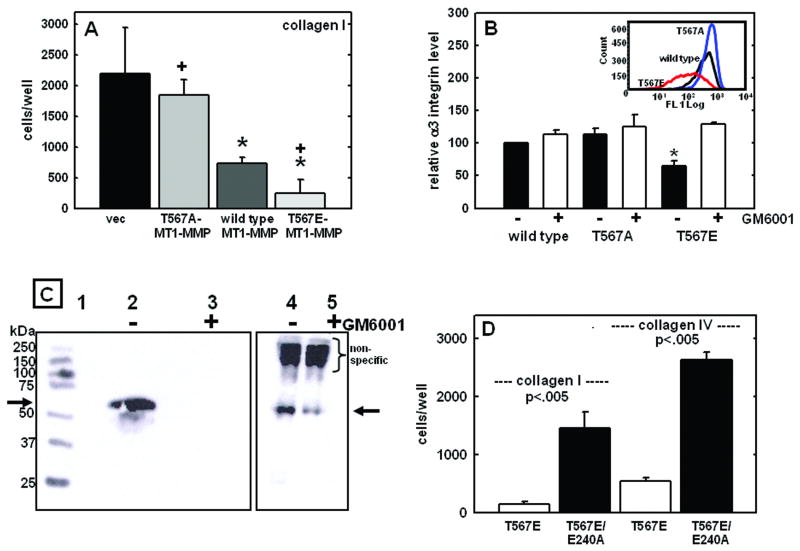
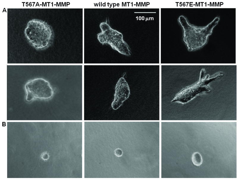
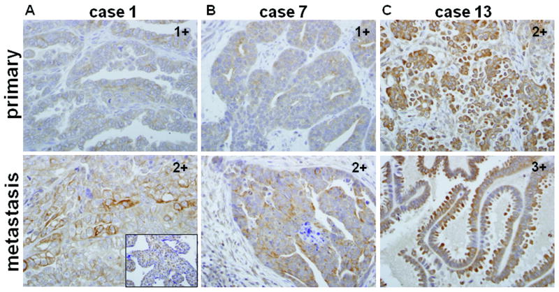
An early event in the metastasis of epithelial ovarian carcinoma is shedding of cells from the primary tumor into the peritoneal cavity followed by diffuse i.p. seeding of secondary lesions. Anchorage-independent metastatic cells are present as both single cells and multicellular aggregates (MCA), the latter of which adhere to and disaggregate on human mesothelial cell monolayers, subsequently forming invasive foci. Although this unique metastatic mechanism presents a distinct set of therapeutic challenges, factors that regulate MCA formation and dissemination have not been extensively evaluated. Proteolytic activity is important at multiple stages in i.p. metastasis, catalyzing migration through the mesothelial monolayer and invasion of the collagen-rich submesothelial matrix to anchor secondary lesions, and acquisition of membrane type 1 matrix metalloproteinase (MT1-MMP; MMP-14) expression promotes a collagen-invasive phenotype in ovarian carcinoma. MT1-MMP is regulated posttranslationally through multiple mechanisms including phosphorylation of its cytoplasmic tail, and the current data using ovarian cancer cells expressing wild-type, phosphomimetic (T567E-MT1-MMP), and phosphodefective (T567A-MT1-MMP) MT1-MMP show that MT1-MMP promotes MCA formation. Confluent T567E-MT1-MMP-expressing cells exhibit rapid detachment kinetics, spontaneous release as cell-cell adherent sheets concomitant with MT1-MMP-catalyzed alpha(3) integrin ectodomain shedding, and robust MCA formation. Expansive growth within three-dimensional collagen gels is also MT1-MMP dependent, with T567E-MT1-MMP-expressing cells exhibiting multiple collagen invasive foci. Analysis of human ovarian tumors shows elevated MT1-MMP in metastases relative to paired primary tumors. These data suggest that MT1-MMP activity may be key to ovarian carcinoma metastatic success by promoting both formation and dissemination of MCAs.


Figures

Figure 1
Figure 1. MT1-MMP expression in MCAs
(A) Expression of MT1-MMP RNA in DOV13 MCAs or 2D cultures. Real time RT-PCR was performed to detect RNA expression. Shown is the average of three independent experiments ± standard deviation. (*p<.05). (B) Expression of MT1-MMP protein. Western blot was performed to detect relative MT1-MMP protein levels with β-tubulin as a loading control. Arrow - 55 kDa active MT1-MMP; arrowhead - 43 kDa MT1-MMP autolysis product. (C) Representative MCA formed by OVCA 433 or DOV13 cells. (D) Expression of MT1-MMP augments MCA formation. Cells were cultured for MCA formation using the hanging drop method and measured using a Zeiss Axiovert Imaging Software. Data shown are expressed in μm, and represent the mean diameter measurement of n=10 MCAs. (+) indicates p<.05 relative to vector-transfected cells; (*) indicates p<.05 relative to cells expressing wild type MT1-MMP. Untransfected DOV13 cells, that express similar levels of endogenous MT1-MMP, are shown for comparison.
Figure 2
Figure 2. MT1-MMP expression promotes detachment of cell-cell adherent monolayers and MCA formation
(A) Confluence initiates detachment of T567E-MT1-MMP-expressing cells. Image shows adherent and detached portions of confluent monolayer with floating aggregate. (B-D) Quantitation of detachment using controlled trypsinization. Kinetics of cell detachment were evaluated by using (B) subconfluent or (C,D) confluent cultures of cells. Experiments were performed in triplicate.
Figure 3
Figure 3. MT1-MMP expression modulates adhesion and α3 integrin ectodomain shedding
(A) Quantitation of adhesion to type I collagen. Results represent averages of three independent experiments. (*) indicates p<.005 relative to vector controls; (+) indicates p<.005 relative to cells expressing wild type MT1-MMP. (B) The effect of MMP inhibition on α3 integrin surface expression. OVCA433 cells were cultured +/− GM6001 (25 uM) overnight prior to harvesting for FACS analysis of integrin α3. (inset) Surface expression of integrin α3 under confluent conditions was assessed by FACS analysis. Blue trace – T567A-MT1-MMP cells; black trace – wild type MT1-MMP cells; red trace – T567E-MT1-MMP cells. (C) Confluent OVCA433 cells expressing T567E-MT1-MMP were cultured in serum-free medium in the absence (lane 2,4) or presence (lane 3,5) of GM6001 (25 uM) overnight. Lanes 2,3 - conditioned media were concentrated and probed with mouse anti-α3 antibody. Lanes 4,5 - immunoprecipitation of unconcentrated conditioned medium with mouse anti-integrin α3 followed by western blotting for integrin α3 extracellular domain. (lane 1 – molecular weight markers in kDa 250, 150, 100, 75, 50, 37, 25). Arrow denotes the migration position of the 70.8 kDa α3 integrin immunoreactive band. (D) Catalytically inactive E240A/T567E-MT1-MMP mutation restores cell adhesion to type I and type IV collagen. Results are averages of three independent experiments.
Figure 4
Figure 4. MT1-MMP promotes matrix-embedded proliferative growth
(A) Cells were seeded at an initial density of 5 × 10⁴ cells/well within 3D collagen gels and photographed after incubation for 6 days. (B) In control experiments, TIMP-2 was copolymerized within the gel at a final concentration of 5ug/ml.
Figure 5
Figure 5. Immunohistochemical analysis of MT1-MMP expression in paired primary ovarian tumors and peritoneal metastatic lesions
(A-C) Examples of paired primary tumor (upper panels) and corresponding metastatic lesion (lower panels) from the same patient stained with MT1-MMP-specific antibodies (1:100 dilution) and peroxidase-conjugated secondary antibody. (inset) - positive control breast carcinoma tissue. Numbers in the upper right corners indicate immunohistological score of each tumor.

References

    1. Jemal A, Siegel R, Ward E, Murry T, Xu J, Thun MJ. Cancer statistics, 2007. CA Cancer J Clin. 2007;57:43–66. - PubMed
    1. Naora H, Montell DJ. Ovarian cancer Metastasis: Integrating insights from disparate model organisms. Nature Rev Cancer. 2005;5:355–366. - PubMed
    1. Ayantunde AA, Parsons SL. Pattern and prognostic factors in patients with malignant ascites. Ann Oncol. 2007;18:945–9. - PubMed
    1. Feldman GB, Knapp RC. Lymphatic drainage of the peritoneal cavity and its significance in ovarian cancer. Am J Obstet Gynecol. 1974;119:991–4. - PubMed
    1. Hudson LG, Zeineldin R, Stack MS. Phenotypic plasticity of neoplastic ovarian epithelium: unique cadherin profiles in tumor progression. Clin Exp Metastasis. 2008;25:643–55. - PMC - PubMed

Publication types