. 2009 Sep 8;106(36):15489-94.
doi: 10.1073/pnas.0904880106. Epub 2009 Aug 25.

Divalent metal transporter 1 (DMT1) regulation by Ndfip1 prevents metal toxicity in human neurons

Affiliations

Divalent metal transporter 1 (DMT1) regulation by Ndfip1 prevents metal toxicity in human neurons

Jason Howitt et al. Proc Natl Acad Sci U S A. .

Abstract

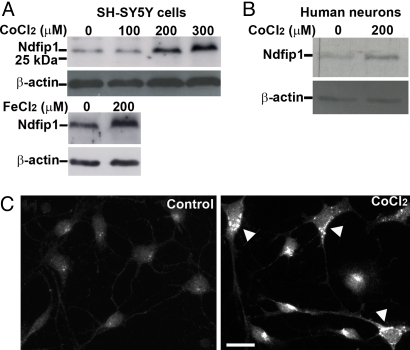
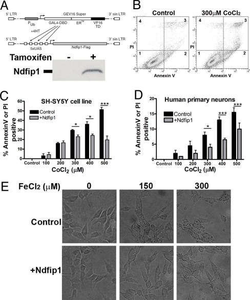
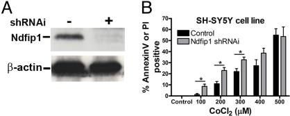
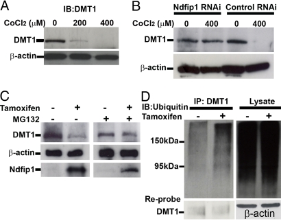
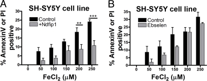
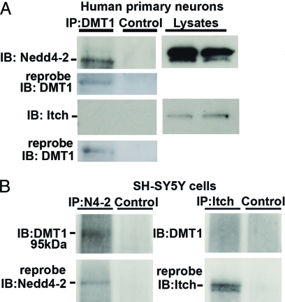
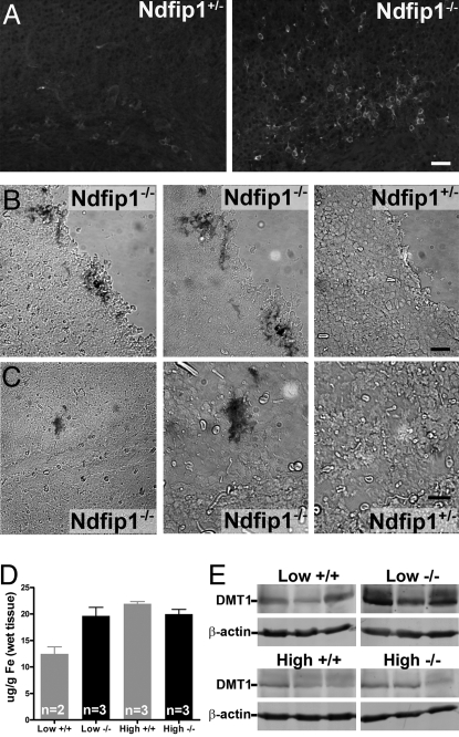
The regulation of metal ion transport within neurons is critical for normal brain function. Of particular importance is the regulation of redox metals such as iron (Fe), where excess levels can contribute to oxidative stress and protein aggregation, leading to neuronal death. The divalent metal transporter 1 (DMT1) plays a central role in the regulation of Fe as well as other metals; hence, failure of DMT1 regulation is linked to human brain pathology. However, it remains unclear how DMT1 is regulated in the brain. Here, we show that DMT1 is regulated by Ndfip1 (Nedd4 family-interacting protein 1), an adaptor protein that recruits E3 ligases to ubiquitinate target proteins. Using human neurons we show the Ndfip1 is upregulated and binds to DMT1 in response to Fe and cobalt (Co) exposure. This interaction results in the ubiquitination and degradation of DMT1, resulting in reduced metal entry. Induction of Ndfip1 expression protects neurons from metal toxicity, and removal of Ndfip1 by shRNAi results in hypersensitivity to metals. We identify Nedd4-2 as an E3 ligase recruited by Ndfip1 for the ubiquitination of DMT1 within human neurons. Comparison of brains from Ndfip1(-/-) with Ndfip1(+/+) mice exposed to Fe reveals that Ndfip1(-/-) brains accumulate Fe within neurons. Together, this evidence suggests a critical role for Ndfip1 in regulating metal transport in human neurons.


Conflict of interest statement

The authors declare no conflict of interest.

Figures

Fig. 1.
Fig. 1.
Ndfip1 is upregulated in neurons in response to metal exposure. (A) SH-SY5Y cell lysates after exposure to increasing concentrations of Co for 18 h results in increased Ndfip1 expression as seen in western blots. A similar result was observed when cells were treated with Fe. (B) Treatment of human primary cortical neurons with Co also upregulates Ndfip1 when treated with 200 μM Co for 18 h. Representative blots from at least three independent experiments shown. (C) Immunocytochemistry with anti-Ndfip1 antibodies indicate basal levels of Ndfip1 expression (control) in cultured human primary cortical neurons (11 DIV) but upregulated (arrowheads) when exposed to 200 μM CoCl2. (Scale bar, 10 μm.)
Fig. 2.
Fig. 2.
Ndfip1 protects human neurons from metal toxicity. (A) Tamoxifen-inducible expression construct of Ndfip1-Flag was created and used in a stable line of SH-SY5Y cells (anti-Flag blot shown). (B) Flow cytometry profile of SH-SY5Y cells treated with Co for 18 h. Different distributions of alive (quadrant 1), apoptotic (quadrant 2), and apoptotic plus necrotic populations (quadrant 3) between control and treated cells are shown. Cells treated with Co are dying through an apoptotic pathway. (C) Flow cytometry analysis of SH-SY5Y cells induced to express exogenous Ndfip1 protein protects against Co toxicity, compared with uninduced controls. (D) Flow cytometry analysis indicates that human embryonic cortical neurons induced to express exogenous Ndfip1 are protected against Co toxicity, compared to uninduced controls. (E) SH-SY5Y cells stained for Fe uptake show that cells induced to express Ndfip1 prevented Fe uptake compared with uninduced controls. Two-way Anova tests (Bonferroni posttests) indicate significant protection by Ndfip1 at higher concentrations of Co in (C) and (D) (P < 0.05* and P < 0.001***, ± SEM). Each flow cytometric analysis represents three independent experiments.
Fig. 3.
Fig. 3.
Removal of Ndfip1 through shRNAi renders cells more susceptible to metal toxicity. (A) Western blot showing Ndfip1 shRNAi constructs used in (B), showing specific reduction in Ndfip1 protein compared to control with non-functional shRNAi. (B) Flow cytometry analysis of SH-SY5Y cells containing shRNAi directed toward Ndfip1 protein are more susceptible to Co toxicity compared with control cells treated with non-functional shRNAi. Two-way Anova tests (Bonferroni posttests) indicate significant cell death in Ndfip1 shRNAi cells at low concentrations of Co when compared to shRNAi control cells (P < 0.05*, ± SEM).
Fig. 4.
Fig. 4.
Ndfip1 co-localizes and interacts directly with the divalent metal transporter DMT1 in cultured human neurons. (A) Confocal images of human brain tissue from 18-week embryos demonstrate over-lapping staining for Ndfip1 and DMT1. (B) Immunoprecipitation of DMT1 from human cortical neurons with or without 200 μM Co treatment shows Ndfip1 to be precipitated (IB: immunoblot of Ndfip1). (C) Conversely, immunoprecipitation with Ndfip1 antibodies resulted in DMT1 protein being precipitated (IB: immunoblot of DMT1), although this band is highly reduced in the Co-treated sample (asterisk marks a non-specific band observed in immunoblots with DMT1 antibody). (Scale bar, 10 μm.)
Fig. 5.
Fig. 5.
DMT1 can be ubiquitinated and is degraded in response to Co toxicity. (A) Western blot of SH-SY5Y cell line treated with varying concentrations of Co for 18 h shows decreasing DMT1 levels at higher Co concentrations. (B) Control shRNAi SH-SY5Y cells also decrease the level of DMT1 at 400 μM CoCl2; however Ndfip1 shRNAi cells do not show a significant decrease in DMT1 levels with 400 μM Co. (C) Ndfip1-inducible SH-SY5Y cells when treated with tamoxifen show a decrease in DMT1 protein levels compared to non-tamoxifen treated cells. Addition of the proteasome inhibitor MG132 prevents the decrease in DMT1 levels in the tamoxifen treated cells. (D) Western blot from inducible Ndfip1 SH-SY5Y cells was immunoprecipitated using anti-DMT1 antibodies and probed for polyubiquitin. Induced cells show an increase in DMT1 ubiquitination. Reprobing of the DMT1 IP lanes with anti-DMT1 showed that both lanes contained DMT1 protein.
Fig. 6.
Fig. 6.
Ndfip1 protects against Fe toxicity. (A) Flow cytometry analysis of SH-SY5Y cells induced to express exogenous Ndfip1 protein protects against Fe toxicity, compared with uninduced controls. (B) Flow cytometry analysis of SH-SY5Y cells treated with Ebselen (1 μM) and given toxic levels of Fe. Two-way Anova tests (Bonferroni posttests) indicate significant protection by Ndfip1 at higher concentrations of Fe in (A) (P < 0.01** and P < 0.001***, ± SEM).
Fig. 7.
Fig. 7.
The E3 ligase Nedd4–2 can interact with DMT1. (A) Human primary neurons in culture for 11 days were immunoprecipitated with DMT1 antibodies and probed with antibodies from the E3 ligases Nedd4–2, or Itch. Only Nedd4–2 was observed to interact with DMT1. Cell lysates indicate that both Nedd4–2 and Itch are present in the neurons. (B) Conversely, immunoprecipitation with Nedd4–2 or Itch antibodies using SH-SY5Y cells shows a positive band for DMT1 with Nedd4–2 immunoprecipitation, but not with Itch. Reprobing demonstrated that both Nedd4–2 and Itch have been precipitated in the blots. Control lanes precipitated with protein A beads show no interactions.
Fig. 8.
Fig. 8.
Loss of Ndfip1 results in increased DMT1 levels and accumulation of Fe in the brain. (A) Ndfip1−/− mice brain sections show increased levels of DMT1 staining when compared with Ndfip1+/− littermates. (B and C) Brains sections from Ndfip1+/− and Ndfip1−/− mice were incubated with FeCl2 for 18 h before staining with Perls Prussian blue for Fe. Ndfip1−/− mice were observed to have significant staining for Fe deposits when compared to Ndfip1+/− littermates at both the cortical surface (B) and within the cortex (C) (left panel, 20× images; two right panels, 40×). (D) ICP-MS analysis of brains of Ndfip1−/− mice fed on a low Fe diet show an accumulation of Fe when compared to wild-type littermates. No difference was observed in the concentration of brain Fe between Ndfip1+/+ and Ndfip1−/− mice when exposed to a high Fe diet. (E) Ndfip1−/− mice when fed a low Fe diet have greater levels of DMT1 when compared to Ndfip1+/+ mice. No difference was observed in the levels of DMT1 in mice fed on a high Fe diet most likely due to transcriptional regulation of the DMT1 gene. (Scale bar, 30 μM.)

References

    1. Bush AI. Metals and neuroscience. Curr Opin Chem Biol. 2000;4:184–191. - PubMed
    1. Zecca L, Youdim MB, Riederer P, Connor JR, Crichton RR. Iron, brain ageing, and neurodegenerative disorders. Nat Rev Neurosci. 2004;5:863–873. - PubMed
    1. Halliwell B. Oxidative stress and neurodegeneration: Where are we now? J Neurochem. 2006;97:1634–1658. - PubMed
    1. Valko M, Morris H, Cronin MT. Metals, toxicity, and oxidative stress. Curr Med Chem. 2005;12:1161–1208. - PubMed
    1. Taylor EM, Crowe A, Morgan EH. Transferrin and iron uptake by the brain: Effects of altered iron status. J Neurochem. 1991;57:1584–1592. - PubMed

Publication types

MeSH terms

Substances