

MI-63: a novel small-molecule inhibitor targets MDM2 and induces apoptosis in embryonal and alveolar rhabdomyosarcoma cells with wild-type p53
- PMID: 19707204
- PMCID: PMC2736841
- DOI: 10.1038/sj.bjc.6605199
MI-63: a novel small-molecule inhibitor targets MDM2 and induces apoptosis in embryonal and alveolar rhabdomyosarcoma cells with wild-type p53
Abstract
Background: Interruption of the role of p53s as a tumour suppressor by MDM2 may be one of the mechanisms by which cancer cells evade current therapy. Blocking the inhibition of wild-type p53 by MDM2 in cancer cells should reactivate p53's tumour suppressor functions and enhance current cancer treatments. MI-63 is a novel non-peptide small molecule that has shown strong binding affinity (K(i)=3 nM) for MDM2; however, its effects on paediatric cancer cells and the specific mechanism of tumour suppressor reactivation have not been evaluated.
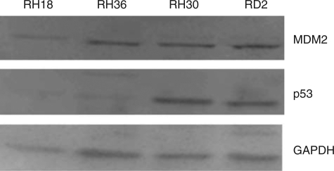
Methods: Rhabdomyosarcoma (RMS), the most common childhood soft tissue sarcoma, expresses either wild-type or mutant p53 protein. We examined the inhibitory effects of MI-63 in embryonal RMS (ERMS) and alveolar RMS (ARMS) cell lines expressing wild-type or mutated p53.
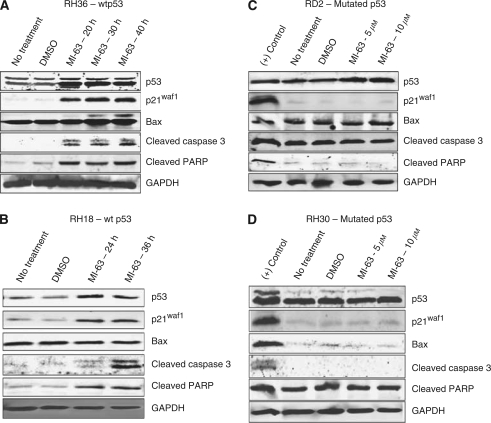
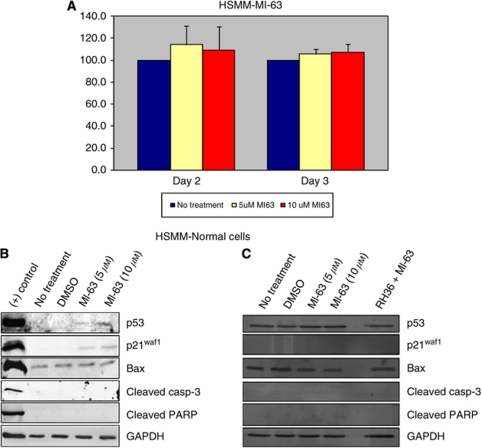
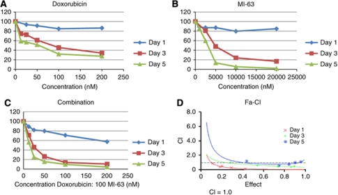
Results: Treatment with MI-63 reduced cell viability by 13.4% and by <1%, respectively, at 72 h in both RH36 and RH18 cell lines expressing wild-type p53. In contrast, RH30 and RD2 cells expressing p53 mutants are resistant to MI-63 treatment. An increased expression of p53, p21(WAF1), and Bax protein was observed after treatment with MI-63 in RMS cells with wild-type p53, and apoptosis was confirmed by cleaved PARP and caspase-3 expression. However, RD2 and RH30 RMS cells, as well as human normal skeletal muscle cells, showed a minimal increase in p53 signalling and no induction of cleaved PARP and caspase-3. MI-63 was compared with Nutlin-3, a known MDM2 inhibitor, and was found to be more potent in the inhibition of cell proliferation/viability. Further, synergy was observed when MI-63 was used in combination with doxorubicin.
Conclusion: These results indicate that MI-63 is a potent therapeutic agent for RMS cells expressing wild-type p53 protein.
Figures


Similar articles
-
Reactivation of p53 by a specific MDM2 antagonist (MI-43) leads to p21-mediated cell cycle arrest and selective cell death in colon cancer.Mol Cancer Ther. 2008 Jun;7(6):1533-42. doi: 10.1158/1535-7163.MCT-08-0140. Mol Cancer Ther. 2008. PMID: 18566224 Free PMC article.
-
MYCN sensitizes neuroblastoma to the MDM2-p53 antagonists Nutlin-3 and MI-63.Oncogene. 2012 Feb 9;31(6):752-63. doi: 10.1038/onc.2011.270. Epub 2011 Jul 4. Oncogene. 2012. PMID: 21725357 Free PMC article.
-
Restoration of p53 pathway by nutlin-3 induces cell cycle arrest and apoptosis in human rhabdomyosarcoma cells.Clin Cancer Res. 2009 Jun 15;15(12):4077-84. doi: 10.1158/1078-0432.CCR-08-2955. Epub 2009 Jun 9. Clin Cancer Res. 2009. PMID: 19509161
-
Spiro-oxindoles as a Promising Class of Small Molecule Inhibitors of p53-MDM2 Interaction Useful in Targeted Cancer Therapy.Top Curr Chem (Cham). 2017 Feb;375(1):3. doi: 10.1007/s41061-016-0089-0. Epub 2016 Dec 9. Top Curr Chem (Cham). 2017. PMID: 27943171 Review.
-
Small-molecule inhibitors of the MDM2-p53 protein-protein interaction to reactivate p53 function: a novel approach for cancer therapy.Annu Rev Pharmacol Toxicol. 2009;49:223-41. doi: 10.1146/annurev.pharmtox.48.113006.094723. Annu Rev Pharmacol Toxicol. 2009. PMID: 18834305 Free PMC article. Review.
Cited by
-
Translating p53 into the clinic.Nat Rev Clin Oncol. 2011 Jan;8(1):25-37. doi: 10.1038/nrclinonc.2010.174. Epub 2010 Oct 26. Nat Rev Clin Oncol. 2011. PMID: 20975744 Review.
-
The Novel Small Molecule STK899704 Promotes Senescence of the Human A549 NSCLC Cells by Inducing DNA Damage Responses and Cell Cycle Arrest.Front Pharmacol. 2018 Apr 16;9:163. doi: 10.3389/fphar.2018.00163. eCollection 2018. Front Pharmacol. 2018. PMID: 29713275 Free PMC article.
-
Stereoselective syntheses of functionalized spirocyclopenteneoxindoles via triphenylphosphine-catalyzed [3+2] cycloaddition reactions.Mol Divers. 2013 Aug;17(3):563-71. doi: 10.1007/s11030-013-9456-8. Epub 2013 Jun 19. Mol Divers. 2013. PMID: 23780558
-
Validation of MdmX as a therapeutic target for reactivating p53 in tumors.Genes Dev. 2011 Aug 15;25(16):1746-57. doi: 10.1101/gad.16722111. Genes Dev. 2011. PMID: 21852537 Free PMC article.
-
A bioengineering method for modeling alveolar Rhabdomyosarcoma and assessing chemotherapy responses.MethodsX. 2021 Jul 27;8:101473. doi: 10.1016/j.mex.2021.101473. eCollection 2021. MethodsX. 2021. PMID: 34430344 Free PMC article.
References
-
- Breneman JC, Lyden E, Pappo AS, Link MP, Anderson JR, Parham DM, Qualman SJ, Wharam MD, Donaldson SS, Maurer HM, Meyer WH, Baker KS, Paidas CN, Crist WM (2003) Prognostic factors and clinical outcomes in children and adolescents with metastatic rhabdomyosarcoma--a report from the Intergroup Rhabdomyosarcoma Study IV. J Clin Oncol 21: 78–84 - PubMed
-
- Chene P (2004) Inhibition of the p53-MDM2 interaction: targeting a protein-protein interface. Mol Cancer Res 2: 20–28 - PubMed
-
- Chou TC, Talalay P (1984) Quantitative analysis of dose-effect relationships: the combined effects of multiple drugs or enzyme inhibitors. Adv Enzyme Regul 22: 27–55 - PubMed
MeSH terms
Substances
LinkOut - more resources
Full Text Sources
Other Literature Sources
Research Materials
Miscellaneous
