

Ultrasound assessment of salivary glands in patients with primary Sjögren's syndrome treated with rituximab: Quantitative and Doppler waveform analysis
- PMID: 19707340
- PMCID: PMC2721315
Ultrasound assessment of salivary glands in patients with primary Sjögren's syndrome treated with rituximab: Quantitative and Doppler waveform analysis
Abstract
Objectives: Noninvasive objective tests are needed to diagnose primary Sjogren's syndrome (pSS) and to evaluate treatment responses. Ultrasound imaging of the salivary glands is rapid and noninvasive. Recent open-label studies suggested that anti-CD20 (rituximab) may be effective in pSS. The purpose of this study was to look for ultrasound evidence of the effects of rituximab in pSS.
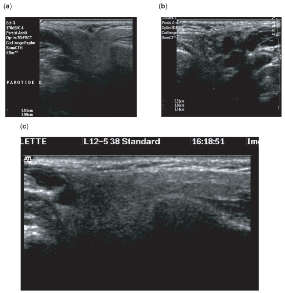
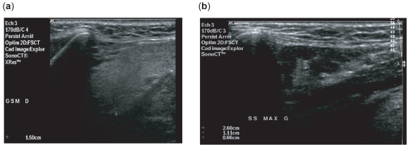
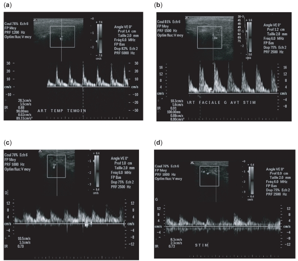
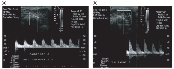
Methods: We compared 16 patients fulfilling the new American-European consensus group criteria for pSS to 9 controls, using B-mode ultrasound features (parenchymal homogeneity and gland size) and Doppler waveform analysis of the transverse facial artery of parotid glands. We compared the same parameters in the patients before and after 12 weeks of intravenous rituximab therapy.
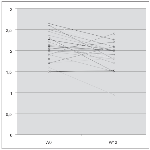
Results: Compared to controls, untreated patients had significant abnormalities in salivary gland structure (p < 0.0001) and parotid size (2.05 +/- 0.33 cm versus 1.70 +/- 0.28 cm; p = 0.001). Doppler waveform analysis showed significant differences before, but not after, lemon stimulation between untreated patients and controls. After rituximab treatment, significant size reductions were noted in the parotids (2.05 +/- 0.3 cm at baseline and 1.86 +/- 0.27 cm at week 12; p = 0.002) and submandibular glands (2.02 +/- 0.54 cm at baseline and 1.66 +/- 0.34 cm at week 12; p = 0.001). Doppler resistive indices after lemon stimulation were significantly increased after rituximab treatment.
Conclusion: Salivary gland measurements and blood inflow responses to salivary stimulation as assessed by ultrasound hold promise as objective noninvasive tools for evaluating rituximab effects in patients with pSS.
Keywords: primary Sjögren’s syndrome; rituximab; ultrasonography.
Figures
References
-
- Ariji Y, Ohki M, Eguchi K, et al. Texture analysis of sonographic features of the parotid gland in Sjögren’s syndrome. Am J Roentgenol. 1996;166:935–41. - PubMed
-
- Ariji Y, Yvasa H, Ariji E. High-frequency color Doppler sonography of the submandibular gland: relationship between salivary secretion and blood flow. Oral Surg Oral Med Oral Pathol Oral Radiol Endosc. 1998;86:476–81. - PubMed
-
- Bradus RJ, Hybarger P, Gooding GA. Parotid gland: US findings in Sjögren syndrome. Work in progress. Radiology. 1988;169:749–51. - PubMed
-
- Carotti M, Salaffi F, Manganelli P, et al. Ultrasonography and colour doppler sonography of salivary glands in primary Sjögren’s sydrome. Clin Rheumatol. 2001;20:213–19. - PubMed
-
- Chikui T, Yonetsu K, Izumi M, et al. Abnormal blood flow to the submandibular glands of patients with Sjögren’s syndrome: Doppler waveform analysis. J Rheumatol. 2000;27:1222–8. - PubMed
LinkOut - more resources
Full Text Sources
