. 2008 Dec;2(4):823-43.
doi: 10.2147/btt.s3770.

Safety and efficacy of enzyme replacement therapy in the nephropathy of Fabry disease

Affiliations

Safety and efficacy of enzyme replacement therapy in the nephropathy of Fabry disease

Fernando C Fervenza et al. Biologics. 2008 Dec.

Abstract

Kidney involvement with progressive loss of kidney function (Fabry nephropathy) is an important complication of Fabry disease, an X-linked lysosomal storage disorder arising from deficiency of alpha-galactosidase activity. Clinical trials have shown that enzyme replacement therapy (ERT) with recombinant human alpha-galactosidase clears globotriaosylceramide from kidney cells, and can stabilize kidney function in patients with mild to moderate Fabry nephropathy. Recent trials show that patients with more advanced Fabry nephropathy and overt proteinuria do not respond as well to ERT alone, but can benefit from anti-proteinuric therapy given in conjunction with ERT. This review focuses on the use of enzyme replacement therapy with agalsidase-alfa and agalsidase-beta in adults with Fabry nephropathy. The current results are reviewed and evaluated. The issues of dosing of enzyme replacement therapy, the use of adjunctive agents to control urinary protein excretion, and the individual factors that affect disease severity are reviewed.

Keywords: Fabry nephropathy; agalsidase; anti-proteinuric therapy; enzyme replacement therapy.


Figures
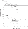
Figure 1
Figure 1
Distribution of proteinuria and eGFR. The median values for eGFR are shown as vertical lines, and the median 24-h urine protein is shown as horizontal lines in both panels. A) Males, n = 300, median eGFR = 81.0 mL/min/1.73 m2, and median proteinuria = 572 mg/24 h. B) Females, n = 306, median eGFR = 88.0 mL/min/1.73 m2, and median proteinuria = 180 mg/24 hr. Data from the Fabry Registry reproduced with permission from Ortiz A, Oliveira JP, Waldek S, et al 2008. Nephropathy in males and females with Fabry disease: cross-sectional description of patients before treatment with enzyme replacement therapy. Nephrol Dial Transplant, 23:1600–7. Copyright © 2008 Oxford University Press.
Figure 2
Figure 2
Effects of agalsidase-beta on rate of loss of kidney function in a 36-year-old male with Fabry nephropathy. Creatinine clearance was measured at the indicated points, and enzyme replacement therapy (1 mg/kg every 2 weeks) was started in early 2001. During the entire follow-up period, there were no major changes in blood pressure or proteinuria (0.4–0.6 g). The patient was started on 5 mg lisinopril in 1994. Because of persistent cough, irbesartan at 150 mg/day was substituted in for lisinopril in December 2001. Reproduced with permission from De Schoenmakere G, Chauveau D, Grunfeld JP. 2003. Enzyme replacement therapy in Anderson-Fabry’s disease: beneficial clinical effect on vital organ function. Nephrol Dial Transplant, 18:33–5. Copyright © 2003 Oxford University Press.
Figure 3
Figure 3
Annualized change in estimated glomerular filtration rate (mL/min/1.73 m2/year ± SD) in patients treated with agalsidase-alfa at 0.2 mg/kg every 2 weeks (left bars) followed by weekly treatment (right bars). Patients are stratified by baseline proteinuria: <0.3 g/day, black bars; >1 g/day, open bars. Adapted with permission from Schiffmann R, Askari H, Timmons M, et al 2007. Weekly enzyme replacement therapy may slow decline of renal function in Fabry patients who are on long-term biweekly dosing. J Am Soc Nephrol, 18:1576–83. Copyright © 2007 American Society of Nephrology.
Figure 4
Figure 4
Annualized progression rate of change in estimated glomerular filtration rate (mL/min/1.73 m2/year ± SD) for a male patient before ERT, and during treatment with agalsidase-alfa at 0.2 mg/kg every 2 weeks, followed by treatment with agalsidase-alfa at 0.4 mg/kg every 2 weeks. The progression slope was reduced in half with the higher dose of ERT. The urine protein excretion averaged 1.98 ± 0.03 (SD) g/24 h before ERT, 2.72 ± 0.52 (SD) g/24 h during ERT at 0.2 mg/kg every other week, and 1.17 ± 0.22 (SD) g/24 h during ERT at 0.4 mg/kg every other week along with enalapril at 5 mg 3 times per day. Adapted with permission Torra R, Algaba F, Ars E, et al 2008. Preservation of renal function in a patient with Fabry nephropathy on enzyme replacement therapy. Clin Nephrol, 69:445–9. Copyright © 2008 Dustri-Verlag.
Figure 5
Figure 5
Annualized progression rate of change in estimated glomerular filtration rate (mL/min/1.73 m2/year ± SD) for a male patient during treatment with agalsidase-alfa at 0.2 mg/kg every 2 weeks, followed by treatment with agalsidase-beta at 1.0 mg/kg every 2 weeks. The progression slope was reduced by 77% with the higher dose of ERT. The average baseline proteinuria was 2.27 ± 0.0.57 (SD) g/day before any treatment, and with added enalapril (10 mg) and losartan (50 mg), was reduced to before any ERT to 1.03 g/day, and was maintained at an average of 0.83 ± 0.54 (SD) g/day during agalsidase-alfa treatment, and 0.56 ± 0.24 (SD) g/day during agalsidase-beta treatment. Adapted with permission from Warnock DG. 2005. Fabry disease: diagnosis and management, with emphasis on the renal manifestations. Curr Opin Nephrol Hypertens, 14:87–95. Copyright © 2005 Lippincott Williams & Wilkins.
Figure 6
Figure 6
Effects of agalsidase-beta on arteriolar intimal and medial, and glomerular capillary endothelial GL-3 deposits. The patient was a 33-year-old male who was diagnosed with Fabry disease on the basis of the kidney biopsy findings. At the time of the initial biopsy, his estimated glomerular filtration rate was 25 mL/min/1.73 m2, and his urine protein excretion was 3.3 g/24 h. His proteinuria was controlled to an average of 0.66 ± 0.44 (SD) g/24 h with 20 mg enalapril and 150 mg irbesartan, and agalsidase-beta treatment was started at 1.0 mg/kg every other week. The kidney biopsy was repeated after 15 months of ERT therapy. A) Renal cortical arteriole, before starting ERT (Masson-trichrome stain). B) Renal cortical arteriole, 15-months after starting ERT (Masson-trichrome stain); note clearing of endothelial and intimal GL-3 deposits. C) Glomerular capillary loop, before starting ERT (electron micrograph); note the dense endothelial deposits with substantial obliteration of the capillary lumen and the “Zebra bodies” in the podocytes. D) Glomerular capillary loop, 15 months after starting ERT (electron micrograph); note that the endothelial deposits have cleared, but “Zebra bodies” persist in the podocytes. The magnification is the same for A and B, but C and D have different magnification factors. For point of reference, the basement membrane thickness is the same for the electron micrographs before and after ERT. Courtesy of William Cook, MD PhD Department of Pathology, University of Alabama at Birmingham.
Figure 7
Figure 7
Glomerular capillary endothelial GL-3 deposits (circled) persist despite prolonged treatment with agalsidase-alfa at 0.2 mg/kg every 2 weeks. A) 38 year old male after 16 months of agalsidase-alfa at 0.2 mg/kg every other week, switched to agalsidase-beta after the biopsy; estimated glomerular filtration rate 47 mL/min/1.73 m2, urine protein = 0.55 g/24 h. This patient’s clinic course is shown in Figure 7. B) 39-year-old male after 24 months of agalsidase-alfa at 0.2 mg/kg every other week, switched to agalsidase-alfa at 0.4 mg/kg every 2 weeks after the biopsy; estimated glomerular filtration rate 34 mL/min/1.73 m2, urine protein = 2.7 g/24 h. This patient’s clinic course is shown in Figure 8. C) 47-year-old male after 60 months of agalsidase-alfa at 0.2 mg/kg every other week, switched to agalsidase-beta at 1.0 mg/kg every 2 weeks after the biopsy; estimated glomerular filtration rate 52 mL/min/1.73 m2, urine protein = 0.41 g/24 h. This patient is the older brother of the patient shown in Figure 7 and Figure 9A. Panel A and C, adapted with permission from Warnock DG. 2005. Fabry disease: diagnosis and management, with emphasis on the renal manifestations. Curr Opin Nephrol Hypertens, 14:87–95. Copyright © 2005. Panel B, adapted with permission from Torra R, Algaba F, Ars E, et al 2008. Preservation of renal function in a patient with Fabry nephropathy on enzyme replacement therapy. Clin Nephrol, 69:445–9. Copyright © 2008 Dustri-Verlag.
Figure 8
Figure 8
Relationship between kidney GL-3 content (nmol/mg protein) and Glomerular Pathology Scores A) and urinary GL-3 excretion (nmol/g creatinine) B) The linear regression shown in panel A is Y = 0.505 (SE 0.0145) X −0.0959 (SE 0.3027); F statistic = 0.0021; r2 = 0.3554. The linear regression shown in panel B is Y = 71.0 (SE 27.5) X −964.8 (SE 572.5); F statistic = 0.0169; r2 = 0.2331. Adapted with permission from Branton MH, Schiffmann R, Sabnis SG, et al 2002. Natural history of Fabry renal disease: influence of alpha-galactosidase A activity and genetic mutations on clinical course. Medicine (Baltimore), 81:122–38. Copyright © 2002 Lippincott Williams & Wilkins.
Figure 9
Figure 9
Relationship between baseline proteinuria and probability of a renal outcome event in the phase III extension study. Baseline proteinuria (expressed as the urinary protein/creatinine ratio) was determined before entry into the double-blinded initial phase of the study. Logistic regression analysis was used to determine the probability of a renal event (defined as 50% increase in serum creatinine compared to pre-treatment value, with the increased value >1.4 mg/dL). Adapted with permission from Germain D, Waldek S, Banikazemi M, et al 2007. Sustained, long-term renal stabilization after 54 months of agalsidase beta therapy in patients with Fabry disease. J Am Soc Nephrol, 18:1547–57. Copyright © 2007 American Society of Nephrology.
Figure 10
Figure 10
Relationship between baseline proteinuria and the rate of loss of estimated glomerular filtration rate (eGFR) in the phase III extension study. The participants were sub-grouped based on their baseline proteinuria, which was determined before entry into the double-blinded initial phase of the study. During the 54-month treatment period, patients (n = 10) with baseline urinary protein/creatinine ratios > 1.0 had a mean rate of decline of eGFR of −7.4 mL/min/1.73 m2/year. In contrast, patients (n = 42) with baseline urinary protein/creatinine ratios < 1.0 had a mean rate of decline of eGFR of −1.0 mL/min/1.73 m2/year. Adapted with permission from Germain D, Waldek S, Banikazemi M, et al 2007. Sustained, long-term renal stabilization after 54 months of agalsidase beta therapy in patients with Fabry disease. J Am Soc Nephrol, 18:1547–57. Copyright © 2007 American Society of Nephrology.

Similar articles

Cited by

References

    1. Aerts JM, Groener JE, Kuiper S, et al. Elevated globotriaosylsphingosine is a hallmark of Fabry disease. Proc Natl Acad Sci U S A. 2008;105:2812–7. - PMC - PubMed
    1. Askari H, Kaneski CR, Semino-Mora C, et al. Cellular and tissue localization of globotriaosylceramide in Fabry disease. Virchows Arch. 2007;451:823–34. - PubMed
    1. Banikazemi M, Bultas J, Waldek S, et al. Agalsidase-beta therapy for advanced Fabry disease: a randomized trial. Ann Intern Med. 2007;146:77–86. - PubMed
    1. Barbey F, Lidove O, Schwarting A. Fabry nephropathy: 5 years of enzyme replacement therapy - a short review. Nephrol Dial Transplant Plus. 2008;1:11–9. - PMC - PubMed
    1. Blom D, Speijer D, Linthorst GE, et al. Recombinant enzyme therapy for Fabry disease: absence of editing of human alpha-galactosidase A mRNA. Am J Hum Genet. 2003;72:23–31. - PMC - PubMed