. 2009:2009:208725.
doi: 10.1155/2009/208725. Epub 2009 Aug 20.

Clinical Applications of FDG PET and PET/CT in Head and Neck Cancer

Affiliations

Clinical Applications of FDG PET and PET/CT in Head and Neck Cancer

Akram Al-Ibraheem et al. J Oncol. 2009.

Abstract

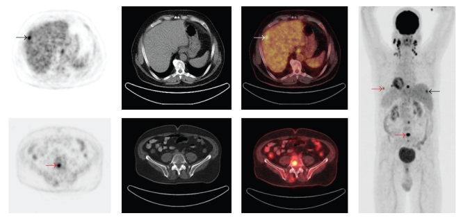
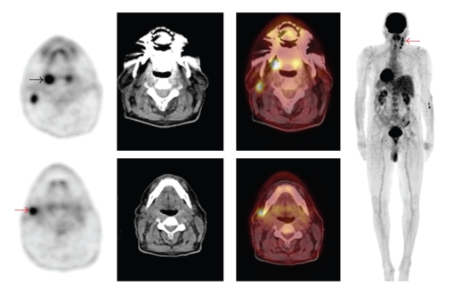
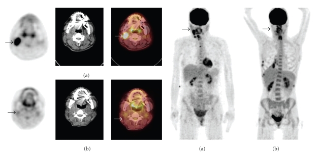
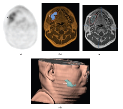
18F-FDG PET plays an increasing role in diagnosis and management planning of head and neck cancer. Hybrid PET/CT has promoted the field of molecular imaging in head and neck cancer. This modality is particular relevant in the head and neck region, given the complex anatomy and variable physiologic FDG uptake patterns. The vast majority of 18F-FDG PET and PET/CT applications in head and neck cancer related to head and neck squamous cell carcinoma. Clinical applications of 18F-FDG PET and PET/CT in head and neck cancer include diagnosis of distant metastases, identification of synchronous 2nd primaries, detection of carcinoma of unknown primary and detection of residual or recurrent disease. Emerging applications are precise delineation of the tumor volume for radiation treatment planning, monitoring treatment, and providing prognostic information. The clinical role of 18F-FDG PET/CT in N0 disease is limited which is in line with findings of other imaging modalities. MRI is usually used for T staging with an intense discussion concerning the preferable imaging modality for regional lymph node staging as PET/CT, MRI, and multi-slice spiral CT are all improving rapidly. Is this review, we summarize recent literature on 18F-FDG PET and PET/CT imaging of head and neck cancer.


Figures

Figure 1
Figure 1
A 61-year-old man with nasopharyngeal SCC and bilateral cervical lymph node metastases underwent PET/CT for staging. Axial PET, CT, PET/CT, and maximum intensity projection (MIP) images are shown. PET/CT revealed focal FDG uptake in the right liver lobe indicating liver metastasis (black, white arrows). PET/CT also revealed multiple focal FDG uptakes in the lumbar spine, sternum, and ribs indicating multiple bone metastases (red arrows). PET/CT was valuable for detection distant metastases.
Figure 2
Figure 2
A 61-year-old man presented with right side cervical lymphadenopathy proved to be carcinoma of unknown primary. Patient underwent PET/CT to reveal primary tumor. Axial PET, CT, PET/CT, and maximum intensity projection (MIP) images are shown. PET/CT showed asymmetrical FDG uptake in the palatine tonsils with intense FDG uptake in the right tonsil (black arrow) as well as multiple hypermetabolic cervical lymph nodes in the right side (red arrows). This patient subsequently underwent surgical resection and histopathology revealed squamous cell carcinoma in the right tonsil. PET/CT was valuable in revealing the primary tumor in this case.
Figure 3
Figure 3
40-year-old women with right side larynx squamous cell carcinoma and a right side cervical lymph node metastases underwent PET/CT imaging before and 1 month after completion of chemoradiotherapy. (a) Pretherapy axial PET, CT, PET/CT, and MIP images reveal intense FDG uptake in the right cervical lymph node (arrows). (b) After treatment axial PET, CT, PET/CT, and MIP images reveal decrease FDG uptake in the corresponding locations (arrows). Appearance was consistent with early response to chemoradiotherapy. FDG PET/CT was valuable in monitoring early response to treatment.
Figure 4
Figure 4
Patient with a squamous cell carcinoma of the right mandible (arrows). (a) The [18F]Galacto-RGD PET shows heterogeneous tracer uptake, which can also be clearly delineated in (b) the image fusion with the corresponding MRI scan. (c) shows the tumour volume in red as defined by MRI. By applying a threshold of SUV 3 and only using pixels with SUVs above this threshold, (d) a subvolume with more intense αvß3 expression can be defined which is shown in the 3D reconstruction (blue line in (c), blue area in (d)).

References

    1. Jemal A, Siegel R, Ward E, et al. Cancer statistics, 2008. CA: Cancer Journal for Clinicians. 2008;58(2):71–96. - PubMed
    1. Baek C-H, Chung MK, Jeong H-S, et al. The clinical usefulness of 18F-FDG PET/CT for the evaluation of lymph node metastasis in periorbital malignancies. Korean Journal of Radiology. 2009;10(1):1–7. - PMC - PubMed
    1. Roh J-L, Yeo N-K, Kim JS, et al. Utility of 2-[18F] fluoro-2-deoxy-d-glucose positron emission tomography and positron emission tomography/computed tomography imaging in the preoperative staging of head and neck squamous cell carcinoma. Oral Oncology. 2007;43(9):887–893. - PubMed
    1. Gordin A, Golz A, Daitzchman M, et al. Fluorine-18 fluorodeoxyglucose positron emission tomography/computed tomography imaging in patients with carcinoma of the nasopharynx: diagnostic accuracy and impact on clinical management. International Journal of Radiation Oncology Biology Physics. 2007;68(2):370–376. - PubMed
    1. Kim MR, Roh J-L, Kim JS, et al. Utility of 18F-fluorodeoxyglucose positron emission tomography in the preoperative staging of squamous cell carcinoma of the oropharynx. European Journal of Surgical Oncology. 2007;33(5):633–638. - PubMed

LinkOut - more resources