

Review
doi: 10.1007/s00412-009-0234-4.
Epub 2009 Aug 26.
H2AX: functional roles and potential applications
Affiliations
- PMID: 19707781
- PMCID: PMC3094848
- DOI: 10.1007/s00412-009-0234-4
Item in Clipboard
Review
H2AX: functional roles and potential applications
Chromosoma.
2009 Dec.
Abstract
Upon DNA double-strand break (DSB) induction in mammals, the histone H2A variant, H2AX, becomes rapidly phosphorylated at serine 139. This modified form, termed gamma-H2AX, is easily identified with antibodies and serves as a sensitive indicator of DNA DSB formation. This review focuses on the potential clinical applications of gamma-H2AX detection in cancer and in response to other cellular stresses. In addition, the role of H2AX in homeostasis and disease will be discussed. Recent work indicates that gamma-H2AX detection may become a powerful tool for monitoring genotoxic events associated with cancer development and tumor progression.
Figures
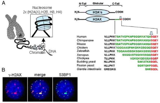
a (left panel) H2AX is a component of chromatin and its fundamental packaging unit, the nucleosome. a (right panel) H2AX is composed of a central globular domain, an N-terminal tail and a unique C-terminal tail consisting of an evolutionarily conserved motif (shown in red) and connected by a linker of variable sequence and length (green). The conserved motif contains the omega-4 serine that is phosphorylated upon DNA DSB formation (arrow). b In response to genotoxic stress and upon DNA DSB formation, the H2AX omega-4 serine is phosphorylated (γ-H2AX), which can be visualized using an anti-γ-H2AX antibody as discrete foci that colocalize with other DNA repair proteins. The images depict a HeLa cell 1 h after exposure to 1 Gy of γ-radiation. Red γ-H2AX, green 53BP1, blue DAPI
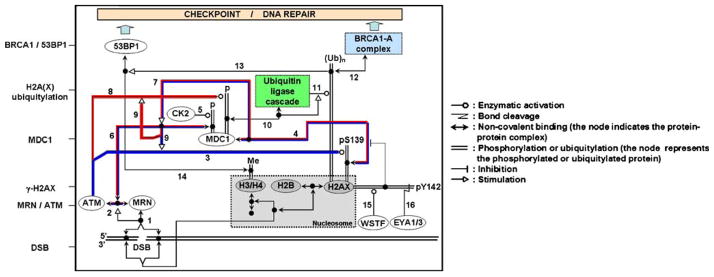
H2AX is a key component of the DNA damage response. This schematic representation illustrates the γ-H2AX-MDC1-BRCA1/53BP1cascade in response to DNA DSB formation after irradiation. Upon DSB formation, the MRN complex (MRE11-RAD50-NBS1) binds to the ends of the DSB (1) and recruits ATM (2). ATM then phosphorylates H2AX on serine 139 to form γ-H2AX (3). This phosphorylation allows the binding of the mediator protein MDC1 (4). The constitutive phosphorylation of MDC1 by CK2 (5) permits the binding of the MRN–ATM complex (via NBS1) to MDC1 (6). The MRN–ATM complex is preferentially recruited at the DSB site because of the presence of the γ-H2AX–MDC1 complex (7). This recruitment of ATM, in turn, enhances the phosphorylation of other proteins at the DSB site, including H2AX (3) and MDC1 (8) itself (feedback loop (9)). MDC1 phosphorylation at the DSB site allows the recruitment of the ubiquitin ligase machinery (10) that will then permit the ubiquitylation H2A and/or H2AX (11). H2A(X) ubiquitylation is necessary for the accumulation of the BRCA1-A complex at break sites via its subunit RAP80 (12). It is generally thought that histone ubiquitylation is necessary for 53BP1 accumulation at the DSB site (13) by providing the chromatin remodeling necessary to expose constitutive H3 and H4 methylated tails (Me) that in turn are recognized by 53BP1 (14). In the absence of DNA damage, H2AX is constitutively phosphorylated by WSTF on tyrosine 142 (15). Following DNA damage, if DNA repair occurs, phosphotyrosine 142 is dephosphorylated by the EYA1/3 phosphatase (16) allowing the binding of MDC1 to γ-H2AX (4). To simplify, the components of the MRN complex, the ubiquitin ligase complex and the BRCA1-A complex are represented by one box each and H2A is not shown. The histones are represented by gray boxes. One isolated node represents another copy of the molecular species that is at the end of the corresponding line. The feedback loops for H2AX and MDC1 phosphorylation are underlined in blue and red, respectively. Symbol conventions (shown at right) are derived from Dr. Kurt Kohn’s molecular interaction maps (Kohn 1999; for further details see http://discover.nci.nih.gov )
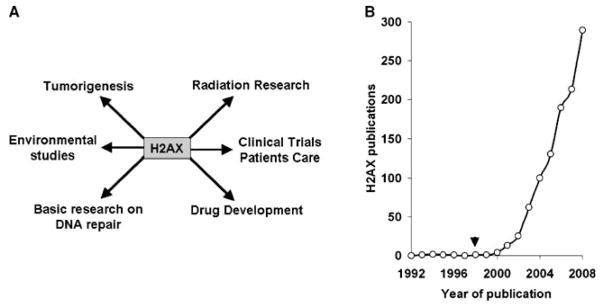
a H2AX is being studied in other areas besides basic research on DNA repair, including drug development, translational studies, radiation research, and environmental studies. As cancer cells and tumors often exhibit high levels of γ-H2AX, it is now considered to be a cancer biomarker. b Since its discovery as a DNA double-strand damage marker in 1998 (arrow), the number of papers published each year since 1992 containing H2AX in the title and/or abstract has continually increased (source: PubMed)
References
-
- Albino AP, Huang X, Jorgensen E, Yang J, Gietl D, Traganos F, Darzynkiewicz Z. Induction of H2AX phosphorylation in pulmonary cells by tobacco smoke: a new assay for carcinogens. Cell Cycle. 2004;3:1062–1068. - PubMed
-
- Bakkenist CJ, Drissi R, Wu J, Kastan MB, Dome JS. Disappearance of the telomere dysfunction-induced stress response in fully senescent cells. Cancer Res. 2004;64:3748–3752. - PubMed
-
- Banath JP, Macphail SH, Olive PL. Radiation sensitivity, H2AX phosphorylation, and kinetics of repair of DNA strand breaks in irradiated cervical cancer cell lines. Cancer Res. 2004;64:7144–7149. - PubMed
-
- Bartkova J, Horejsi Z, Koed K, et al. DNA damage response as a candidate anti-cancer barrier in early human tumorigenesis. Nature. 2005;434:864–870. - PubMed
Publication types
MeSH terms
Substances
Grants and funding
LinkOut - more resources
Full Text Sources
Other Literature Sources
