. 2009 Nov;66(21):3469-86.
doi: 10.1007/s00018-009-0133-0. Epub 2009 Aug 26.

Molecular models of the open and closed states of the whole human CFTR protein

Affiliations

Molecular models of the open and closed states of the whole human CFTR protein

Jean-Paul Mornon et al. Cell Mol Life Sci. 2009 Nov.

Abstract

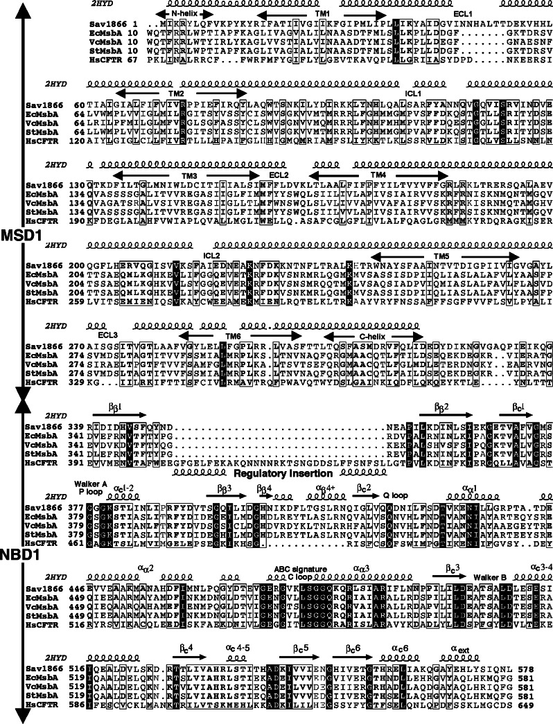
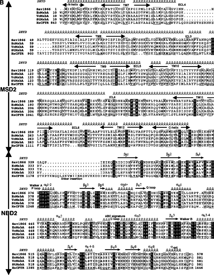
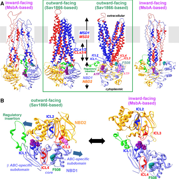
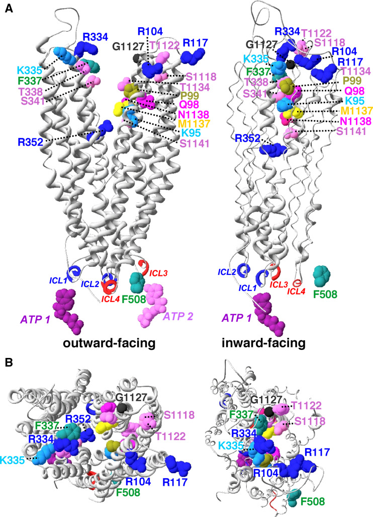
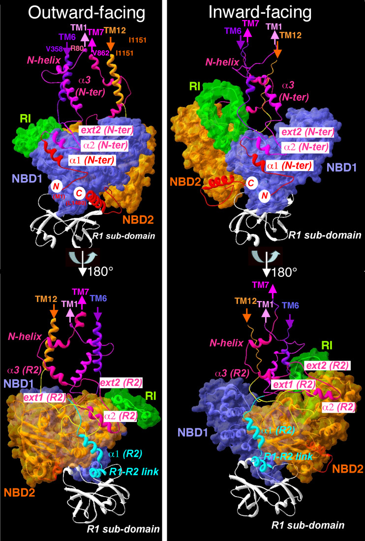
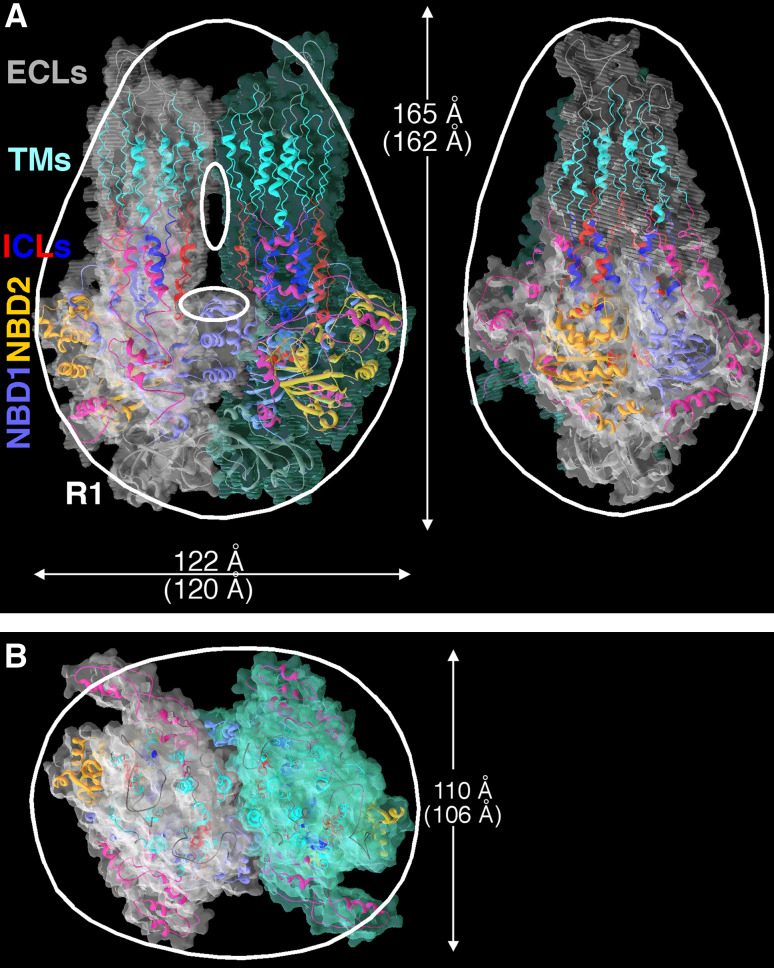
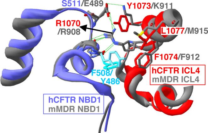
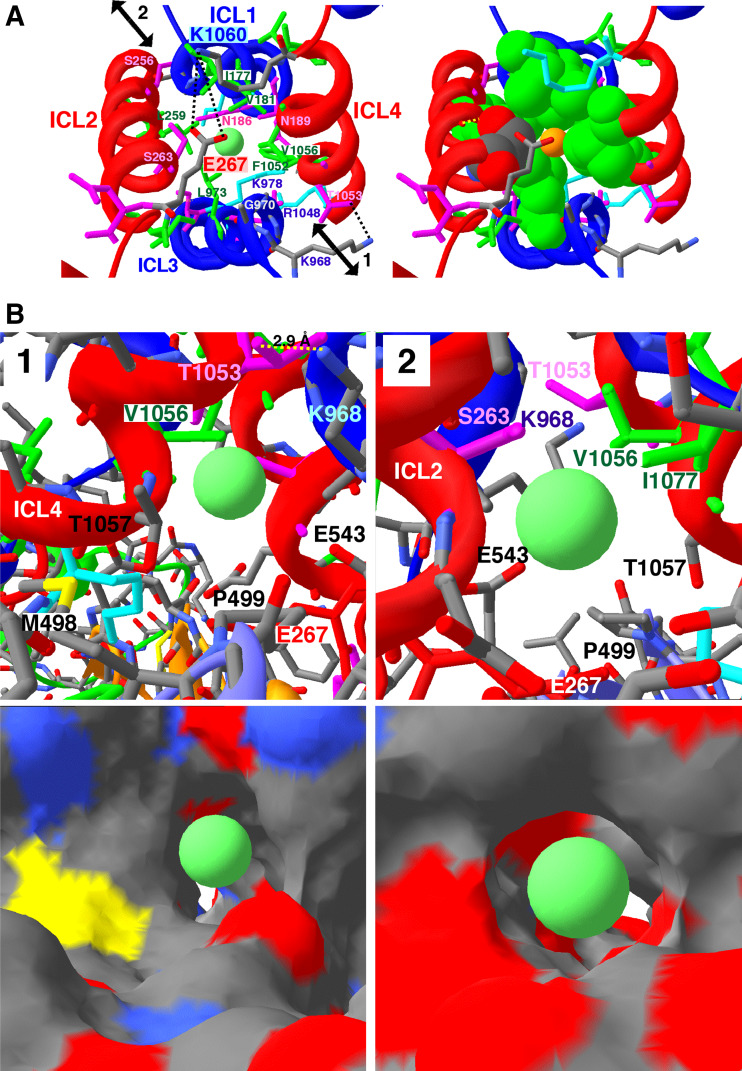
Cystic fibrosis transmembrane conductance regulator (CFTR), involved in cystic fibrosis (CF), is a chloride channel belonging to the ATP-binding cassette (ABC) superfamily. Using the experimental structure of Sav1866 as template, we previously modeled the human CFTR structure, including membrane-spanning domains (MSD) and nucleotide-binding domains (NBD), in an outward-facing conformation (open channel state). Here, we constructed a model of the CFTR inward-facing conformation (closed channel) on the basis of the recent corrected structures of MsbA and compared the structural features of those two states of the channel. Interestingly, the MSD:NBD coupling interfaces including F508 (DeltaF508 being the most common CF mutation) are mainly left unchanged. This prediction, completed by the modeling of the regulatory R domain, is supported by experimental data and provides a molecular basis for a better understanding of the functioning of CFTR, especially of the structural features that make CFTR the unique channel among the ABC transporters.


Figures

Fig. 1
Fig. 1
Alignment of the human CFTR MSDs and NBDs sequence with sequences of bacterial ABC exporters, whose 3D structures are known in different conformations [S. aureus Sav1866 (pdb 2hyd) and Escherichia coli, Vibrio cholerae, and Salmonella typhimurium MsbA (pdb 3b5w, 3b5x, and 3b60, respectively)]. a MSD1/NBD1; b MSD2/NBD2. The MsbA sequences were added to the Sav1866/CFTR alignment, which is identical to that previously reported [19]. The observed secondary structures of Sav1866 are indicated on top of its sequence. The MSD and NBD secondary structures are labeled according to the labeling previously reported for CFTR [12, 19]. The α helices of the CFTR NBD1-specific regulatory insertion (RI) are reported below its sequence. Identities are shown in white on a black background, whereas similarities are boxed. This figure was made using ESPript [65]
Fig. 1
Fig. 1
Alignment of the human CFTR MSDs and NBDs sequence with sequences of bacterial ABC exporters, whose 3D structures are known in different conformations [S. aureus Sav1866 (pdb 2hyd) and Escherichia coli, Vibrio cholerae, and Salmonella typhimurium MsbA (pdb 3b5w, 3b5x, and 3b60, respectively)]. a MSD1/NBD1; b MSD2/NBD2. The MsbA sequences were added to the Sav1866/CFTR alignment, which is identical to that previously reported [19]. The observed secondary structures of Sav1866 are indicated on top of its sequence. The MSD and NBD secondary structures are labeled according to the labeling previously reported for CFTR [12, 19]. The α helices of the CFTR NBD1-specific regulatory insertion (RI) are reported below its sequence. Identities are shown in white on a black background, whereas similarities are boxed. This figure was made using ESPript [65]
Fig. 2
Fig. 2
The CFTR MSD1:NBD1:MSD2:NBD2 assembly, in the outward- and inward-facing conformations. a Overall architecture. Two orthogonal views using a ribbon representation are shown for each of the two models. MSD1 and NBD1 are colored dark and light blue, respectively, whereas MSD2 and NBD2 are colored red and orange, respectively. The NBD1-specific regulatory insertion (RI) is shown in green. F508 (in green) is shown in a van der Waals representation, as well as the ATP molecules (dark and light pink for the ATP molecules bound to the non-canonical (NBD1) and canonical (NBD2) sites, respectively). Although the inward-facing conformation of MsbA is free from ATP molecules, we chose to leave one ATP molecule in the non-canonical ATP-binding site in the model of the inward-facing conformation of CFTR (closed channel), as ATP stays for a long time at this site. The position of the membrane lipid bilayer is indicated in grey. MSD membrane-spanning domain, NBD nucleotide-binding domain, ICL intracellular loop. b The NBD1:NBD2 assembly (viewed from the membrane). The NBD1/NBD2 heterodimer is shown, as well as the coupling helices of the MSDs intracellular loops (ICL1–ICL4) which make contacts with the NBDs. The transition between the two forms of the NBD1:NBD2 assembly is obtained by an almost horizontal rigid-body translation (sliding, see blue arrows) of NBD2, the regulatory insertion likely playing the role of a “safety catch” which hinders the complete disassembly of the two NBDs. ATP (dark pink) is expected to be bound in the non-conventional binding site (NBD1) in both conformers, whereas the conventional binding site is only occupied by ATP (light pink) in the outward-facing model (open channel). However, one can note that in the inward-facing configuration, both ATP molecules can be loaded in the canonical and non-canonical binding sites of the NBDs with no steric hindrance, in close contact with each other, their O3′ atoms being separated by only 4.5 Å. Such a situation might actually occur during the transition between the closed, inward-facing and the open, outward-facing conformations
Fig. 3
Fig. 3
Overall architecture of the CFTR chloride channel, in the outward-facing and inward-facing configurations (open and closed channel, respectively). Two orthogonal views of the MSDs are shown, with the coupling helices of the four ICLs colored in blue (MSD1 ICL1 and ICL2) and red (MSD2 ICL3 and ICL4). a Longitudinal side views, b top views from the extracellular side. As in Fig. 2, two ATP molecules are shown in the outward-facing conformation, whereas only one (in the non-conventional binding site: NBD1) is shown for the inward-facing conformation. Critical amino acid residues lining the channel pore are shown in a van der Waals representation
Fig. 4
Fig. 4
The R domain model, in the outward-facing (left) and inward-facing (right) configuration. Two opposed side views at 180° (upper panels vs lower panels) of NBD1 (in blue, with its regulatory insertion (RI) in green) and NBD2 (in orange) showing the R1 sub-domain (white) as well as the helices and extended strands of R2 sub-domain and of the CFTR N-terminus (which was used as template for modeling the R2 sub-domain, see text). The N-terminal and C-terminal ends of CFTR are indicated in red font on a white circle background
Fig. 5
Fig. 5
Compatibility of a dimeric assembly of the whole CFTR model in its closed, inward-facing configuration with ellipsoidal structures observed for the CFTR dimer by electron microscopy. a Two orthogonal side views; b view from the extracellular side. The contours of the ellipsoïdal particle are shown in white, as deduced from Fig. 8 of the article by Mio et al. [28]. The estimated dimensions of a dimeric assembly of our CFTR model are indicated above those observed by electron microscopy, which are shown between parentheses. For the two upper orthogonal side views, the structures encountered from the top to the bottom are colored as follows: ECLs in white, TMs in light blue, ICLs in dark blue and red, NBD1 in blue and NBD2 in orange. The N- and C-terminal segments, as well as linkers, are in pink. At the bottom, the R1 domains are shown in white. The interface between each monomer mainly includes, from top to bottom, contacts between TM10 and TM10, αα2 of NBD1 and R2, as well as R1 and R1. Glycans are localized on asparagine residues N894 and N900, in the first ECL of the second MSD (at top of the particle)
Fig. 6
Fig. 6
Comparison of our outward-facing Sav1866-based 3D model of human CFTR [19] with the experimental 3D structure of mouse P-gp/MDR in its inward-facing configuration [22]: superimposition of the NBD1/ICL4 interface. The 3D crystal structure of mouse P-gp/MDR (pdb 3g5u) was energetically refined in a similar way to our CFTR model. The human CFTR NBD1 and ICL4 structures are shown in blue and red, respectively, whereas the corresponding segments of mouse P-gp (MDR) are shown in grey (ribbon representation). In particular, critical bonds between the CFTR ILC4 residue R1070 (NE, NH1, and NH2 atoms) and the CFTR NBD1 residues I507 (CO)/F508 (CO)/S511 (OG1) may also be found in MDR between the side chain atoms of R908 (which corresponds to CFTR R1070) and R485 (CO)/Y486 (CO)/E489 (OE1). An additional bond can be observed in MDR between the ICL4 K911 and the NBD1 E489
Fig. 7
Fig. 7
A putative cytoplasmic entry to the CFTR channel. a Views of the ICLs bundle region seen from the NBD1:NBD2 interface (not shown for sake of clarity) and along the pseudo two-fold axis, with E267 in the foreground. The side chain of this residue may lock the putative CFTR channel at its basis. E267 weakly interacts with its symmetrical amino acid K1060 (distance between the two amino acids ~4.8 Å), nearly at the centre of the pore which has a diameter of 5–8 Å, a size at least equal to (or greater than) that of the selectivity filter of the ClC channel [66]. Lysine and arginine residues are shown in blue, hydrophobic residues in green, and hydrophilic ones in pink. However, as illustrated in ESM: Fig. S11, E267 might jump to another conformer, where it may weakly interact with the side chain of K968. Chloride ions may thus either freely run from the funnel (arrows 1 and 2), described in more details in b, to the open CFTR channel (and vice versa), or alternatively access with more difficulties the CFTR channel through the ICLs bottom, when this access is not locked. Left a chloride ion (green) is shown at the level of a hydrophobic ring (I177, V181, L259, S263, G970, L973, F1052, V1056). One can note that the side chain oxygen of S263 binds to the V260 (not indicated) main chain carbonyl, as it is often the case within helices, and thus only exposes its Cβ atom to the channel. The hydrophobic ring is followed by a more hydrophilic one (including S185, N186, K190, N974, S977, K978, K1040), where the channel becomes larger. Right hydrophobic surfaces shown in green with the chloride ion in yellow and S263 in standard colors (oxygen in red, carbon in grey). b Symmetrical views (1 and 2) of the putative access [for chloride ions coming from (or moving to) the cytoplasmic milieu] to the beginning of the putative CFTR channel pore. This access, which is located between the helices of the ICLs bundle, consists of an almost linear horizontal funnel (~20 Å long) running at the basis of ICLs and nearly perpendicular to the vertical pseudo two-fold axis of CFTR. The directions of views 1 and 2 are indicated by arrows in (a). The top views show the atomic environment of the funnel, the bottom views the corresponding molecular surfaces (red oxygen, blue nitrogen, yellow sulfur, grey carbon atoms). A chloride ion (green ball) was positioned at both entrances of the funnel. In both bottom views, the small white areas near the center correspond to the protein exterior at the other side of the funnel

Similar articles

Cited by

References

    1. Gadsby DC, Vergani P, Csanády L. The ABC protein turned chloride channel whose failure causes cystic fibrosis. Nature. 2006;440:477–483. doi: 10.1038/nature04712. - DOI - PMC - PubMed
    1. Rowe SM, Miller S, Sorscher EJ. Cystic fibrosis. N Engl J Med. 2005;352:1992–2001. doi: 10.1056/NEJMra043184. - DOI - PubMed
    1. Biemans-Oldehinkel E, Doeven MK, Poolman B. ABC transporter architecture and regulatory roles of accessory domains. FEBS Lett. 2006;580:1023–1035. doi: 10.1016/j.febslet.2005.11.079. - DOI - PubMed
    1. Anderson MP, Gregory RJ, Thompson S, Souza DW, Paul S, Mulligan RC, Smith AE, Welsh MJ. Demonstration that CFTR is a chloride channel by alteration of its anion selectivity. Science. 1991;253:202–205. doi: 10.1126/science.1712984. - DOI - PubMed
    1. Bear CE, Li CH, Kartner N, Bridges RJ, Jensen TJ, Ramjeesingh M, Riordan JR. Purification and functional reconstitution of the cystic fibrosis transmembrane conductance regulator (CFTR) Cell. 1992;68:809–818. doi: 10.1016/0092-8674(92)90155-6. - DOI - PubMed

Publication types

MeSH terms

Substances

LinkOut - more resources