. 2010 Mar;30(2):199-218.
doi: 10.1007/s10571-009-9443-x. Epub 2009 Aug 26.

Neural stem/progenitor cells derived from the embryonic dorsal telencephalon of D6/GFP mice differentiate primarily into neurons after transplantation into a cortical lesion

Affiliations

Neural stem/progenitor cells derived from the embryonic dorsal telencephalon of D6/GFP mice differentiate primarily into neurons after transplantation into a cortical lesion

Iva Prajerova et al. Cell Mol Neurobiol. 2010 Mar.

Abstract

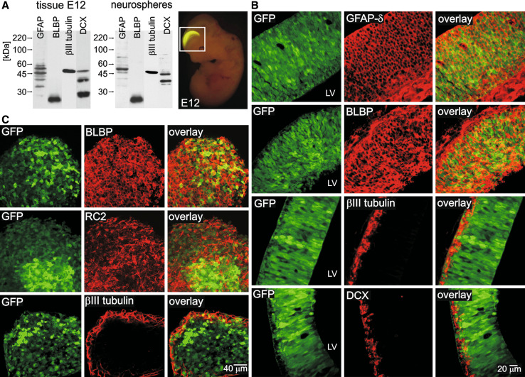
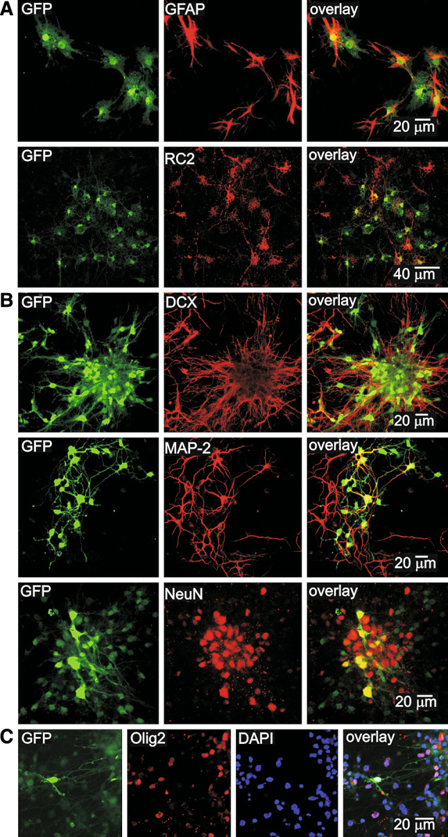
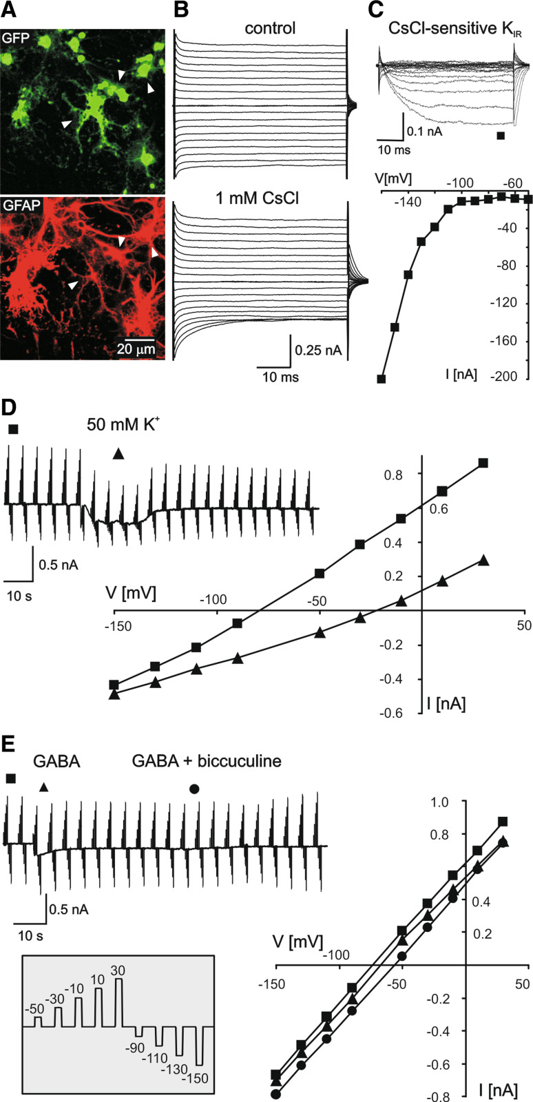
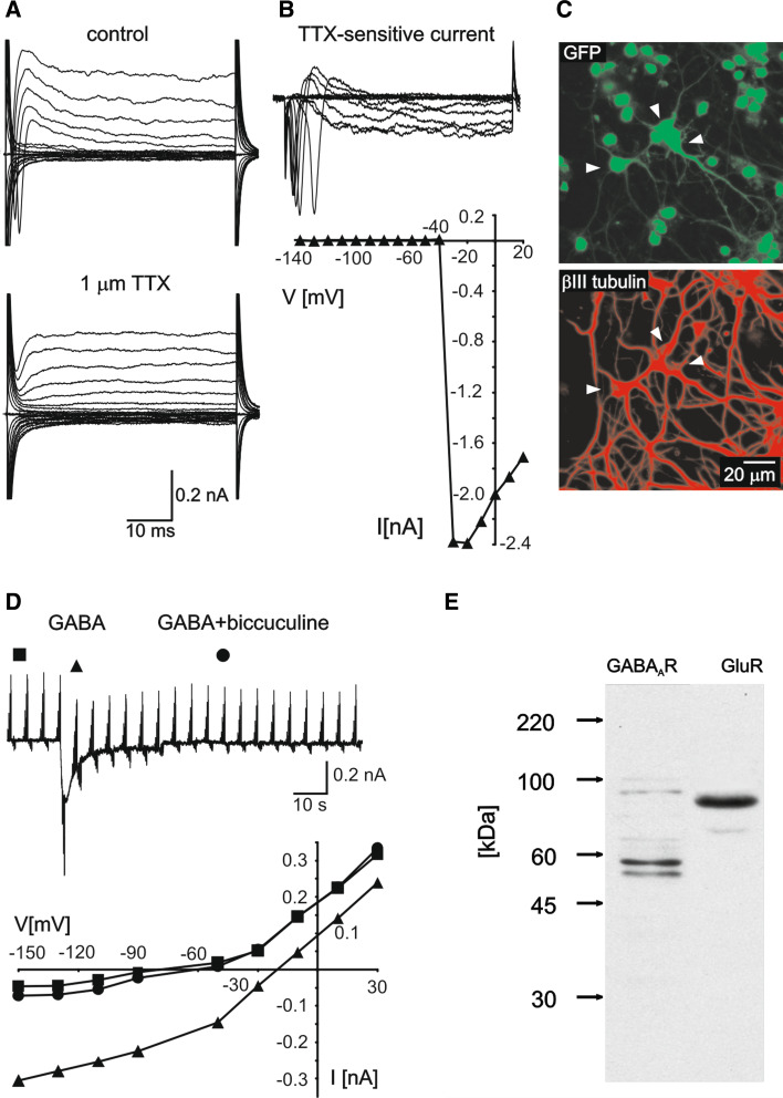
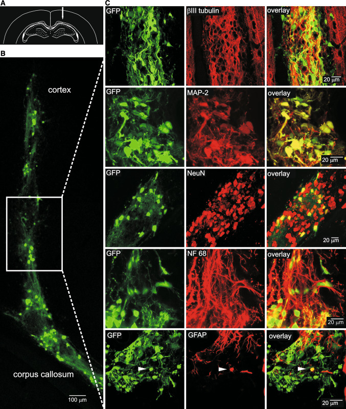
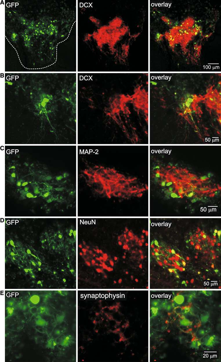
D6 is a promoter/enhancer of the mDach1 gene that is involved in the development of the neocortex and hippocampus. It is expressed by proliferating neural stem/progenitor cells (NSPCs) of the cortex at early stages of neurogenesis. The differentiation potential of NSPCs isolated from embryonic day 12 mouse embryos, in which the expression of green fluorescent protein (GFP) is driven by the D6 promoter/enhancer, has been studied in vitro and after transplantation into the intact adult rat brain as well as into the site of a photochemical lesion. The electrophysiological properties of D6/GFP-derived cells were studied using the whole-cell patch-clamp technique, and immunohistochemical analyses were carried out. D6/GFP-derived neurospheres expressed markers of radial glia and gave rise predominantly to immature neurons and GFAP-positive cells during in vitro differentiation. One week after transplantation into the intact brain or into the site of a photochemical lesion, transplanted cells expressed only neuronal markers. D6/GFP-derived neurons were characterised by the expression of tetrodotoxin-sensitive Na(+)-currents and K (A)- and K (DR) currents sensitive to 4-aminopyridine. They were able to fire repetitive action potentials and responded to the application of GABA. Our results indicate that after transplantation into the site of a photochemical lesion, D6/GFP-derived NSPCs survive and differentiate into neurons, and their membrane properties are comparable to those transplanted into the non-injured cortex. Therefore, region-specific D6/GFP-derived NSPCs represent a promising tool for studying neurogenesis and cell replacement in a damaged cellular environment.


Figures

Fig. 1
Fig. 1
Characterisation of D6/GFP-derived NSPCs in the embryonic telencephalon and in the neurospheres. a Sagital view of a D6/GFP E12 embryo showing fluorescence in the dorsal part of the developing cortex (right). Western blot analysis of isolated tissue and derived neurospheres, harvested 1 week after cultivation, showing GFAP, BLBP, βIII tubulin and DCX in both samples (left). b Enlargement of the brain section in (a, right), showing the localisation of GFAP-δ, BLBP, βIII tubulin and DCX in the sagital sections of an E12 D6/GFP telencephalon. c Immunocytochemical analysis of neurospheres showing the radial glia markers BLBP (top) and RC2 (middle) and the neuronal progenitor marker βIII tubulin (bottom). LV lateral ventricle
Fig. 2
Fig. 2
Immunocytochemical characterisation of D6/GFP NSPCs 1 week after the onset of in vitro differentiation. a Large flat cells of the underlying layer positive for GFAP (top) and RC2 (bottom). b Clusters of cells with neuronal morphology positive for the neuronal markers DCX (top), MAP-2 (middle) and NeuN (bottom). c A detailed image of a GFP-positive cell stained for Olig2. Yellow colour indicates double-stained cells
Fig. 3
Fig. 3
Electrophysiological characterisation of passive cells, 1 week after the onset of in vitro differentiation. a Immunocytochemical identification of a recorded cell (indicated by arrowhead, top), showing positive staining for GFAP (bottom). b Typical current pattern of a large flat D6/GFP-derived cell obtained by hyper- and depolarising the cell from a holding potential of −70 mV to potentials ranging from −160 to +20 mV, under control conditions (top) and after the application of 1 mM CsCl (bottom); c the resulting current/voltage relationship of the CsCl-sensitive current. d A typical inward current and depolarisation evoked by the application of a 50 mM K+-solution (left) and the resulting current/voltage relationship (right) for control ACF (filled squares) and 50 mM K+ solution (filled triangles), showing the shift of V rev. e A typical inward current evoked by the application of 100 μM GABA completely blocked by the application of 100 μM biccuculine (left) and the resulting current/voltage relationship (right) for control ACF (filled squares), GABA-containing ACF (filled circles) and GABA applied together with biccuculine (filled triangles). The currents shown in d, e were obtained by clamping the cell membrane potential to different values, by rectangular voltage steps, from a holding potential of −70 mV to potentials ranging from −150 to +30 mV (see the inset in e)
Fig. 4
Fig. 4
Membrane properties of D6/GFP-derived neuronal cells, 1 week after the onset of in vitro differentiation. a A typical membrane current pattern of a GFP/βIII tubulin-positive cell prior to (top) and after (bottom) the application of 1 μM TTX, obtained by hyper- and depolarising the cell membrane from a holding potential of −70 mV to potentials ranging from −160 to +20 mV. b The TTX-sensitive current was obtained by subtracting the currents after TTX application from the control currents. The resulting current/voltage relationship for the TTX-sensitive current is shown below. c The morphology and immunocytochemical identification of a recorded neuronal cell (indicated by an arrowhead, top) showing positive staining for βIII tubulin (bottom). d A typical inward current evoked by the application of 100 μM GABA blocked by 100 μM biccuculine (top) and the resulting current/voltage relationship (bottom) for control ACF (filled squares), GABA-containing ACF (filled triangles) and GABA applied together with biccuculine (filled circles) showing the shift of V rev. e Western blot analysis showing the expression of GABAA and glutamate receptors in cell culture
Fig. 5
Fig. 5
Voltage-dependent K+ currents of D6/GFP-derived neuronal cells and their pharmacological properties. a Current patterns obtained by hyper- and depolarising the cell from a holding potential of −50 mV to potentials ranging from −140 to +40 mV (left) and the isolated K DR current, obtained by passive-current subtraction (right). b Current patterns obtained by hyper- and depolarising the cell from a holding potential of −50 mV to potentials ranging from −140 to +40 mV, after a hyperpolarising prepulse to −110 mV (left), and the isolated K A current (right) obtained by subtracting the currents shown in (a, left) from those shown in (b, left). c Current pattern shown in a after the application of 5 mM 4-AP (left) and the isolated K DR current (right). d Current pattern shown in b after the application of 5 mM 4-AP (left) and the isolated K A current (right). e Current/voltage relationships for the subtracted K A and K DR currents in control ACF (empty squares and triangles) and after the application of 5 mM 4-AP (filled squares and triangles)
Fig. 6
Fig. 6
D6-GFP NSPCs cells survive and give rise to neurons 1 week after transplantation into the non-injured rat cortex. a A schematic image of the injection site in the cortex. b Coronal section of an adult rat cortex and corpus callosum showing the injection site filled with D6-GFP cells, 1 week after transplantation. c Higher magnification images of the injection site showing GFP-positive cells immunostained for the neuronal markers βIII tubulin, MAP-2, NeuN and NF 68. Note that D6/GFP cells surviving in the brain were positive predominantly for neuronal markers, while GFP/GFAP double stained cells were rare (an example is indicated by arrowhead) and displayed undifferentiated morphology. Yellow colour indicates double-stained cells
Fig. 7
Fig. 7
D6-GFP NSPCs cells give rise to neurons 1 week after transplantation into the site of a photochemical lesion. a Overview of the photochemical lesion filled with GFP-positive cells 1 week after transplantation and immunostained for DCX. be Higher magnification images of the interior of the lesion showing GFP-positive cells immunostained for DCX (b), MAP-2 (c), NeuN (d) and synaptophysin (e). Yellow colour indicates double-stained cells
Fig. 8
Fig. 8
One week after transplantation into the intact or injured rat brain, D6/GFP-derived neurons display characteristics of mature neurons. a A typical membrane current pattern of an in vivo D6/GFP-derived neuron obtained by hyper- and depolarising the cell membrane from a holding potential of −70 mV to potentials ranging from −160 to +20 mV. Note the repetitive firing of APs during continuous current injection—see the inset. b The morphology and immunohistochemical identification of a recorded D6/GFP-derived neuron (indicated by an arrowhead, top), 1 week after transplantation into the intact rat cortex, showing positive staining for NeuN (bottom). c A typical inward current evoked by the application of 100 μM GABA blocked by 100 μM biccuculine (top) and the resulting current/voltage relationship (bottom) for control (filled squares), GABA (filled triangles) and GABA applied together with biccuculine (filled circles). d The characteristics of the action potential (AP) in D6/GFP-derived neuronal cells differentiated in vitro and D6/GFP-derived neurons differentiated after transplantation into the intact and injured rat brain. Asterisks indicate significant differences between cells differentiated in vitro and cells differentiated in vivo. * P < 0.05; ** P < 0.01, *** P < 0.001

Similar articles

Cited by

References

    1. Abematsu M, Kagawa T, Fukuda S, Inoue T, Takebayashi H, Komiya S, Taga T (2006) Basic fibroblast growth factor endows dorsal telencephalic neural progenitors with the ability to differentiate into oligodendrocytes but not gamma-aminobutyric acidergic neurons. J Neurosci Res 83(5):731–743 - PubMed
    1. Akesson E, Piao JH, Samuelsson EB, Holmberg L, Kjaeldgaard A, Falci S, Sundstrom E, Seiger A (2007) Long-term culture and neuronal survival after intraspinal transplantation of human spinal cord-derived neurospheres. Physiol Behav 92(1–2):60–66 - PubMed
    1. Anderova M, Antonova T, Petrik D, Neprasova H, Chvatal A, Sykova E (2004) Voltage-dependent potassium currents in hypertrophied rat astrocytes after a cortical stab wound. Glia 48(4):311–326 - PubMed
    1. Anderova M, Kubinova S, Jelitai M, Neprasova H, Glogarova K, Prajerova I, Urdzikova L, Chvatal A, Sykova E (2006) Transplantation of embryonic neuroectodermal progenitor cells into the site of a photochemical lesion: immunohistochemical and electrophysiological analysis. J Neurobiol 66(10):1084–1100 - PubMed
    1. Andreasen M, Skov J, Nedergaard S (2007) Inwardly rectifying K(+) (Kir) channels antagonize ictal-like epileptiform activity in area CA1 of the rat hippocampus. Hippocampus 17(11):1037–1048 - PubMed

Publication types

MeSH terms

LinkOut - more resources