. 2009 Oct 23;284(43):29945-55.
doi: 10.1074/jbc.M109.055251. Epub 2009 Aug 26.

A novel class of cyclin-dependent kinase inhibitors identified by molecular docking act through a unique mechanism

Affiliations

A novel class of cyclin-dependent kinase inhibitors identified by molecular docking act through a unique mechanism

Patrick Corsino et al. J Biol Chem. .

Abstract

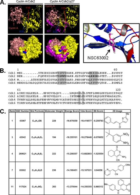
The cyclin-dependent kinase (Cdk) family is emerging as an important therapeutic target in the treatment of cancer. Cdks 1, 2, 4, and 6 are the key members that regulate the cell cycle, as opposed to Cdks that control processes such as transcription (Cdk7 and Cdk9). For this reason, Cdks 1, 2, 4, and 6 have been the subject of extensive cell cycle-related research, and consequently many inhibitors have been developed to target these proteins. However, the compounds that comprise the current list of Cdk inhibitors are largely ATP-competitive. Here we report the identification of a novel structural site on Cdk2, which is well conserved between the cell cycle Cdks. Small molecules identified by a high throughput in silico screen of this pocket exhibit cytostatic effects and act by reducing the apparent protein levels of cell cycle Cdks. Drug-induced cell cycle arrest is associated with decreased Rb phosphorylation and decreased expression of E2F-dependent genes. Multiple lines of evidence indicate that the primary mechanism of action of these compounds is the direct induction of Cdk1, Cdk2, and Cdk4 protein aggregation.


Figures

FIGURE 1.
FIGURE 1.
Identification of a novel Cdk2 binding pocket and interacting molecules by high-throughput in silico screening. In A: left, visualization of the Cyclin A-Cdk2 and Cyclin A-Cdk2-p27 crystal structure with PyMOL (yellow, Cdk2; salmon, Cyclin A; and purple, p27; tyrosine 15 and arginine 36 are colored red and blue, respectively, as a point of reference). Right: ribbon view of Cdk2 with NSC63002 docked inside the pocket. B, sequence alignments of human Cdk1, Cdk2, Cdk4, and Cdk6. Shaded regions represent amino acid residues lining the pocket targeted by molecular docking. C, a selection of the most effective compounds identified by molecular docking as determined by [3H]thymidine incorporation.
FIGURE 2.
FIGURE 2.
NSC compounds inhibit cell proliferation. A, BT549 breast cancer, and HCT116 colon cancer cells were treated with increasing doses of NSC43067, NSC43042, and NSC63002. After a 24-h incubation, the cells were pulsed with [3H]thymidine for 2 h to measure DNA synthesis. All compounds caused a dose-dependent decrease in cell proliferation in both cell lines. B, BT549 cells, or in part C, HCT116 cells, were treated with increasing doses of NSC43042 and NSC63002. After 24 h, the nuclei of the cells were stained with propidium iodide and subjected to analysis by flow cytometry. Cells arrested in the G1, S, and G2 phases of the cell cycle were observed depending on the compound and the concentration. Left, data from triplicate samples plotted by cell cycle phase; right, individual histograms of compound and vehicle-treated cells.
FIGURE 3.
FIGURE 3.
NSC compounds affect cell cycle Cdk levels and activity. A, BT549 cells were treated with 100 μm or 200 μm NSC43067, NSC269621, or NSC63002 for 24 or 48 h. Protein from the cells was extracted with 0.1% Triton X-100 extraction buffer, normalized, and subjected to immunoblot analysis. The levels of Cdk1, Cdk2, and Cdk4, but not actin or protein phosphatase 2A (PP2A), decrease sharply after treatment with the NSC compounds. Cyclins A, D1, and E decreased to some extent. B, Mv1Lu mink lung epithelial cells overexpressing D1K2, E2F1, or empty vector were treated with either 0.2% DMSO or 200 μm NSC63002 for 24 h. The presence of higher levels of E2F1 or a constitutively active form of Cdk2 (D1K2) partially reverses the effects of the compounds on the levels of E2F-dependent gene products. C, Cyclin E-Cdk2 complexes were preincubated with 200 μm NSC43042 or NSC63002 overnight, then added to Rb substrate with [32P]ATP to measure kinase activity in vitro. Compound treatment decreases Cdk2 kinase activity relative to control.
FIGURE 4.
FIGURE 4.
The decrease in Cdk levels occurs on the same time frame as the decrease in cell proliferation. A, BT549 cells were treated with 0.2% DMSO or 200 μm NSC43042 or NSC63002 for 1, 2, 4, or 24 h. Immunoblot analysis reveals a decrease in the levels of Cdk1 as early as 4 h, with Cdk2 and Cdk4 decreasing by 24 h. B, BT549 cells were treated with 0.2% DMSO or 200 μm NSC43042 or NSC63002 for 4, 8, 12, and 24 h. [3H]Thymidine incorporation analysis reveals that the decrease in Cdk levels occurs on the same time scale as the inhibition of cell proliferation.
FIGURE 5.
FIGURE 5.
The decrease in Cdk levels is a result of protein aggregation. A, 293A cells were transiently transfected with either pcDNA3 vector or Cdk4/pcDNA3 and treated with 0.2% DMSO or 200 μm NSC63002 for 24 h. Immunoblot analysis reveals a marked increase in high molecular mass bands in samples treated with NSC63002. B, BT549 cells were treated with 0.2% DMSO or 200 μm NSC63002 for 24 h. Cell extracts were centrifuged at either 16,000 × g for 20 min or 150,000 × g for 1 h. Immunoblot analysis reveals that more Cdk 1, 2, and 4 pelleted by high speed centrifugation from samples treated with 200 μm NSC63002 than the DMSO-treated samples or the lower speed centrifugation. C, BT549 cells were treated with 0.2% DMSO or 200 μm NSC63002 for 24 h. Immunofluorescence analysis reveals the formation of Cdk4 aggregates in samples treated with NSC63002. D, 293A cells stably expressing a GFP-Cdk4 fusion protein were treated with 0.2% DMSO or 200 μm NSC63002 for 24 h. Fluorescence microscopy reveals the formation of Cdk4 aggregates in NSC compound-treated cells. E, immunoblot analysis of the 293A cells overexpressing GFP-Cdk4 confirms the presence of full-length GFP-Cdk4 and no significant degradation products.
FIGURE 6.
FIGURE 6.
The effects of the compounds are reversible. A, 293A/GFP-Cdk4 cells were treated with 200 μm NSC63002 for 18 h, and visualized by time-lapse microscopy with images taken every 30 min. Aggregates appear to form between 6 and 9 h (earlier in other cells, see supplemental Fig. S3) with most cells containing some degree of Cdk4 aggregation by 18 h. B, 293A/GFP-Cdk4 cells pre-treated for 12 h with 200 μm NSC63002 were re-fed with fresh media and followed by time-lapse microscopy for a further 16 h with images taken every 60 min. By the final image at 15 h, every aggregate appears to have disappeared. C, BT549 cells were treated with 100 μm NSC43042 or NSC63002 for the 2, 4, 8, 12, or 24 h. Cell proliferation was measured by [3H]thymidine incorporation. D, similar to part C, cells were treated for 2, 4, 8, 12, or 24 h, except they were re-fed with fresh media for a further 24 h, before measuring cell proliferation by [3H]thymidine incorporation. Compared with part C part D reveals that cells re-fed after compound treatment recover their proliferative capacity.
FIGURE 7.
FIGURE 7.
Aggregated Cdks associate with chaperone proteins and markers for aggresomes. A, 293A/GFP-Cdk4 cells were treated with 200 μm NSC43042 or NSC63002 for 24 h. Compound-treated cells display larger and more frequent foci of Cdk4 that co-localized with the chaperone protein Hsp70. B, BT549 cells were treated with 200 μm NSC43042 or NSC63002 for 24 h. Cell lysates were centrifuged at 150,000 × g for 1 h, and both supernatant and pellet were processed for Western analysis. An increase in Hsc70, Hsp70, and Hsp90 was observed in the pellet of compound-treated samples. C, 293A/GFP-Cdk4 cells were treated with 200 μm NSC63002 for 24 h. Treated cells display larger foci of Cdk4 protein that co-localized with the aggresome marker γ-tubulin.

Similar articles

Cited by

References

    1. Deshpande A., Sicinski P., Hinds P. W. (2005) Oncogene 24, 2909–2915 - PubMed
    1. Sánchez I., Dynlacht B. D. (2005) Semin. Cell Dev. Biol. 16, 311–321 - PubMed
    1. Benson C., White J., De Bono J., O'Donnell A., Raynaud F., Cruickshank C., McGrath H., Walton M., Workman P., Kaye S., Cassidy J., Gianella-Borradori A., Judson I., Twelves C. (2007) Br. J. Cancer 96, 29–37 - PMC - PubMed
    1. Christian B. A., Grever M. R., Byrd J. C., Lin T. S. (2007) Curr. Opin. Oncol. 19, 573–578 - PubMed
    1. Singh R. P., Deep G., Blouin M. J., Pollak M. N., Agarwal R. (2007) Carcinogenesis 28, 2567–2574 - PubMed

Publication types

MeSH terms