. 2009 Nov;83(21):11244-53.
doi: 10.1128/JVI.01413-09. Epub 2009 Aug 26.

Analysis of protein levels of 24 cytokines in scrapie agent-infected brain and glial cell cultures from mice differing in prion protein expression levels

Affiliations

Analysis of protein levels of 24 cytokines in scrapie agent-infected brain and glial cell cultures from mice differing in prion protein expression levels

Déborah Tribouillard-Tanvier et al. J Virol. 2009 Nov.

Abstract

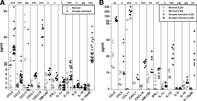
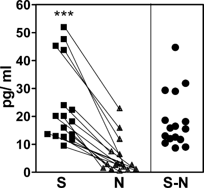
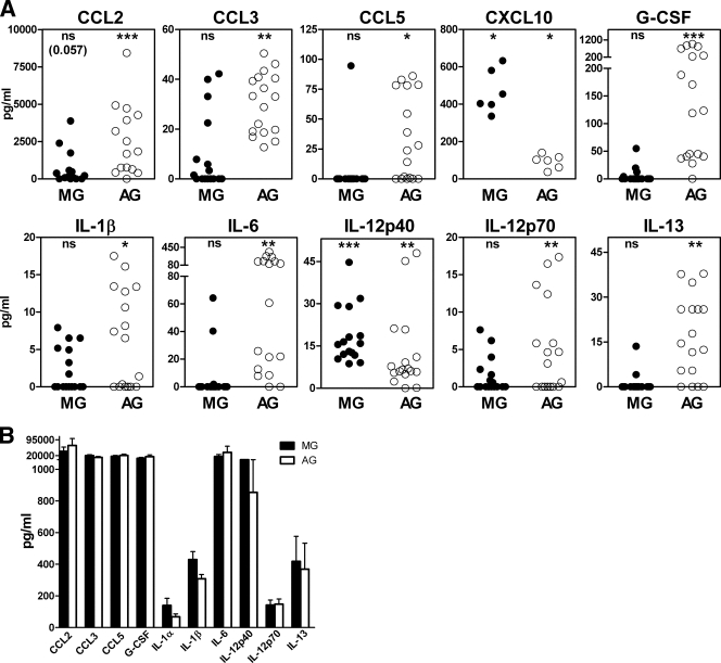
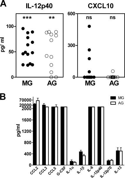
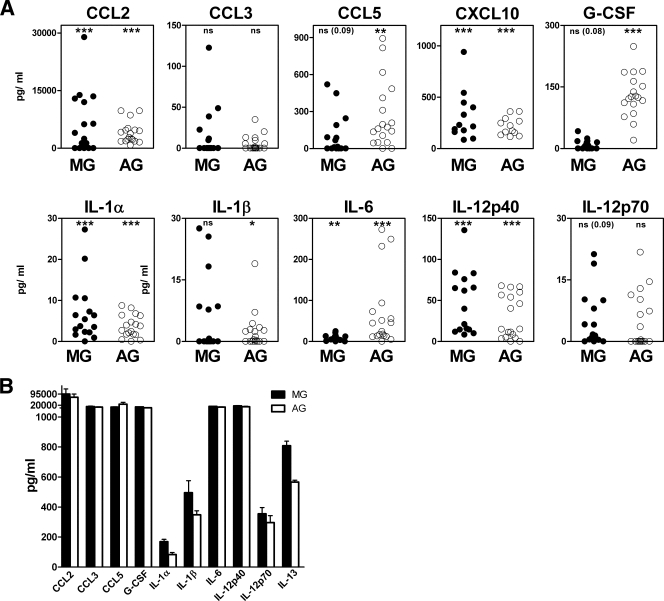
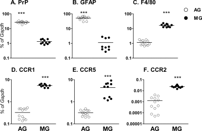
Activation of microglia and astroglia is seen in many neurodegenerative diseases including prion diseases. Activated glial cells produce cytokines as a protective response against certain pathogens and as part of the host inflammatory response to brain damage. In addition, cytokines might also exacerbate tissue damage initiated by other processes. In the present work using multiplex assays to analyze protein levels of 24 cytokines in scrapie agent-infected C57BL/10 mouse brains, we observed elevation of CCL2, CCL5, CXCL1, CXCL10, granulocyte-macrophage colony-stimulating factor (GM-CSF), gamma interferon (IFN-gamma), interleukin 1alpha (IL-1alpha), IL-1beta, IL-6, and IL-12p40. Scrapie agent-infected wild-type mice and transgenic mice expressing anchorless prion protein (PrP) had similar cytokine responses in spite of extensive differences in neuropathology. Therefore, these responses may be primarily a reaction to brain damage induced by prion infection rather than specific inducers of a particular type of pathology. To study the roles of astroglia and microglia in these cytokine responses, primary glial cultures were exposed to scrapie agent-infected brain homogenates. Microglia produced only IL-12p40 and CXCL10, whereas astroglia produced these cytokines plus CCL2, CCL3, CCL5, CXCL1, G-CSF, IL-1beta, IL-6, IL-12p70, and IL-13. Glial cytokine responses from wild-type mice and transgenic mice expressing anchorless PrP differed only slightly, but glia from PrP-null mice produced only IL-12p40, indicating that PrP expression was required for scrapie agent induction of other cytokines detected. The difference in cytokine response between microglia and astroglia correlated with 20-fold-higher levels of PrP expression in astroglia versus microglia, suggesting that high-level PrP expression on astroglia might be important for induction of certain cytokines.


Figures

FIG. 1.
FIG. 1.
Cytokine expression in scrapie agent-infected mouse brain. (A) Wild-type C57BL/10SnJ mice. Brain homogenates from 21 scrapie agent-infected mice and 14 uninfected age-matched mice were tested for cytokine levels by multiplex bead arrays. The cytokine levels of scrapie agent-infected mice are shown as solid circles, and the cytokine levels of healthy uninfected (normal) brains are open circles. Each symbol represents the value for one mouse. CCL2, CCL5, CXCL1, CXCL10, GM-CSF, IFN-γ, IL-1α, IL-1β, IL-6, and IL-12p40 were expressed at significantly higher levels in the scrapie agent-infected brains than in the healthy brains. Statistical significance by the Mann-Whitney test is indicated as follows: *, P < 0.05; ***, P < 0.001; ns, not significant. Other cytokines tested, but not detected, in wild-type mouse brains were CCL3, CCL4, CCL11, G-CSF, IL-2, IL-3, IL-4, IL-5, IL-9, IL-10, IL-12p70, IL-13, IL-17, and TNF. (B) Transgenic mice expressing anchorless PrP. Brain homogenates from homozygous transgenic scrapie agent-infected mice (lines 23 [L23] and 44 [L44]) were obtained at 350 to 400 dpi and were tested for cytokine levels by multiplex bead arrays. Uninfected age-matched transgenic mice were used as controls. The cytokine levels of scrapie agent-infected mice are shown as solid symbols, and the cytokine levels of healthy uninfected (normal) brains are shown as open symbols. Each symbol represents the value for one mouse. CCL2, CCL5, CCL11, CXCL1, CXCL10, GM-CSF, IFN-γ, IL-1α, IL-1β, and IL-12p40 were expressed at significantly higher levels in the scrapie agent-infected mice than in the uninfected mice. Statistical significance by the Mann-Whitney test is indicated as follows: *, P < 0.05; **, P < 0.005; ***, P < 0.001; ns, not significant. Other cytokines tested, but not detected, in brains were CCL3, CCL4, G-CSF, IL-2, IL-3, IL-4, IL-5, IL-6, IL-9, IL-10, IL-12p70, IL-13, IL-17, and TNF.
FIG. 2.
FIG. 2.
In vitro stimulation of IL-12p40 in microglial cells by scrapie agent-infected and healthy uninfected brain homogenates. S (▪) is the cytokine level in glial culture supernatant 24 h after stimulation with scrapie agent-infected brain homogenate minus the background cytokine level detected in scrapie agent-infected brain homogenate prior to use as a stimulant. N (▴) is the cytokine level in glial culture supernatant 24 h after stimulation with healthy brain homogenate minus the background cytokine level detected in healthy brain homogenate prior to use as a stimulant. Values for paired cultures in each experiment are linked by lines. SN subtracted values for each pair are shown in the right-hand side of the figure (•). The S and N values were significantly different (P < 0.001) by the Mann-Whitney matched-pair test (***). Data shown in Fig. 3A, 4A, and 5A are SN values.
FIG. 3.
FIG. 3.
Cytokine expression in C57BL/10SnJ glial cells exposed to scrapie agent-infected brain homogenate. (A) Exposure to scrapie agent-infected or healthy uninfected brain homogenates. Purified microglia (MG) or astroglia (AG) were stimulated by overlay with scrapie agent-infected or healthy brain homogenates. Supernatants were analyzed by multiplex assay 24 h after stimulation. The data show the scrapie agent-induced cytokine level as explained in Materials and Methods. Statistical analysis was done using the Wilcoxon matched-pair test comparing the S and N values for cultures from 16 independent experiments for each cytokine (6 experiments for CXCL10). Subtracted values for scrapie agent-infected brain stimulation minus healthy brain stimulation (SN) are shown (see the legend to Fig. 2 and Materials and Methods for details). The cytokine levels from microglia are shown as solid circles, and the cytokine levels from astroglia are shown as open circles. MG produced IL-12p40 and CXCL10, and AG produced these two cytokines plus CCL2, CCL3, CCL5, G-CSF, IL-1β, IL-6, IL-12p70, IL-13, and CXCL1 (not shown). Statistical significance by the Mann-Whitney matched-pair test is indicated as follows: *, P < 0.05; **, P < 0.005; ***, P < 0.001; ns, not significant. (B) Stimulation of glia by LPS. Supernatants of microglia and astroglia cultures stimulated for 24 h with LPS at a concentration of 1 μg/ml were analyzed for cytokines using Bio-Plex multiple-cytokine assay kits. The values represent the means plus standard errors of the means (error bars) for seven different experiments.
FIG. 4.
FIG. 4.
Scrapie agent-induced cytokine profiles in astroglia and microglia from PrP-null mice. Supernatants of PrP-null glial cell cultures were analyzed for cytokines by using Bio-Plex multiple-cytokine assay kits 24 h after overlay with scrapie agent-infected or healthy uninfected brain homogenates. Scrapie agent-induced cytokine level is shown as explained in Materials and Methods. Statistical analysis was done using the Wilcoxon matched-pair test comparing the S and N values for cultures from 14 independent experiments for each cytokine. Subtracted values for scrapie agent-infected brain stimulation minus healthy brain stimulation (SN) are shown (see the legend to Fig. 2 and Materials and Methods for details). (A) Only IL-12p40 was significantly elevated in astoglia and microglia. Twenty-three other cytokines were found to be negative; CXCL10/IP-10 is shown as an example. Statistical significance by the Mann-Whitney matched-pair test is indicated as follows: ns, not significant; **, P < 0.005; ***, P < 0.001). (B) LPS stimulation. The levels of cytokines produced by microglia (MG) and astroglia (AG) are shown. The values represent the means plus standard errors of the means (error bars) for seven different experiments after stimulation for 24 h with LPS at a concentration of 1 μg/ml. Ten cytokines are shown; however, all 24 cytokines tested were produced at elevated levels.
FIG. 5.
FIG. 5.
(A) Scrapie agent-induced cytokine profiles in astroglia and microglia from transgenic mice expressing anchorless PrP. Supernatants of glial cell cultures from transgenic mice were analyzed for amounts of cytokines by using the Bio-Plex multiple-cytokine assay kits 24 h after overlay with brain homogenates. Stimulated culture supernatants of microglia (MG) and astroglia (AG) were analyzed for scrapie agent-induced cytokine level as described in Materials and Methods. Statistical analysis was done using the Wilcoxon matched-pair test comparing the S and N values for cultures from 18 independent experiments for each cytokine. Subtracted values for scrapie agent-infected brain stimulation minus healthy uninfected brain stimulation (SN) are shown (see the legend to Fig. 2 and Materials and Methods for details). After scrapie agent stimulation, astroglia significantly produced CCL2, CCL5, CXCL10, G-CSF, IL-1α, IL-1β, IL-6, and IL-12p40. Statistical significance by the Mann-Whitney matched-pair test is indicated as follows: ns, not significant; *, P < 0.05; **, P < 0.005; ***, P < 0.001). Other cytokines were not significantly elevated. Microglia significantly produced CCL2, CXCL10, IL-1α, IL-6, and IL-12p40 after scrapie agent stimulation. Other cytokines were not significantly elevated. (B) LPS stimulation. Supernatants of MG and AG cultures stimulated for 24 h with LPS at a concentration of 1 μg/ml were analyzed for cytokines using Bio-Plex multiple-cytokine assay kits. The values represent the means plus standard errors of the means (error bars) for eight different experiments.
FIG. 6.
FIG. 6.
Expression of PrP mRNA in microglia and astroglia. Expression of mRNA encoding PrP and five other genes in microglia (MG) and astroglia (AG) was determined using quantitative real-time reverse transcriptase PCR. Data are shown as the percent difference relative to glyceraldehyde-3-phosphate dehydrogenase (GAPDH) mRNA expression for the following genes: PrP (A), GFAP (B), F4/80 (C), CCR1 (D), CCR5 (E), and CCR2 (F). Two different litters of mice were analyzed in separate experiments with six wells of PCR per experiment. Statistical analysis was performed by a one-tailed Mann-Whitney test (***, P < 0.001). Microglia expressed approximately 20-fold-less PrP mRNA than astroglia did. Microglia expressed significantly higher levels of chemokine receptors CCR1, CCR2, and CCR5 than astroglia did. Each symbol shows the amount of mRNA from one mouse, and the black bar shows the mean value.

Similar articles

Cited by

References

    1. Baker, C. A., and L. Manuelidis. 2003. Unique inflammatory RNA profiles of microglia in Creutzfeldt-Jakob disease. Proc. Natl. Acad. Sci. USA 100:675-679. - PMC - PubMed
    1. Bell, M. D., R. Lopez-Gonzalez, L. Lawson, D. Hughes, I. Fraser, S. Gordon, and V. H. Perry. 1994. Upregulation of the macrophage scavenger receptor in response to different forms of injury in the CNS. J. Neurocytol. 23:605-613. - PubMed
    1. Beringue, V., M. Demoy, C. I. Lasmezas, B. Gouritin, C. Weingarten, J. P. Deslys, J. P. Andreux, P. Couvreur, and D. Dormont. 2000. Role of spleen macrophages in the clearance of scrapie agent early in pathogenesis. J. Pathol. 190:495-502. - PubMed
    1. Boillee, S., C. Vande Velde, and D. W. Cleveland. 2006. ALS: a disease of motor neurons and their nonneuronal neighbors. Neuron 52:39-59. - PubMed
    1. Brandner, S. 2003. CNS pathogenesis of prion diseases. Br. Med. Bull. 66:131-139. - PubMed

Publication types