. 2009 Nov;83(21):10877-91.
doi: 10.1128/JVI.00525-09. Epub 2009 Aug 26.

The Epstein-Barr virus protein kinase BGLF4 and the exonuclease BGLF5 have opposite effects on the regulation of viral protein production

Affiliations

The Epstein-Barr virus protein kinase BGLF4 and the exonuclease BGLF5 have opposite effects on the regulation of viral protein production

Regina Feederle et al. J Virol. 2009 Nov.

Abstract

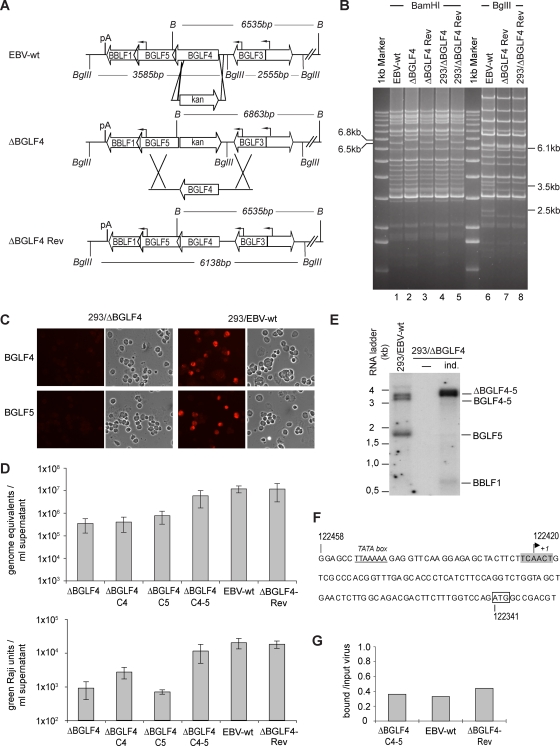
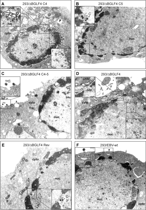
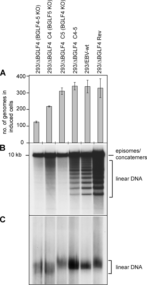
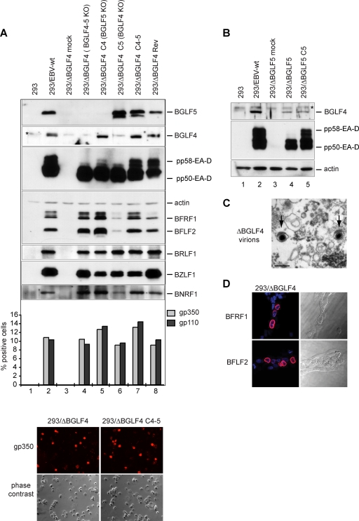
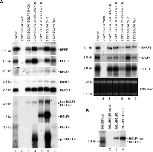
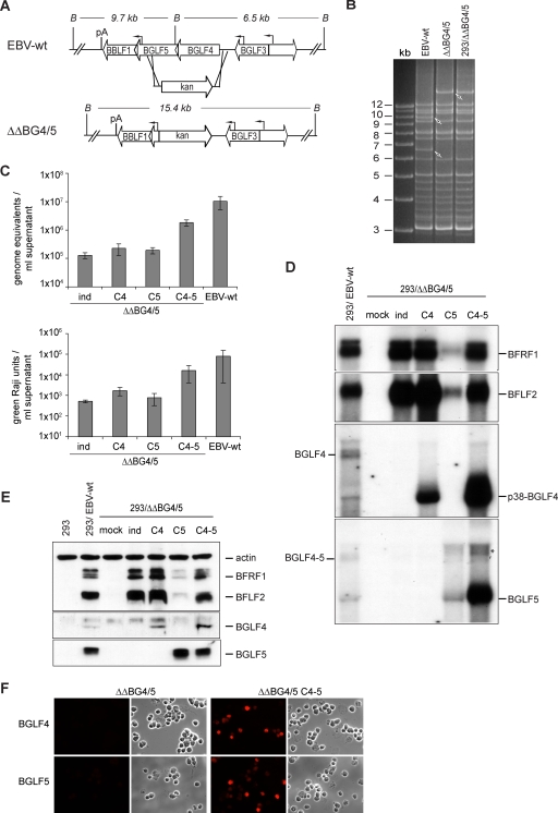
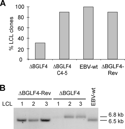
The Epstein-Barr virus BGLF4 and BGLF5 genes encode a protein kinase and an alkaline exonuclease, respectively. Both proteins were previously found to regulate multiple steps of virus replication, including lytic DNA replication and primary egress. However, while inactivation of BGLF4 led to the downregulation of several viral proteins, the absence of BGLF5 had the opposite effect. Using recombinant viruses that lack both viral enzymes, we confirm and extend these initial observations, e.g., by showing that both BGLF4 and BGLF5 are required for proper phosphorylation of the DNA polymerase processivity factor BMRF1. We further found that neither BGLF4 nor BGLF5 is required for baseline viral protein production. Complementation with BGLF5 downregulated mRNA levels and translation of numerous viral genes, though to various degrees, whereas BGLF4 had the opposite effect. BGLF4 and BGLF5 influences on viral expression were most pronounced for BFRF1 and BFLF2, two proteins essential for nuclear egress. For most viral genes studied, cotransfection of BGLF4 and BGLF5 had only a marginal influence on their expression patterns, showing that BGLF4 antagonizes BGLF5-mediated viral gene shutoff. To be able to exert its functions on viral gene expression, BGLF4 must be able to escape BGLF5's shutoff activities. Indeed, we found that BGLF5 stimulated the BGLF4 gene's transcription through an as yet uncharacterized molecular mechanism. The BGLF4/BGLF5 enzyme pair builds a regulatory loop that allows fine-tuning of virus protein production, which is required for efficient viral replication.


Figures

FIG. 1.
FIG. 1.
Design, construction, and initial characterization of a ΔBGLF4 null mutant and its revertant. (A) Schematic representation of the strategy followed to construct the ΔBGLF4 mutant. The figure also depicts some of the viral genes present on the BamHI G fragment. A PCR-amplified DNA fragment containing the kan restriction gene flanked by oligonucleotides homologous to the BGLF4 locus was subjected to homologous recombination with the EBV wild-type genome. Recombination led to a BamHI G fragment size shift from 6,535 bp to 6,863 bp and resulted in the disruption of the BGLF4 gene open reading frame with the exception of its last 50 codons. Exactly the reverse strategy was followed to construct the ΔBGLF4-Rev revertant virus. The targeting vector consisted of the wild-type BGLF4 gene sequence recombined with the mutant BGLF4 virus genome. The BglII restriction site located between the BGLF3 and BGLF4 genes was excised from this targeting vector; digestion with BglII can therefore distinguish between DNA from the wild-type and revertant viruses. B, BamHI; pA, poly(A) site. (B) ΔBGLF4 and ΔBGLF4-Rev genome restriction fragment analysis. The ΔBGLF4 and the ΔBGLF4-Rev genome restriction patterns were determined at different steps of mutant construction to ensure integrity and exactness of the viral recombinants. Bacterial DNAs of these EBV genomes cloned into E. coli were digested with BamHI and separated on an agarose gel. EBV-wt DNA provided a positive control. The ΔBGLF4 mutant showed an altered 6.8-kb BamHI G fragment that replaced the wild-type 6.5-kb BamHI G fragment (lane 2), whereas the ΔBGLF4-Rev revertant had regained the wild-type allele (lane 3). Recombinant genome structure was reassessed after stable transfection into 293 cells and rescue in E. coli (293/ΔBGLF4 and 293/ΔBGLF4-Rev). The ΔBGLF4-Rev DNA was further digested with BglII restriction enzymes. This analysis confirmed the integrity and the absence of the BglII restriction site in the ΔBGLF4-Rev genome (lanes 7 and 8). (C) Induced 293/ΔBGLF4 cells are negative for both BGLF4 and BGLF5. Induced 293/ΔBGLF4 cells were immunostained with polyclonal antibodies specific for BGLF4 or BGLF5 (top). Induced 293/EBV-wt cells were used as positive controls (bottom). The cell morphology under phase-contrast illumination is also shown. (D) Viral titers in various induced cell lines. Viral genome DNA equivalents per ml of supernatant (physical titers) were quantified by qPCR amplification of the viral BALF5 gene (top) or by infection of Raji cells at limiting dilutions (functional titers that represent the number of infectious viruses per ml of supernatant) (bottom). Shown are mean values from five independent experiments. (E) Northern blot analysis of BamHI G transcripts. mRNA from noninduced (−) and induced (ind.) 293/ΔBGLF4 cells and induced 293/EBV-wt cells was hybridized with a BGLF5-specific probe. Induced 293/ΔBGLF4 cells produce a 328-bp-larger transcript as a consequence of the exchange between the BGLF4 gene and the kan gene. (F) Sequence of the BGLF5 gene upstream region. The transcription start site (+1) within a putative initiator element (highlighted in gray) was determined by 5′ RACE using 293/EBV-wt RNA. The predicted TATA box (underlined) and the BGLF5 ATG (box) are indicated. Numbers refer to B95.8 coordinates. (G) Complemented viruses display normal binding properties. B cells were exposed to supernatants from induced 293/EBV-wt, 293/ΔBGLF4-Rev, or 293/ΔBGLF4-C4-5 cells at an MOI of 10 genome equivalents per cell. Unbound viruses were washed off, and the number of viruses bound per cell was determined by qPCR. The ratios between bound viruses and input virus load are indicated.
FIG. 2.
FIG. 2.
Electron micrographs of induced producer cells that carry various recombinants. (A) Induced 293/ΔBGLF4-C4 cells contained immature nucleocapsids (A and B capsids) lacking an electron-dense DNA core (inset I). The inner nuclear membrane was markedly thickened and showed accumulation of electron-dense material between outer and inner nuclear membranes. Images of extensive membrane folding in the perinuclear space are shown in inset II. (B) Induced 293/ΔBGLF4-C4 cells showed intranuclear capsid maturation. The nuclear membrane displayed normal morphological features. Intracytoplasmic or extracellular virions were not visible. (C) Induced and complemented 293/ΔBGLF4-C4-5 cells showed evidence of intranuclear maturation, with numerous DNA-filled nucleocapsids displaying an electron-dense core. Further, intracytoplasmic and extracellular virions that had successfully undergone primary and secondary egress were identified (inset). (D) Induced 293/ΔBGLF4 cells that lack BGLF4 and BGLF5 displayed morphological features that were very similar to those observed in 293/ΔBGLF4-C4 cells. Micrographs revealed an obvious capsid maturation block (inset I) and morphological nuclear membrane abnormalities (inset II). (E and F) 293/ΔBGLF4-Rev and 293/EBV-wt cells showed normal virus maturation, as described for panel C. cyto, cytoplasm; nuc, nucleus; ONM, outer nuclear membrane; INM, inner nuclear membrane; A, B, and C, A-, B-, and C-type capsids. Inset bars, 200 nm.
FIG. 3.
FIG. 3.
Role of BGLF4 and BGLF5 in viral lytic DNA replication. (A) Viral DNA replication in induced cells was quantitated by qPCR amplification. Mean values and standard deviations from three independent experiments are presented. (B) Southern blot analysis of BamHI-cleaved DNA fragments hybridized with a terminal repeat-specific probe. The 10-kb fragment results from restriction of complete BamHI Nhet fragments that are present only in nonlinear genomes, i.e., circular genomes or genome concatemers. In contrast, the smaller fragments are generated by restriction of single unit length linear genomes. (C) Gardella gel electrophoresis coupled to Southern blot analysis using a nonrepetitive gp350-specific probe. This assay allows distinction between circular and unit length linear DNA molecules. Note the faster electrophoresis migration pattern in induced 293/ΔBGLF4 and 293/ΔBGLF4-C4 cells.
FIG. 4.
FIG. 4.
Viral protein expression patterns in the various mutants. (A) Western blot analysis and immunostains of viral proteins in mutant producer cell lines and their wild-type parent or revertant. The expression of members from all three classes of viral proteins in cells induced by transfection of BZLF1 is shown. Induced cells were analyzed 1 day (immediate early BZLF1 and BRLF1 genes), 2 days (early EA-D/BMRF1, BGLF4, BGLF5, BFRF1, and BFLF2 genes), or 3 days (late BNRF1, gp110, and gp350 genes) after induction. Actin was stained as a uniform loading control. The BZLF1 staining identifies a stronger signal in cells cotransfected with BZLF1 and an empty pCDNA vector than in cells cotransfected with BZLF1 and BGLF4 and/or BGLF5. This, however, did not impede activation of the lytic program, as shown by the very similar BRLF1 levels in all transfected-cell populations. The immunoblot against EA-D/BMRF1 recognizes the variably phosphorylated forms of this protein. Expression of gp110 and gp350 is difficult to evaluate by Western blotting and was instead assessed by immunofluorescence. The percentages of positive cells are given. Also shown is an example of an immunostain against gp350 as well as a phase-contrast picture. The BGLF4 antibody shows a nonspecific signal (indicated by an asterisk) that migrates directly above the BGLF4-specific band. This cross-reactivity was observed with several different BGLF4-specific antibodies (11, 31), suggesting that BGLF4 shares immunogenic epitopes with a cellular protein. Cross-reactivity was not visible in immunostains. (B) Western blot analysis of BGLF4 and EA-D/BMRF1 in 293/ΔBGLF5 cells. (C) Supernatant from induced 293/ΔBGLF4 cells was pelleted by ultracentrifugation and after fixation was examined by electron microscopy. The picture shows rare examples of mature virions with electron-dense cores (arrows). Bar, 200 nm. (D) Immunostains of induced 293/ΔBGLF4 cells with BFLF2- and BFRF1-specific antibodies. Nuclei were visualized by chromatin staining with Hoechst 33258 (blue), and fluorescent signals were recorded with a confocal microscope. Nomarski pictures revealing fine morphological features of induced cells are shown at the right. Note the obvious thickening of the nuclear membranes in the induced cells.
FIG. 5.
FIG. 5.
Total mRNA expression of a panel of viral genes in the various mutants. The mRNA expression profile was assessed by Northern blot analysis using specific probes. (A) Induced 293/ΔBGLF4 cells transfected with the indicated plasmids were analyzed 1 day (immediate early BZLF1 and BRLF1 genes; left), 2 days (early EA-D/BMRF1, BGLF4, BGLF5, BFRF1, and BFLF2 genes; left), or 3 days (late BNRF1, gp110, and gp350 genes; right) after induction. Ethidium bromide (EtBr) staining of the denaturing gel was used as a loading control. Hybridization with a BGLF5-specific probe identified a 1.7-kb mRNA (BGLF5), a 3.4-kb mRNA (BGLF4-5), and a 3.7-kb mRNA in cells where the BGLF4 gene was replaced by the kan resistance gene (kan-BGLF5) (see Fig. 1A). An unspecific band (*) is observed in cells transfected with the BGLF5 expression plasmid. The BGLF4-specific probe detects only the 3.4-kb mRNA or the gene expressed from the BGLF4 expression plasmid (p38-BGLF4). (B) BGLF4 mRNA expression in induced 293/ΔBGLF5 cells. A BGLF4-specific probe detects the 3.4-kb BGLF4 mRNA in EBV-wt cells and a 3.7-kb transcript in ΔBGLF5 cells carrying the kan resistance gene (BGLF4-kan).
FIG. 6.
FIG. 6.
Construction and analysis of a BGLF4-BGLF5 double mutant. (A) Schematic map of the BGLF4-BGLF5 genes in EBV-wt and in the double-mutant (ΔΔBG4/5) virus after homologous recombination with the kan resistance gene targeting vector. The BamHI cleavage sites and the expected fragment sizes are given. B, BamHI; pA, poly(A) site. (B) BamHI restriction fragment analysis of EBV-wt and ΔΔBG4/5 genomes after construction in E. coli or after rescue from stably transfected 293 cells (293/ΔΔBG4/5). (C) Viral titers in various induced cell lines. Viral genome DNA equivalents per ml of supernatant as measured by qPCR (top) or the numbers of infectious viruses per ml of supernatant as measured by Raji cell infection (bottom) are presented. Values are means from seven different mutant clones and four different EBV-wt clones. Complementation with either BGLF4 or BGLF5 alone was performed with one 293/ΔΔBG4/5 cell clone. All data were obtained from three independent experiments. (D) Northern blot analysis of BFRF1, BFLF2, BGLF4, and BGLF5 mRNA expression in induced and complemented 293/ΔΔBG4/5 cells and 293/EBV-wt cells. *, unspecific band observed in cells transfected with the BGLF5 expression plasmid (see Fig. 5A). (E) Western blot analysis of BFRF1, BFLF2, BGLF4, and BGLF5 protein expression in induced and complemented 293/ΔΔBG4/5 cells. Protein extracts from HEK293 cells and 293/EBV-wt cells served as negative and positive controls, respectively. (F) Immunostaining of induced or complemented 293/ΔΔBG4/5 cells with BGFL4- and BGLF5-specific antibodies confirms the absence of both proteins in 293/ΔΔBG4/5 cells.
FIG. 7.
FIG. 7.
The BGLF4 and BGLF5 double null mutant retains EBV B-cell-transforming properties. (A) BGLF4 and BGLF5 are not required for maintenance of the transformed phenotype. Resting B cells were exposed to supernatants from 293/ΔBGLF4, 293/ΔBGLF4-C4-5, 293/ΔBGLF4-Rev, and 293/EBV-wt cells at an MOI of 10 genome equivalents per cell. B-cell transformation efficiency was assessed by counting the wells containing outgrown cell clones. (B) Southern blot analysis of LCLs obtained by infection of resting B cells with supernatants from induced 293/ΔBGLF4, 293/ΔBGLF4-Rev, and 293/EBV-wt cells. Genomic DNA was cleaved with BamHI, separated by gel electrophoresis, and hybridized with a BGLF3-specific probe that allows distinction between wild-type and recombinant genomes (the wild-type BamHI G fragment is 6.5 kb, whereas the recombinant fragment is 6.8 kb).

Similar articles

Cited by

References

    1. Asai, R., A. Kato, K. Kato, M. Kanamori-Koyama, K. Sugimoto, T. Sairenji, Y. Nishiyama, and Y. Kawaguchi. 2006. Epstein-Barr virus protein kinase BGLF4 is a virion tegument protein that dissociates from virions in a phosphorylation-dependent process and phosphorylates the viral immediate-early protein BZLF1. J. Virol. 80:5125-5134. - PMC - PubMed
    1. Baer, R., A. T. Bankier, M. D. Biggin, P. L. Deininger, P. J. Farrell, T. J. Gibson, G. Hatfull, G. S. Hudson, S. C. Satchwell, C. Seguin, P. S. Tufnell, and B. G. Barell. 1984. DNA sequence and expression of the B95-8 Epstein-Barr virus genome. Nature 310:207-211. - PubMed
    1. Chen, H. F., M. Sauter, P. Haiss, and N. Muller-Lantzsch. 1991. Immunological characterization of the Epstein-Barr virus phosphoprotein PP58 and deoxyribonuclease expressed in the baculovirus expression system. Int. J. Cancer 48:879-888. - PubMed
    1. Chen, M. R., S. J. Chang, H. Huang, and J. Y. Chen. 2000. A protein kinase activity associated with Epstein-Barr virus BGLF4 phosphorylates the viral early antigen EA-D in vitro. J. Virol. 74:3093-3104. - PMC - PubMed
    1. Countryman, J., H. Jenson, R. Seibl, H. Wolf, and G. Miller. 1987. Polymorphic proteins encoded within BZLF1 of defective and standard Epstein-Barr viruses disrupt latency. J. Virol. 61:3672-3679. - PMC - PubMed

MeSH terms

LinkOut - more resources