. 2009 Nov;83(21):10857-68.
doi: 10.1128/JVI.01191-09. Epub 2009 Aug 26.

Bimolecular complementation of paramyxovirus fusion and hemagglutinin-neuraminidase proteins enhances fusion: implications for the mechanism of fusion triggering

Affiliations

Bimolecular complementation of paramyxovirus fusion and hemagglutinin-neuraminidase proteins enhances fusion: implications for the mechanism of fusion triggering

Sarah A Connolly et al. J Virol. 2009 Nov.

Abstract

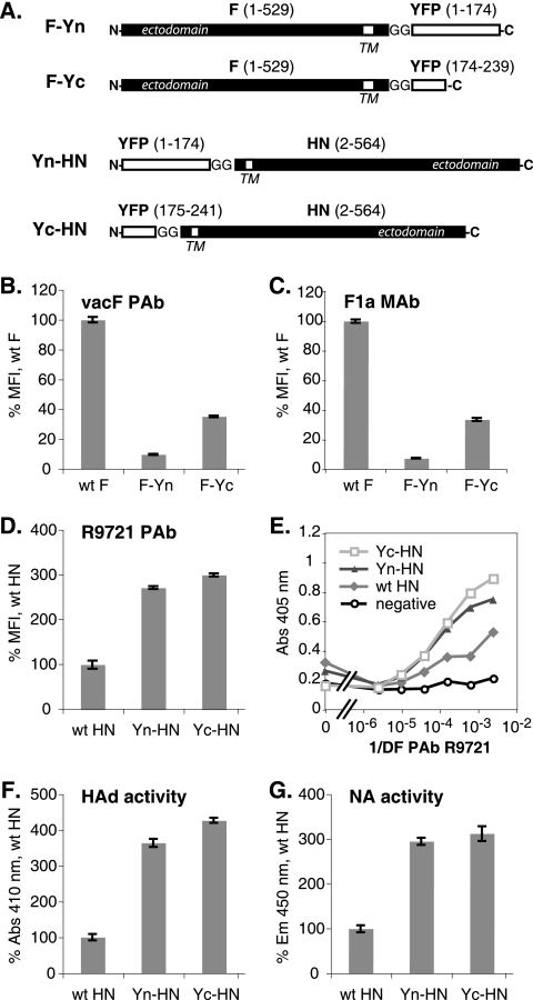
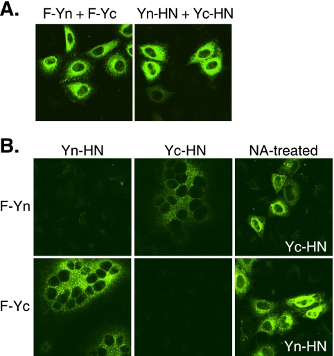
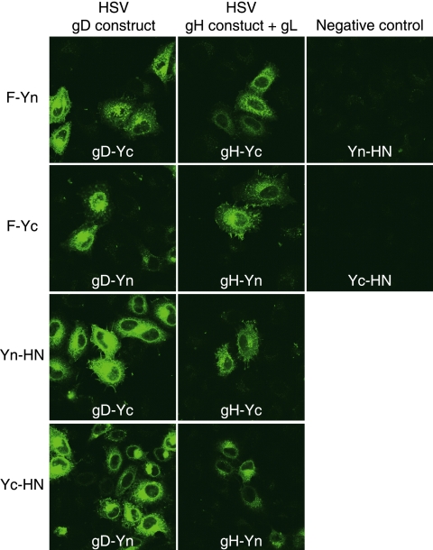
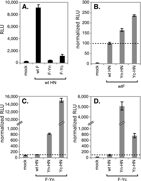
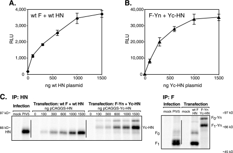
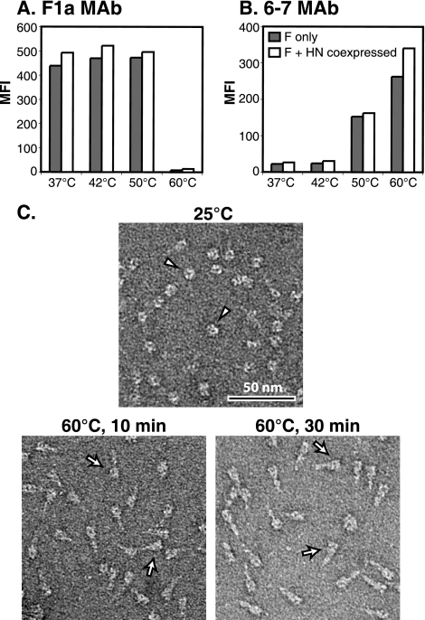
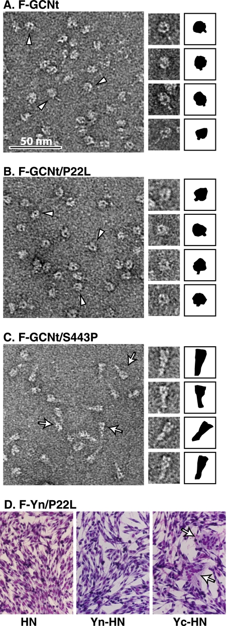
For paramyxoviruses, entry requires a receptor-binding protein (hemagglutinin-neuraminidase [HN], H, or G) and a fusion protein (F). Like other class I viral fusion proteins, F is expressed as a prefusion metastable protein that undergoes a refolding event to induce fusion. HN binding to its receptor triggers F refolding by an unknown mechanism. HN may serve as a clamp that stabilizes F in its prefusion state until HN binds the target cell (the "clamp model"). Alternatively, HN itself may undergo a conformational change after receptor binding that destabilizes F and causes F to trigger (the "provocateur model"). To examine F-HN interactions by bimolecular fluorescence complementation (BiFC), the cytoplasmic tails of parainfluenza virus 5 (PIV5) F and HN were fused to complementary fragments of yellow fluorescent protein (YFP). Coexpression of the BiFC constructs resulted in fluorescence; however, coexpression with unrelated BiFC constructs also produced fluorescence. The affinity of the two halves of YFP presumably superseded the F-HN interaction. Unexpectedly, coexpression of the BiFC F and HN constructs greatly enhanced fusion in multiple cell types. We hypothesize that the increase in fusion occurs because the BiFC tags bring F and HN together more frequently than occurs in a wild-type (wt) scenario. This implies that normally much of wt F is not associated with wt HN, in conflict with the clamp model for activation. Correspondingly, we show that wt PIV5 fusion occurs in an HN concentration-dependent manner. Also inconsistent with the clamp model are the findings that BiFC F does not adopt a postfusion conformation when expressed in the absence of HN and that HN coexpression does not provide resistance to the heat-induced triggering of F. In support of a provocateur model of F activation, we demonstrate by analysis of the morphology of soluble F trimers that the hyperfusogenic mutation S443P has a destabilizing effect on F.


Figures

FIG. 1.
FIG. 1.
Expression of BiFC constructs. (A) Schematic diagram of F and HN BiFC constructs. The N- and C-terminal halves of YFP were added to the cytoplasmic tails of W3A F and HN via a diglycine linker. TM, transmembrane domain. (B to D) Levels of surface expression of the F and HN constructs on transfected HeLa-CD4-LTR-β-gal cells were determined by flow cytometry using the anti-F PAb vacF, the anti-F MAb F1a, or the anti-HN PAb R9721. The mean fluorescence intensity (MFI) is shown as a percentage of wt protein levels. (E) Levels of surface expression of the HN constructs on transfected Vero cells were determined by cell-based ELISA using the PAb R9721. Abs, absorbance; 1/DF, reciprocal of dilution factor. (F) The HAd activities of the HN constructs were determined by measuring RBC binding to transfected HeLa-CD4-LTR-β-gal cells by spectroscopy. (G) The NA activities of the HN constructs on transfected HeLa-CD4-LTR-β-gal cells were determined using a fluorimetric assay. Em, emission.
FIG. 2.
FIG. 2.
BiFC analyses. Transfected HeLa-CD4-LTR-β-gal cells expressing the F and HN constructs were fixed at 20 h posttransfection and examined for YFP fluorescence. (A) BiFC analyses of F or HN oligomers. (B) BiFC analyses of F and HN complexes. NA-treated samples were incubated with NA starting at 4 h posttransfection.
FIG. 3.
FIG. 3.
BiFC with heterologous glycoproteins. Transfected HeLa-CD4-LTR-β-gal cells expressing combinations of PIV5 or HSV glycoprotein constructs were fixed at 20 h posttransfection and examined for YFP fluorescence. For the negative controls, cells were transfected with pairs of noncomplementary YFP halves.
FIG. 4.
FIG. 4.
BiFC constructs mediate enhanced syncytium formation. BHK or Vero cells were transfected with plasmids encoding the F and HN constructs, and the cells were imaged 24 h posttransfection. Syncytium formation is graded as follows from least to most: −, +, ++, and +++. Arrows indicate syncytia.
FIG. 5.
FIG. 5.
BiFC constructs mediate enhanced fusion. Vero cells were transfected with plasmids encoding F, HN, and luciferase under the control of the T7 polymerase promoter. Cells were overlaid with BSR-T7 cells at 20 h posttransfection. After 7 h, the cells were lysed and the luciferase activity (expressed in RLU) was determined. (A) Comparison of the degrees of fusion mediated by wt F, F-Yn, and F-Yc when the proteins are coexpressed with wt HN. (B to D) Degrees of fusion mediated by wt F (B), F-Yn (C), and F-Yc (D) when the proteins are coexpressed with wt HN, Yn-HN, or Yc-HN. For each F construct, the data were normalized by setting the RLU obtained after coexpression with wt HN at 100%. All samples were analyzed in parallel, and the standard deviations of results for triplicate samples are shown.
FIG. 6.
FIG. 6.
F-mediated fusion is dependent on the HN concentration. (A and B) Vero cells were transfected with plasmids encoding luciferase (400 ng/well) and wt F or F-Yn (400 ng/well) and increasing amounts of plasmid encoding HN or Yc-HN. Cells were overlaid with BSR-T7 cells at 20 h posttransfection. After 7 h, the cells were lysed and the luciferase activity (expressed in RLU) was determined. The standard deviations of results for triplicate samples are shown. (C) Vero cells were transfected as described in the legend to panels A and B or infected with PIV5 at 3 PFU/cell for comparison. Cells were radiolabeled, surface proteins were biotinylated, and surface-expressed F or HN was immunoprecipitated (IP) and visualized by SDS-PAGE. For the anti-F IP, the amount of HN or Yc-HN DNA used in cotransfection was 1,500 ng/well.
FIG. 7.
FIG. 7.
HN coexpression does not alter the heat-induced triggering of F. (A and B) Transfected HeLa-CD4-LTR-β-gal cells expressing F only (gray bars) or both F and HN (white bars) were heated to the indicated temperatures for 10 min at 24 h posttransfection. Cell surface reactivity with MAb 6-7 or MAb F1a was analyzed by flow cytometry. MFI, mean fluorescence intensity. (C) A soluble form of PIV5 F (F-GCNt) was expressed in insect cells by a recombinant baculovirus, and secreted F protein was purified. Trimers were visualized by EM before or after being heated to 60°C for 10 or 30 min.
FIG. 8.
FIG. 8.
BiFC constructs trigger postfusion-specific MAb 6-7 reactivity after fusion. HeLa-CD4-LTR-β-gal cells were transfected with F-Yn and Yc-HN constructs and incubated overnight in the presence or absence of NA to block syncytium formation. Cells were stained with MAb 6-7 or F1a and imaged by confocal microscopy.
FIG. 9.
FIG. 9.
P22L is a stabilizing mutation, and S443P is a destabilizing mutation. (A to C) Wild-type (A), P22L mutant (B), or S443P mutant (C) soluble forms of PIV5 F (F-GCNt) were expressed and purified using baculovirus. Trimers were visualized by EM, and those with prefusion (arrowheads) or postfusion (arrows) morphology are noted. The shapes of individual trimers are depicted in black to the right of the images. (D) BHK cells were transfected with plasmids encoding the F-Yn P22L mutant and wt HN, Yn-HN, or Yc-HN. Cells were imaged 24 h posttransfection.

References

    1. Aguilar, H. C., Z. A. Ataman, V. Aspericueta, A. Q. Fang, M. Stroud, O. A. Negrete, R. A. Kammerer, and B. Lee. 2009. A novel receptor-induced activation site in the Nipah virus attachment glycoprotein (G) involved in triggering the fusion glycoprotein (F). J. Biol. Chem. 284:1628-1635. - PMC - PubMed
    1. Aguilar, H. C., K. A. Matreyek, C. M. Filone, S. T. Hashimi, E. L. Levroney, O. A. Negrete, A. Betrolotti-Ciarlet, D. Y. Choi, I. McHardy, J. A. Fulcher, S. V. Su, M. C. Wolf, L. Kohatsu, L. G. Baum, and B. Lee. 2006. N-glycans on Nipah virus fusion protein protect against neutralization but reduce membrane fusion and viral entry. J. Virol. 80:4878-4889. - PMC - PubMed
    1. Atanasiu, D., J. C. Whitbeck, T. M. Cairns, B. Reilly, G. H. Cohen, and R. J. Eisenberg. 2007. Bimolecular complementation reveals that glycoproteins gB and gH/gL of herpes simplex virus interact with each other during cell fusion. Proc. Natl. Acad. Sci. USA 104:18718-18723. - PMC - PubMed
    1. Bishop, K. A., A. C. Hickey, D. Khetawat, J. R. Patch, K. N. Bossart, Z. Zhu, L. F. Wang, D. S. Dimitrov, and C. C. Broder. 2008. Residues in the stalk domain of the Hendra virus G glycoprotein modulate conformational changes associated with receptor binding. J. Virol. 82:11398-11409. - PMC - PubMed
    1. Bishop, K. A., T. S. Stantchev, A. C. Hickey, D. Khetawat, K. N. Bossart, V. Krasnoperov, P. Gill, Y. R. Feng, L. Wang, B. T. Eaton, L. F. Wang, and C. C. Broder. 2007. Identification of Hendra virus G glycoprotein residues that are critical for receptor binding. J. Virol. 81:5893-5901. - PMC - PubMed

Publication types