. 2009 Oct;136(19):3247-56.
doi: 10.1242/dev.039859. Epub 2009 Aug 26.

Fine-tuning of secondary arbor development: the effects of the ecdysone receptor on the adult neuronal lineages of the Drosophila thoracic CNS

Affiliations

Fine-tuning of secondary arbor development: the effects of the ecdysone receptor on the adult neuronal lineages of the Drosophila thoracic CNS

Heather L D Brown et al. Development. 2009 Oct.

Abstract

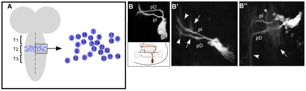
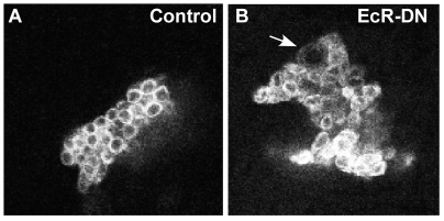
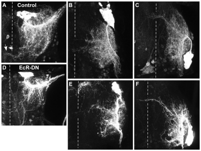
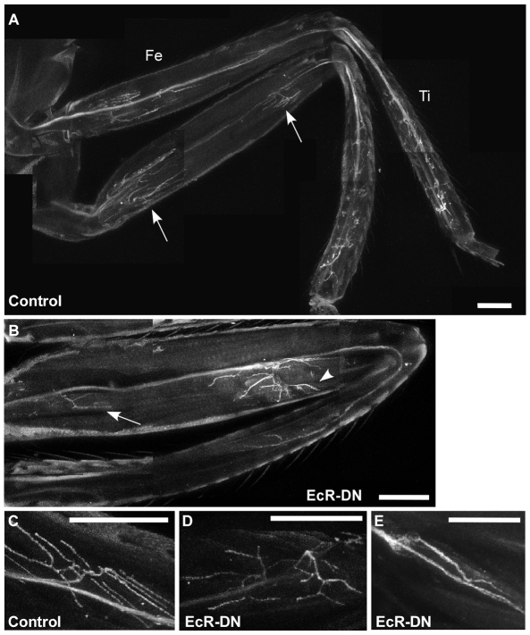
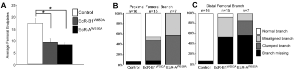
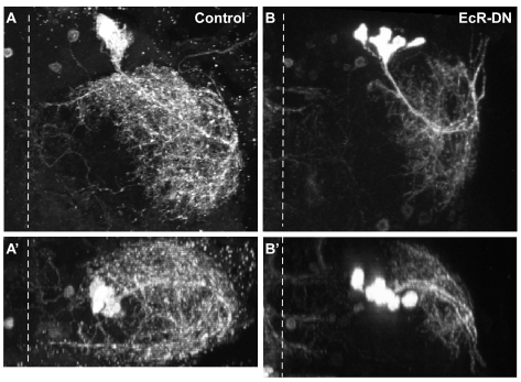
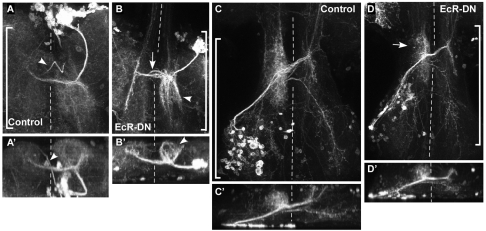
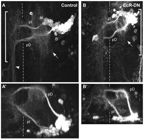
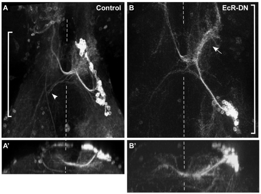
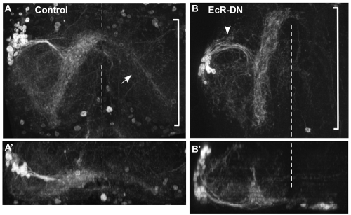
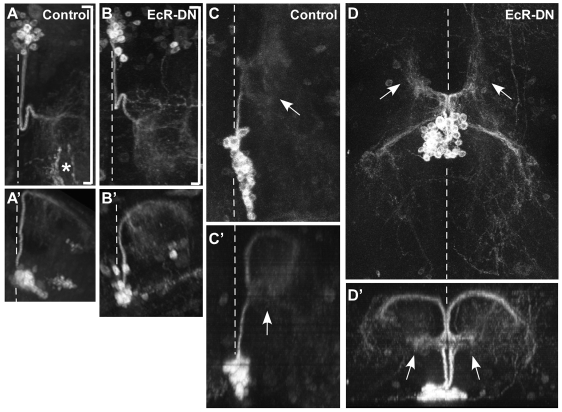
The adult central nervous system (CNS) of Drosophila is largely composed of relatively homogenous neuronal classes born during larval life. These adult-specific neuron lineages send out initial projections and then arrest development until metamorphosis, when intense sprouting occurs to establish the massive synaptic connections necessary for the behavior and function of the adult fly. In this study, we identified and characterized specific lineages in the adult CNS and described their secondary branch patterns. Because prior studies show that the outgrowth of incumbent remodeling neurons in the CNS is highly dependent on the ecdysone pathway, we investigated the role of ecdysone in the development of the adult-specific neuronal lineages using a dominant-negative construct of the ecdysone receptor (EcR-DN). When EcR-DN was expressed in clones of the adult-specific lineages, neuroblasts persisted longer, but we saw no alteration in the initial projections of the lineages. Defects were observed in secondary arbors of adult neurons, including clumping and cohesion of fine branches, misrouting, smaller arbors and some defasciculation. The defects varied across the multiple neuron lineages in both appearance and severity. These results indicate that the ecdysone receptor complex influences the fine-tuning of connectivity between neuronal circuits, in conjunction with other factors driving outgrowth and synaptic partnering.


Figures

Fig. 1.
Fig. 1.
Postembryonic neuron lineage development in the CNS. (A) Neuroblast (NB) map of the second thoracic (T2) hemineuromere. Thoracic neuromeres T1 and T3 lack one or two of the NBs present in the T2 set. (B-B″) Examples of confocal projections of MARCM clones showing the time course of development of neurons produced by NB 6. (B) A lineage 6 clone in T3 at pupariation, showing the two axon bundles emerging from the cell cluster; one projects across the posterior intermediate (pI) commissure and the other across the posterior dorsal (pD) commissure. Below is a cross-section schematic of the thoracic segmental neuropil showing the bundle trajectories relative to the major commissures (see Truman et al., 2004). (B′) A lineage 6 clone in T2 at 24 hours APF (raised at 29°C) showing sprouting of terminal (arrowheads) and interstitial (arrows) arbors from both bundles. (B″) Both hemilineages of lineage 6 have well-developed ipsilateral arbors (arrow) and contralateral projections in the adult. Contralateral axons that cross in the pD commissure have posterior projecting terminal arbors (arrowhead).
Fig. 2.
Fig. 2.
Expression levels of EcR-DN constructs in MARCM neuroblast clones from pupae at 24 hours APF (raised at 29°C). (A,A′) CD8::GFP expression (A) and EcR-B1 immunoreactivity (A′) in a MARCM clone expressing CD8::GFP and EcR-B1W650A. (B,B′) CD8::GFP expression (B) and EcR-A immunoreactivity (B′) in a MARCM neuroblast clone expressing CD8::GFP and EcR-AW650A. EcR-A immunoreactivity is seen in all adult-specific neurons, but it is enhanced in the neurons within the clone.
Fig. 3.
Fig. 3.
Neuroblast persistence in MARCM clones expressing EcR-DN. (A) A MARCM clone from a control pupa at 24 hours APF (raised at 29°C). No neuroblast is present. (B) A MARCM clone expressing EcR-DN at 24 hours APF (raised at 29°C), showing a persistent neuroblast (arrow), identified by its large size and position at the apex of the cell cluster.
Fig. 4.
Fig. 4.
Expression of EcR-DN severely affects the dendrites of lineage 15 in the adult CNS. (A-C) Dorsal projections of confocal stacks showing the adult morphology of lineage 15 motoneurons in T1 (A), T2 (B) and T3 (C) of control animals. Arrow indicates small contralateral branch. (D-F) Projections of lineage 15 clones expressing EcR-DN, in T1 (D), T2 (E) and T3 (F). Clones of lineage 15 expressing EcR-DN have reduced and clumped dendritic arbors. The dashed line indicates the midline in this and subsequent figures.
Fig. 5.
Fig. 5.
Expression of EcR-DN results in disrupted peripheral projections of lineage 15 in the leg. (A) Legs from control adults showing branching of GFP-labeled axons from lineage 15 MARCM clones in T2 and T3. Arrows point to the two major branch elaboration sites, proximally and distally within the femur (Fe). Axon branches in the tibia (Ti) are less stereotyped than those in the femur. (B) A lineage 15 clone expressing EcR-DN B1W650A has a greatly reduced proximal branch (arrow) and a misaligned distal branch in the femur (arrowhead). (C) Enlarged distal arbor of lineage 15 in the femur of a control animal, showing parallel endplates oriented along the muscle fibers. (D) The distal arbor in the femur of an animal expressing EcR-B1W650A in lineage 15 shows a star-shaped arbor that is not oriented properly along the muscle fibers. (E) The proximal arbor in a femur of an animal expressing EcR-B1W650A in lineage 15 shows reduced, clumped processes. Scale bars: 0.1 mm.
Fig. 6.
Fig. 6.
Quantification of lineage 15 secondary branch defects in the femur. (A) The average number of femoral endplates in controls (white), clones expressing B1W650A (gray) and clones expressing AW650A (black). (B,C) Percentage of femurs that are missing branches (black), have clumped branches (dark gray), have misaligned branches (light gray), or have normal branches (white) in lineage 15 controls, and in clones expressing B1W650A or AW650A. (B) Analysis of femoral proximal branches; (C) analysis of femoral distal branches.
Fig. 7.
Fig. 7.
Expression of EcR-DN severely affects the dendrites of lineage 24 in the adult CNS. Dorsal (top) and transverse (bottom) projections of lineage 24 MARCM clones. (A,A′) A control clone in T1 has a diffuse dendritic arbor innervating the leg neuropil. (B,B′) A lineage 24 clone expressing EcR-DN has stunted dendritic arbors.
Fig. 8.
Fig. 8.
Expression of EcR-DN causes severe effects on lineage 7 arbors in the adult CNS. Dorsal (top) and transverse (bottom) projections of lineage 7 MARCM clones. Brackets indicate the portion of the confocal stack used to create the transverse projection in this and subsequent figures. (A,A′) A control clone in T1 extends across the aI (anterior intermediate) commissure, forming dorsally projecting arbors on both sides of the midline. Arrowhead points to the axon bundle from an adjacent lineage 2 clone. (B,B′) A clone of lineage 7 expressing EcR-DN has a clustered ipsilateral arbor in dense strands (arrowhead), a contralateral arbor that is longitudinally compressed and projects anteriorly, and a defasciculated primary neurite bundle (arrow). (C,C′) A T3 lineage 7 clone in a control animal. (D,D′) A T3 lineage 7 clone expressing EcR-DN. The ipsilateral arbor is compressed (arrow). The posterior contralateral axon appears unaffected by EcR-DN.
Fig. 9.
Fig. 9.
EcR-DN reduces lineage 6 arbors in the adult CNS. Dorsal (top) and transverse (bottom) projections of lineage 6 MARCM clones. (A,A′) A T1 control clone showing the two hemilineage clusters. One sends a dorsal axon bundle with a large ipsilateral arbor (arrow) before crossing the pD commissure and extending a compact bundle into the T2 neuromere (arrowhead). The other cluster has a smaller ipsilateral arbor and a more diffuse contralateral projection that is primarily in T1. (B,B′) A T1 lineage 6 clone expressing EcR-DN. The ipsilateral arbors of both clusters are reduced and somewhat clumped (arrow), while the contralateral projection shows a high degree of disorganization of the T2 projection.
Fig. 10.
Fig. 10.
EcR-DN expression affects the dendritic arbor of lineage 18 in the adult CNS. Dorsal (top) and transverse (bottom) projections of lineage 18 MARCM clones. (A,A′) A ventral projection of a T3 lineage 18 clone in a control CNS that extends across the aI commissure, and then extends dorsally projecting arbors on either side of the midline and a smaller arbor into the contralateral leg neuropil (arrowhead). The cell bodies have been pushed apart during metamorphosis. (B,B′) A ventral projection of a T3 lineage 18 clone expressing EcR-DN. Arrow indicates the reduced and compacted ipsilateral arbor.
Fig. 11.
Fig. 11.
EcR-DN expression in lineage 9 results in minor defasciculation of the primary bundle. Dorsal (top) and transverse (bottom) projections of lineage 9 MARCM clones. (A,A′) A T3 clone of lineage 9 in a control CNS. Lineage 9 projects around the anterior edge of the leg neuropil to form an arbor along the ventral side. A small arbor extends contralaterally across the aV commissure (arrow). (B,B′) A T3 clone of lineage 9 expressing EcR-DN, in which the primary bundle is defasciculated (arrowhead).
Fig. 12.
Fig. 12.
EcR-DN expression does not affect lineage 2 arborizations in the adult CNS. Dorsal (top) and transverse (bottom) projections of lineage 2 MARCM clones. (A,A′) A T1 clone of lineage 2 in control animals. Lineage 2 has a characteristic `jog' as it extends dorsally through the neuropil before forming a diffuse ipsilateral arbor in the flight neuropil. Asterisk marks the projection of an afferent peripheral neuron clone not associated with lineage 2. (B,B′) A T1 clone of lineage 2 expressing EcR-DN appears to be unaffected by EcR-DN expression. (C,C′) A T2 clone of lineage 2 in a control animal. Unlike in T1, lineage 2 in T2 and T3 shows interstitial sprouting (arrow) in addition to its terminal arbor. (D,D′) T2 clones of lineage 2 expressing EcR-DN. Lineage 2 in both hemineuromeres of T2 is marked, and results in identical lineage 2 clones on either side of the midline. Arrows indicate the interstitial arbors of both clones.

Similar articles

Cited by

References

    1. Baek, M. and Mann, R. S. (2009). Lineage and birth date specify motor neuron targeting and dendritic architecture in adult Drosophila. J. Neurosci. 29, 6904-6916. - PMC - PubMed
    1. Bai, J., Uehara, Y. and Montell, D. J. (2000). Regulation of invasive cell behavior by taiman, a Drosophila protein related to AIB1, a steroid receptor coactivator amplified in breast cancer. Cell 103, 1047-1058. - PubMed
    1. Berreur, P., Porcheron, P., Moriniere, M., Berreur-Bonnenfant, J., Belinski-Deutsch, S., Busson, D. and Lamour-Audit, C. (1984). Ecdysteroids during the third larval instar in 1(3)ecd-1ts, a temperature-sensitive mutant of Drosophila melanogaster. Gen. Comp. Endocrinol. 54, 76-84. - PubMed
    1. Britton, J. S. and Edgar, B. A. (1998). Environmental control of the cell cycle in Drosophila: nutrition activates mitotic and endoreplicative cells by distinct mechanisms. Development 125, 2149-2158. - PubMed
    1. Brown, H. L., Cherbas, L., Cherbas, P. and Truman, J. W. (2006). Use of time-lapse imaging and dominant negative receptors to dissect the steroid receptor control of neuronal remodeling in Drosophila. Development 133, 275-285. - PubMed

Publication types

MeSH terms