. 2009 Oct;136(19):3215-22.
doi: 10.1242/dev.038893. Epub 2009 Aug 26.

Suppression of Erk signalling promotes ground state pluripotency in the mouse embryo

Affiliations

Suppression of Erk signalling promotes ground state pluripotency in the mouse embryo

Jennifer Nichols et al. Development. 2009 Oct.

Abstract

Embryonic stem (ES) cells can be derived and propagated from multiple strains of mouse and rat through application of small-molecule inhibitors of the fibroblast growth factor (FGF)/Erk pathway and of glycogen synthase kinase 3. These conditions shield pluripotent cells from differentiation-inducing stimuli. We investigate the effect of these inhibitors on the development of pluripotent epiblast in intact pre-implantation embryos. We find that blockade of Erk signalling from the 8-cell stage does not impede blastocyst formation but suppresses development of the hypoblast. The size of the inner cell mass (ICM) compartment is not reduced, however. Throughout the ICM, the epiblast-specific marker Nanog is expressed, and in XX embryos epigenetic silencing of the paternal X chromosome is erased. Epiblast identity and pluripotency were confirmed by contribution to chimaeras with germline transmission. These observations indicate that segregation of hypoblast from the bipotent ICM is dependent on FGF/Erk signalling and that in the absence of this signal, the entire ICM can acquire pluripotency. Furthermore, the epiblast does not require paracrine support from the hypoblast. Thus, naïve epiblast and ES cells are in a similar ground state, with an autonomous capacity for survival and replication, and high vulnerability to Erk signalling. We probed directly the relationship between naïve epiblast and ES cells. Dissociated ICM cells from freshly harvested late blastocysts gave rise to up to 12 ES cell clones per embryo when plated in the presence of inhibitors. We propose that ES cells are not a tissue culture creation, but are essentially identical to pre-implantation epiblast cells.


Figures
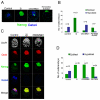
Fig. 1.
Fig. 1.
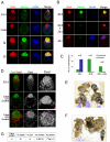
Effect of FGF/Erk and Gsk3 inhibition on inner cell mass development. (A) Confocal images of mouse embryos grown from the 8-cell stage (E2.5) for 3 days in control medium, in 3i (medium supplemented with Chir99021, PD184352 and SU5402), medium supplemented with Chir99021 alone, or medium supplemented with PD184352 and SU5402. Embryos were immunostained using antibodies raised against Nanog (green) and Gata4 (blue). (B) Bar chart showing cell numbers of epiblast (Nanog positive, green) and hypoblast (Gata4 positive, blue) of embryos cultured in the conditions shown in A. Bars indicate the mean ± s.d. (C) Confocal images of embryos grown from the 8-cell stage for 2 days in control medium, in 2i (medium supplemented with PD0325901 and Chir99021), or medium supplemented with PD0325901. Embryos were immunostained using antibodies raised against Oct4 (red), Nanog (green) and Gata4 (blue) and nuclei were counterstained with DAPI. (D) Bar chart showing cell numbers of epiblast (green) and hypoblast (blue) of embryos cultured in the conditions shown in C. Bars indicate the mean ± s.d.
Fig. 2.
Fig. 2.
Formation of functional epiblast in embryos cultured in ground state conditions. (A) Confocal images of mouse embryos freshly isolated at 4.5 dpc (E4.5), embryos cultured for 2 days from E3.75 in control medium, in 3i or in 2i. Embryos were immunostained using antibodies against Oct4 (red), Nanog (green) and Gata4 (blue). (B) Confocal images of embryos grown from the 8-cell stage (E2.5) for 4 days in 2i, for 2 days in 2i then a further 2 days in control medium, or in control medium for 4 days. Embryos were immunostained as in A. (C) Bar chart showing cell numbers of epiblast (Oct4 positive, Gata4 negative, green) and hypoblast (Gata4 positive, blue) of embryos cultured in the conditions shown in B. Bars indicate the mean ± s.d. (D) Confocal images of embryos freshly isolated at E4.5, embryos cultured from the 8-cell stage (E2.5) for 3 days in 2i, or from E3.5 for 2 days in 2i. Embryos were immunostained using antibodies against Eed (green) and Gata4 (red). Scale bars: 20 μm. (E) Mice generated from injection of isolated epiblast cells from embryos grown for 3 days in 2i. Donor cells were from 129/Sv embryos (agouti coat colour); host blastocysts were of the C57BL/6/O1a strain (black coat colour). (F) One of the female chimaeras and her C57BL/6/O1a mate with their offspring comprising seven agouti pups, demonstrating transmission through the germline of the donor cell genotype, and one black offspring produced from the germ cells of the host embryo. (G) Summary of blastocyst injection experiments to test the developmental capacity of epiblast cells from embryos cultured in ground state culture conditions. Asterisk indicates that nine mice were tested for germline transmission.
Fig. 3.
Fig. 3.
Effect of 2i on trophectoderm development. (A) Confocal images of mouse embryos cultured from the zygote stage for 6 days in control medium or 2i. Embryos were stained for Oct4 (red), Nanog (green) and Gata4 (blue) and nuclei counterstained with DAPI. (B) Confocal images of embryos cultured from zygotes for 6 days in control medium, Chir99021, 2i or PD0325901. Embryos were immunostained using antibodies against Cdx2 (red), Nanog (green) and Gata4 (blue) and nuclei counterstained with DAPI. (C) Bar chart showing cell numbers of epiblast (Nanog positive, green), hypoblast (Gata4 positive, blue) and trophectoderm (Cdx2 positive, red) of embryos cultured in the conditions shown in B. Bars indicate the mean ± s.d.
Fig. 4.
Fig. 4.
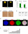
Effect of LIF on epiblast cells in embryos cultured in 2i and explanted. (A) Confocal images of mouse embryos cultured from the 8-cell stage for 2 days in 2i or 2i+LIF. Embryos were immunostained using antibodies against Oct4 (red), Nanog (green) and Gata4 (blue). (B) Bar chart showing cell numbers of epiblast (green) and hypoblast (blue) of embryos cultured in the conditions shown in A. Bars indicate the mean ± s.d. (C) (Left) Bright-field image of a single epiblast cell immediately after isolation from an embryo cultured for 3 days in 2i+LIF, plated into one well of a 96-well plate. (Right) Bright-field image of a colony produced from the same cell after growth in 2i+LIF for 8 days. Scale bars: 20 μm. (D) Summary of colonies produced from single epiblast cells plated into 2i or 2i+LIF. (E) Confocal images of a colony grown from a single epiblast cell plated into 2i+LIF for 8 days, as shown in C. Colonies were immunostained using antibodies against Oct4 (red) and Nanog (green) and nuclei were counterstained with DAPI. (F) Bar chart showing the number of ES cell clones produced per embryo from single ICM cells isolated from freshly flushed F1 embryos at E4.5.

Similar articles

Cited by

References

    1. Arman, E., Krausz-Haffner, R., Chen, Y., Heath, J. K. and Lonai, P. (1998). Targeted disruption of fibroblast growth factor (FGF) receptor 2 suggests a role for FGF signaling in pregastrulation mammalian development. Proc. Natl. Acad. Sci. USA 95, 5082-5087. - PMC - PubMed
    1. Bain, J., Plater, L., Elliott, M., Shpiro, N., Hastie, C. J., McLauchlan, H., Klevernic, I., Arthur, J. S., Alessi, D. R. and Cohen, P. (2007). The selectivity of protein kinase inhibitors: a further update. Biochem. J. 408, 297-315. - PMC - PubMed
    1. Batlle-Morera, L., Smith, A. and Nichols, J. (2008). Parameters influencing derivation of embryonic stem cells from murine embryos. Genesis 46, 758-767. - PubMed
    1. Bradley, A., Evans, M. J., Kaufman, M. H. and Robertson, E. (1984). Formation of germ-line chimaeras from embryo-derived teratocarcinoma cell lines. Nature 309, 255-256. - PubMed
    1. Brons, I. G., Smithers, L. E., Trotter, M. W., Rugg-Gunn, P., Sun, B., Chuva de Sousa Lopes, S. M., Howlett, S. K., Clarkson, A., Ahrlund-Richter, L., Pedersen, R. A. et al. (2007). Derivation of pluripotent epiblast stem cells from mammalian embryos. Nature 448, 191-195. - PubMed

Publication types

MeSH terms

Substances