. 2009 Aug 26;29(34):10582-7.
doi: 10.1523/JNEUROSCI.1736-09.2009.

Beta-amyloid monomers are neuroprotective

Affiliations

Beta-amyloid monomers are neuroprotective

Maria Laura Giuffrida et al. J Neurosci. .

Abstract

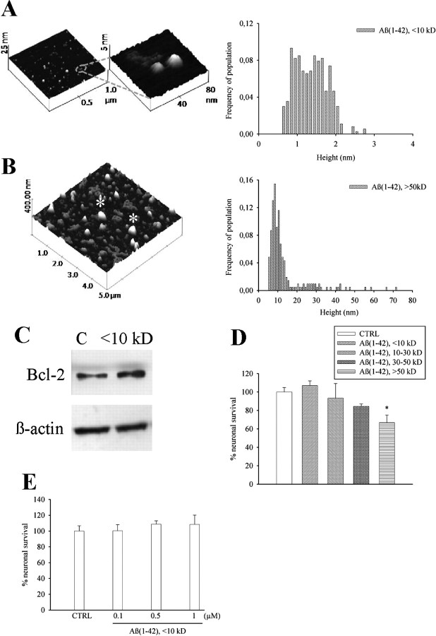
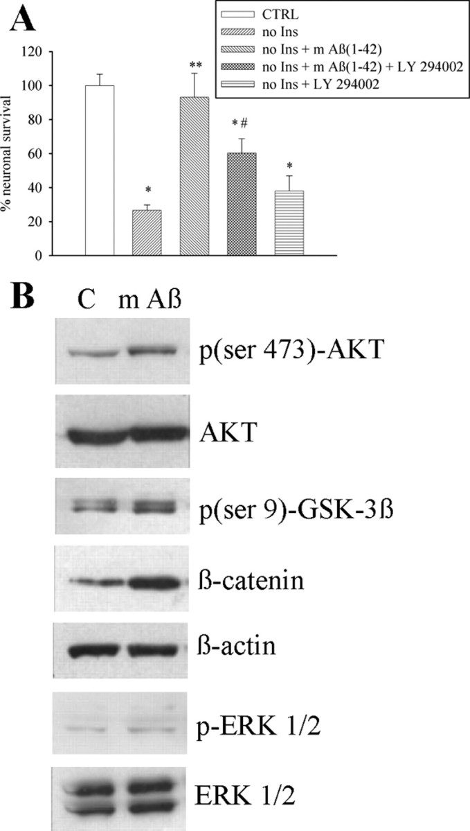
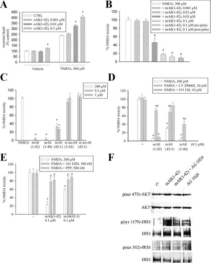
The 42-aa-long beta-amyloid protein--Abeta(1-42)--is thought to play a central role in the pathogenesis of Alzheimer's disease (AD) (Walsh and Selkoe, 2007). Data from AD brain (Shankar et al., 2008), transgenic APP (amyloid precursor protein)-overexpressing mice (Lesné et al., 2006), and neuronal cultures treated with synthetic Abeta peptides (Lambert et al., 1998) indicate that self-association of Abeta(1-42) monomers into soluble oligomers is required for neurotoxicity. The function of monomeric Abeta(1-42) is unknown. The evidence that Abeta(1-42) is present in the brain and CSF of normal individuals suggests that the peptide is physiologically active (Shoji, 2002). Here we show that synthetic Abeta(1-42) monomers support the survival of developing neurons under conditions of trophic deprivation and protect mature neurons against excitotoxic death, a process that contributes to the overall neurodegeneration associated with AD. The neuroprotective action of Abeta(1-42) monomers was mediated by the activation of the PI-3-K (phosphatidylinositol-3-kinase) pathway, and involved the stimulation of IGF-1 (insulin-like growth factor-1) receptors and/or other receptors of the insulin superfamily. Interestingly, monomers of Abeta(1-42) carrying the Arctic mutation (E22G) associated with familiar AD (Nilsberth et al., 2001) were not neuroprotective. We suggest that pathological aggregation of Abeta(1-42) may also cause neurodegeneration by depriving neurons of the protective activity of Abeta(1-42) monomers. This "loss-of-function" hypothesis of neuronal death should be taken into consideration when designing therapies aimed at reducing Abeta burden.


Figures

Figure 1.
Figure 1.
1-42 monomers: separation, characterization, and lack of neurotoxicity in culture. A, B, AFM images of low-mass (<10 kDa, A) and high-mass (>50 kDa, B) Aβ1-42 species isolated from a single suspension by cutoff filters. The respective frequencies of species in the two samples are shown on the right side. The monomer fraction consisted primarily of small globules 1.3 nm in height (mean ± SD: 1.36 ± 0.42, n = 365). In contrast, the oligomer fraction consisted of larger globules 13 nm in height (mean ± SD: 13.5 ± 10.8, n = 207). In B, the asterisk indicates structures derived from the aggregation of several oligomer species which were excluded from the statistics. C, Representative Western blot image of Bcl-2 bands in control cultures (C) and cultures treated with <10 kDa Aβ1-42 for 6 h. β-Actin bands are shown for control of loading. Quantitation of Bcl-2/β-actin ratios was as follows: control (C) = 0.7 ± 0.1; <10 kDa Aβ1-42 = 1.2 ± 0.03* (means ± SEM of three independent experiments; *significantly different from control at p < 0.05 by Student's t test). Viability of pure cortical neurons, as measured by MTT assay, following 48 h treatment with different Aβ1-42 fractions (all at 0.1 μm) or different concentrations of <10 kDa Aβ1-42, is shown in D and E, respectively. Values are means ± SEM of eight determinations from two independent experiments. *p < 0.05 (one-way ANOVA + Fisher's LSD) compared with control (CTRL).
Figure 2.
Figure 2.
1-42 monomers support neuronal survival via the activation of the PI-3-K pathway. A, Viability of pure cortical neurons, as measured by MTT assay, insulin-deprived since plating for 1 week (no Ins), in the absence or presence of 0.1 μm1-42 monomers [m Aβ(1-42)]. Where required, the PI-3-K inhibitor, LY294002 (10 μm), was added two times (at plating and after 48 h). Values are means ± SEM of six determinations from two independent experiments. *,**,#Different from control (CTRL) (*), no Ins (**), or no Ins + m Aβ1-42 (#) at p < 0.05 by one-way ANOVA + Fisher's LSD test. B, Representative Western blot images of the PI-3-K-activated form of the serine-threonine kinase AKT [p(ser 473)-AKT], of the corresponding inactivated GSK-3β [p(ser 9)-GSK-3β], and of β-catenin levels in control cultures (C) and cultures treated with 0.1 μm monomeric Aβ1-42 (m Aβ) for 10 min. Levels of phosphorylated ERK1/2 (pERK1/2) in the same cultures are also shown. Total AKT levels, β-actin bands, or total ERK levels are shown for control of loading. Quantitation of p(ser 473)-AKT/AKT ratios was as follows: control (C) = 0.26 ± 0.04; mAβ1-42 = 0.65 ± 0.1* (means ± SEM of three independent experiments; *significantly different from control at p < 0.05 by Student's t test). Quantitation of p(ser 9)-GSK-3β/GSK-3β ratios was as follows: control (C) = 0.88 ± 0.15; mAβ1-42 = 1.44 ± 0.08* (means ± SEM of three independent experiments; *significantly different from control at p < 0.05 by Student's t test). Quantitation of β-catenin/β-actin ratios was as follows: control (C) = 0.47 ± 0.05; mAβ1-42 = 1.3 ± 0.09* (means ± SEM of three independent experiments; *significantly different from control at p < 0.05 by Student's t test). Quantitation of pERK 1/2/ERK 1/2 ratios was as follows: control (C) = 0.6 ± 0.08; mAβ1-42 = 0.55 ± 0.12. (means ± SEM of three independent experiments).
Figure 3.
Figure 3.
1-42 monomers protect neurons against NMDA toxicity. A, NMDA-induced toxicity in mixed cortical cultures is potentiated by oligomeric Aβ1-42 [oAβ(1-42)] and (B) prevented by monomeric Aβ1-42 [mAβ(1-42)]. Toxicity was induced by a 10 min pulse with 300 μm NMDA and assessed by trypan blue staining (set to 100%) 24 h later. In the prepulse condition, Aβ1-42 monomers were applied 24 h before the NMDA pulse and cultures were extensively washed before the experiment. In the postpulse condition, Aβ1-42 monomers were applied soon after the excitotoxic pulse and kept for 24 h into the medium. Both in A and in B, values are means ± SEM of 9–18 determinations from three-six independent experiments. A, *Significantly different from the respective control (CTRL) at p < 0.05 (one-way ANOVA + Fisher's LSD test). B, *Significantly different from NMDA at p < 0.001 (one-way ANOVA + Fisher's LSD test). C, NMDA-induced toxicity in mixed cortical cultures is attenuated by the monomeric forms of Aβ1-42, Aβ1-40, and Aβ42-1, but not by Aβ1-42 or Aβ42-1 containing the Arctic mutation (m arcAβ). Values are means ± SEM of 12–18 determinations from three-six independent experiments. *Significantly different from NMDA at p < 0.001 (one-way ANOVA + Fisher's LSD test). D, The PI-3-K inhibitor LY 294002 (10 μm) prevents the neuroprotective activity of monomeric forms of Aβ1-42, Aβ1-40, and Aβ42-1. Values are means ± SEM of 8 determinations from two independent experiments. *,#Significantly different from NMDA (*) or from the respective Aβ conditions (#) at p < 0.05 (one-way ANOVA + Fisher's LSD test). [UO126] = 10 μm. E, The selective inhibitor of the insulin receptor superfamily, AG1024, and the preferential IGF-1 receptor inhibitor, picropodophyllin (PPP), prevent the neuroprotective activity of monomeric forms of Aβ1-42 and Aβ42-1. Values are means ± SEM of eight determinations from two independent experiments. *,#Significantly different from NMDA (*) or from the respective Aβ conditions (#) at p < 0.05 (one-way ANOVA + Fisher's LSD test). F, Representative immunoblots of p(ser 473)-AKT, p(tyr 1179)-IRS1, and p(ser 302)-IRS1 in pure neuronal cultures treated with 0.1 μm monomeric Aβ1-42 [mAβ(1-42)] for 5 min both in the absence and in the presence of 100 nm AG1024. Quantitation of p(ser 473)AKT/AKT ratios was as follows: control (C) = 0.5 ± 0.04; mAβ1-42 = 1.37 ± 0.1*; mAβ1-42 + AG1024 = 0.7 ± 0.08**; AG1024 = 0.55 ± 0.06 [means ± SEM of three independent experiments; *,**significantly different from control (*) or mAβ1-42 alone (**) at p < 0.05 by one-way ANOVA + Fisher's LSD test]. Quantitation of p(tyr 1179)-IRS1/IRS1 ratios was as follows: control (C) = 0.42 ± 0.12; mAβ1-42 = 1.31 ± 0.03*; mAβ1-42 + AG1024 = 0.63 ± 0.02**; AG1024 = 0.65 ± 0.08 [means ± SEM of three independent experiments; *,**significantly different from control (*) or mAβ1-42 alone (**) at p < 0.05 by one-way ANOVA + Fisher's LSD test]. Quantitation of p(ser 302)-IRS1/IRS1 ratios was as follows: control (C) = 0.39 ± 0.09; mAβ1-42 = 0.85 ± 0.06*; mAβ1-42 + AG1024 = 0.28 ± 0.11**; AG1024 = 0.46 ± 0.08 [means ± SEM of three independent experiments; *,**significantly different from control (*) or mAβ1-42 (**) alone at p < 0.05 by one-way ANOVA + Fisher's LSD test].

Similar articles

Cited by

References

    1. Adams JM, Cory S. The Bcl-2 protein family: arbiters of cell survival. Science. 1998;281:1322–1326. - PubMed
    1. Avruch J. Insulin signal transduction through protein kinase cascades. Mol Cell Biochem. 1998;182:31–48. - PubMed
    1. Brody DL, Magnoni S, Schwetye KE, Spinner ML, Esparza TJ, Stocchetti N, Zipfel GJ, Holtzman DM. Amyloid-beta dynamics correlate with neurological status in the injured human brain. Science. 2008;321:1221–1224. - PMC - PubMed
    1. Copani A, Koh JY, Cotman CW. Beta-amyloid increases neuronal susceptibility to injury by glucose deprivation. Neuroreport. 1991;2:763–765. - PubMed
    1. Copani A, Condorelli F, Caruso A, Vancheri C, Sala A, Giuffrida Stella AM, Canonico PL, Nicoletti F, Sortino MA. Mitotic signaling by beta-amyloid causes neuronal death. FASEB J. 1999;13:2225–2234. - PubMed

Publication types

MeSH terms