. 2009 Aug 26;29(34):10627-37.
doi: 10.1523/JNEUROSCI.0620-09.2009.

The p75 neurotrophin receptor promotes amyloid-beta(1-42)-induced neuritic dystrophy in vitro and in vivo

Affiliations

The p75 neurotrophin receptor promotes amyloid-beta(1-42)-induced neuritic dystrophy in vitro and in vivo

Juliet K Knowles et al. J Neurosci. .

Abstract

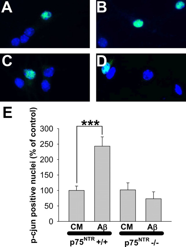
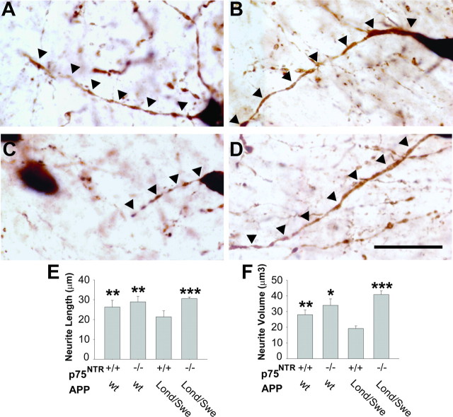
Oligomeric forms of amyloid-beta (Abeta) are thought to play a causal role in Alzheimer's disease (AD), and the p75 neurotrophin receptor (p75(NTR)) has been implicated in Abeta-induced neurodegeneration. To further define the functions of p75(NTR) in AD, we examined the interaction of oligomeric Abeta(1-42) with p75(NTR), and the effects of that interaction on neurite integrity in neuron cultures and in a chronic AD mouse model. Atomic force microscopy was used to ascertain the aggregated state of Abeta, and fluorescence resonance energy transfer analysis revealed that Abeta oligomers interact with the extracellular domain of p75(NTR). In vitro studies of Abeta-induced death in neuron cultures isolated from wild-type and p75(NTR-/-) mice, in which the p75(NTR) extracellular domain is deleted, showed reduced sensitivity of mutant cells to Abeta-induced cell death. Interestingly, Abeta-induced neuritic dystrophy and activation of c-Jun, a known mediator of Abeta-induced deleterious signaling, were completely prevented in p75(NTR-/-) neuron cultures. Thy1-hAPP(Lond/Swe) x p75(NTR-/-) mice exhibited significantly diminished hippocampal neuritic dystrophy and complete reversal of basal forebrain cholinergic neurite degeneration relative to those expressing wild-type p75(NTR). Abeta levels were not affected, suggesting that removal of p75(NTR) extracellular domain reduced the ability of excess Abeta to promote neuritic degeneration. These findings indicate that although p75(NTR) likely does not mediate all Abeta effects, it does play a significant role in enabling Abeta-induced neurodegeneration in vitro and in vivo, establishing p75(NTR) as an important therapeutic target for AD.


Figures

Figure 1.
Figure 1.
Aβ oligomers interact with the extracellular domain of p75NTR. For FRET analysis, the indicated proteins linked to either fluorescein or Cy3B were incubated at 500 nm in PBS solution containing 0.7% BSA. The intensity of fluorescent emissions from 500 to 750 nm was measured after excitation at λ = 490 nm (y-axis = fluorescent activity in arbitrary units). Conditions in which FRET occurred are indicated by bold lines. Incubation of fluorescein-labeled Streptavidin, NGF, and Aβ alone demonstrated no FRET. Incubation of fluorescein-labeled Aβ with denatured p75NTR-Cy3B or BSA-Cy3B also demonstrated no FRET. As a positive control, fluorescein-labeled Streptavidin (SA-FITC) was reacted with Cy3B-labeled biotinylated IgG (IgG-Bt Cy3B), resulting in a 25% energy transfer as indicated by decreased fluorescein fluorescence at 520 nm, accompanied by increased Cy3B fluorescence at 570 nm (bold line). Incubation of fluorescein-NGF or fluorescein-Aβ with Cy3B-p75NTR exhibited FRET (24% energy transfer for NGF-p75 and 50% for Aβ-p75NTR, both lines shown in bold) and, thereby, NGF and Aβ interaction with p75NTR. The relatively high transfer energy (50%) in the Aβ-p75NTR condition raised the possibility of Aβ-FITC self-quenching due to potential Aβ self-aggregation. To further assess Aβ-p75NTR interaction, a solution containing Aβ-Cy3B and p75NTR-FITC (fluorophore reversal) was submitted to FRET analysis and demonstrated 20% energy transfer (data not shown), confirming a molecular interaction between p75NTR and Aβ. Data are representative of three separate experiments.
Figure 2.
Figure 2.
p75NTR modulates Aβ-induced neuron death. p75NTR+/+ or p75NTR−/− neurons, 6–7 DIV, were treated with increasing doses of Aβ for 72 h then fixed in fresh paraformaldehyde. A, B, Hippocampal (A) or basal forebrain (B) neurons were photographed under phase contrast microscopy and survival was quantified based on morphological criteria. p75NTR+/+ and p75NTR−/− neurons each exhibited dose-dependent decreases in survival. In both hippocampal and basal forebrain cultures, p75NTR−/− neurons showed significantly less Aβ-induced death than p75NTR+/+ neurons at multiple Aβ doses, with 25–40% more neurons surviving (indicated by asterisks; see Materials and Methods for explanation). Hippocampal neuron data represent n = 46–56 fields from six separate cultures (p75NTR+/+) or 30–40 fields from four separate cultures (p75NTR−/−). Basal forebrain neuron data represent n = 28–38 fields from four separate p75NTR+/+ and p75NTR−/− cultures. C, To confirm the morphologic findings described in A, B, hippocampal neuron death was quantified by TUNEL analysis. p75NTR+/+ and p75NTR−/− hippocampal neurons each exhibited increases in death proportional to the dose of Aβ; however, at multiple doses of Aβ, p75NTR−/− neurons exhibited only 60–80% of death exhibited by p75NTR+/+. Data represent n = 80–160 fields from seven separate cultures (p75NTR+/+) or 70–89 fields from six separate cultures (p75NTR−/−). D, To confirm the modulatory effect of p75NTR on Aβ-induced neuron death using an independent method, 6–7 DIV p75NTR+/+ or p75NTR−/− hippocampal neurons were treated with fresh CM alone, or in the presence or absence of Aβ, p75NTR antibody, or control IgG. p75NTR antibody (but not IgG control) prevented entirely Aβ-induced neuron death when treated with 5 μm Aβ, whereas the protective effect was significantly diminished when neurons were treated with 10 μm Aβ. Data represent the means of n = 20–30 fields from six independent experiments.
Figure 3.
Figure 3.
p75NTR mediates Aβ-induced neuritic dystrophy. A–D, p75NTR+/+ or p75NTR−/− hippocampal neurons, 21 DIV, were treated with fresh CM alone or with CM containing 5 μm Aβ for 48 h, then fixed in fresh paraformaldehyde and stained for MAP-2. Neurons were photographed with fluorescence microscopy. In the presence of CM alone, p75NTR+/+ (A) and p75NTR−/− (C) neurites were indistinguishable and without excessive tortuosity. In the presence of Aβ, p75NTR+/+ neurons exhibited pronounced neuritic dystrophy and shrinkage (B), whereas the appearance of dystrophy in p75NTR −/− neurons was greatly diminished (D). E, Quantitation of neurite tortuosity revealed a significant ∼1.3-fold increase in mean differential curvature in p75NTR+/+ neurons treated with Aβ, whereas this effect was absent in p75NTR−/− neurons. Data represent the means from n = 25–31 fields from three separate p75NTR+/+ cultures and four p75NTR−/− cultures. F, Quantitation of neurite volume normalized to neurite length demonstrated that treatment of p75NTR+/+ neurons with Aβ resulted in a 42% decrease in neurite volume, whereas this effect was absent in p75NTR−/− cultures. Data represent the means from 12 to 14 fields from three separate cultures for each genotype.
Figure 4.
Figure 4.
p75NTR mediates Aβ-induced c-Jun activation. A–D, p75NTR+/+ or p75NTR−/− hippocampal neurons, 6–7 DIV, were treated with CM or CM containing 5 μm Aβ for 10–12 h, then fixed in fresh paraformaldehyde and immunostained for phospho-c-Jun and DAPI. Relative to CM alone (A), Aβ-treatment of p75NTR+/+ cells resulted in a higher frequency of p-c-Jun-positive nuclei (B). In contrast, cultures of p75NTR−/− neurons treated with CM (C) or Aβ (D) appeared similar. E, Quantitative analysis revealed that Aβ induced a significant ∼2.5-fold increase in p-c-Jun-positive nuclei of p75NTR+/+ neurons, whereas this effect was entirely absent in p75NTR−/− neurons. Data represent the mean of n = 40–50 fields per experimental condition from three independent experiments. A total of 300–600 neurons were analyzed per condition; in the presence of CM alone, 5–10% of both p75NTR+/+ and p75NTR−/− neurons exhibited p-c-jun-positive nuclei.
Figure 5.
Figure 5.
Neuritic dystrophy but not amyloid deposition in Thy1-hAPPLond/Swe mice is mediated by p75NTR. Dystrophic neurites were visualized with anti-APP 8E5 antibody staining in APPLond/Swe, p75NTR+/+ and p75NTR−/− mice. Upper left, APPLond/Swe, p75NTR+/+ mice exhibit prominent neuritic dystrophy. Upper right, Costaining of the identical area with thioflavin-S shows localization of dystrophic neurites with amyloid plaque. Lower left, Neuritic dystrophy appears diminished in p75NTR−/− mice. Lower right, Costaining with thioflavin-S demonstrates no gross change in plaque number or size in p75NTR−/− mice. Images were acquired at 63×. Scale bars, 25 μm. Quantitation of amyloid load by hippocampal and cortical plaque area measurement and ELISA quantitation of soluble Aβ(1-42) concentration in hippocampal and cortical extracts from APPLond/Swe, p75NTR+/+ and p75NTR−/− mice revealed no differences (see Results for ELISA data).
Figure 6.
Figure 6.
BFCN neuritic dystrophy in Thy1-hAPPLond/Swe mice is mediated by p75NTR. A–D, Representative 100× images of ChAT-stained neurites projecting from basal forebrain cholinergic neurons from APPwt, p75NTR+/+ (A), APPwt, p75NTR−/− (B), APPLond/Swe, p75NTR+/+ (C), and APPLond/Swe, p75NTR−/− (D) mice are shown. Scale bar, 30 μm. Many neurites from APPLond/Swe, p75NTR+/+ mice exhibited degenerative changes, including interrupted segments, decreased length, and decreased volume, whereas these effects were not apparent in APPLond/Swe, p75NTR−/− mice. E, F, Neurite trees projecting from BFCNs were manually traced using Neurolucida software. Relative to APPwt, p75NTR+/+ mice, BFCN neurites of APPLond/Swe mice expressing wild-type p75NTR exhibited decreased length (E) and volume (F). However, in APPLond/Swe mice expressing p75NTR−/−, these changes were entirely absent. Asterisks (see Materials and Methods for explanation) represent statistically significant comparisons relative to APPLond/Swe, p75NTR+/+ mice. Data represent the mean ± SEM from n = 6 APPwt, p75NTR+/+ mice, 4 APPwt, p75NTR−/− mice, 7 APPLond/Swe, p75NTR+/+ mice, and 5 APPLond/Swe, p75NTR−/− mice.
Figure 7.
Figure 7.
A–D, Loss of cortical cholinergic projection fibers in Thy1-hAPPLond/Swe mice is mediated by p75NTR. Relative to APPwt, p75NTR+/+ (A) and APPwt, p75NTR−/− mice (B), ChAT-stained fibers in the cingulate cortex of APPLond/Swe, p75NTR+/+ mice (C) appeared diminished, whereas this effect was not apparent in APPLond/Swe, p75NTR−/− mice (D). Images were acquired at 100×. Scale bar, 20 μm. E, Compared with APPwt mice, cortical cholinergic fiber density was reduced by ∼50% in APPLond/Swe, p75NTR+/+ mice but not in APPLond/Swe, p75NTR−/− mice. Data represent the mean ± SEM percent area relative to wild-type mice, n = 5 APPwt, p75NTR+/+ mice, 5 APPwt, p75NTR−/− mice, 8 APPLond/Swe, p75NTR+/+ mice, and 5 APPLond/Swe, p75NTR−/− mice. Asterisks represent statistically significant comparisons relative to APPLond/Swe, p75NTR+/+ mice (see Materials and Methods for explanation).

Similar articles

Cited by

References

    1. Amendola J, Durand J. Morphological differences between wild-type and transgenic superoxide dismutase 1 lumbar motoneurons in postnatal mice. J Comp Neurol. 2008;511:329–341. - PubMed
    1. Barker PA. p75NTR is positively promiscuous; novel partners and new insights. Neuron. 2004;42:529–533. - PubMed
    1. Bozyczko-Coyne D, O'Kane TM, Wu ZL, Dobrzanski P, Murthy S, Vaught JL, Scott RW. CEP-1347/KT-7515, an inhibitor of SAPK/JNK pathway activation, promotes survival and blocks multiple events associated with Abeta-induced cortical neuron apoptosis. J Neurochem. 2001;77:849–863. - PubMed
    1. Braak H, Alafuzoff I, Arzberger T, Kretzschmar H, Del Tredici K. Staging of Alzheimer disease-associated neurofibrillary pathology using paraffin sections and immunocytochemistry. Acta Neuropathol. 2006;112:389–404. - PMC - PubMed
    1. Brendza RP, Bacskai BJ, Cirrito JR, Simmons KA, Skoch JM, Klunk WE, Mathis CA, Bales KR, Paul SM, Hyman BT, Holtzman DM. Anti-Abeta antibody treatment promotes the rapid recovery of amyloid-associated neuritic dystrophy in PDAPP transgenic mice. J Clin Invest. 2005;115:428–433. - PMC - PubMed

Publication types

MeSH terms