

Combined application of BDNF to the eye and brain enhances ganglion cell survival and function in the cat after optic nerve injury
- PMID: 19710411
- PMCID: PMC2869067
- DOI: 10.1167/iovs.09-3740
Combined application of BDNF to the eye and brain enhances ganglion cell survival and function in the cat after optic nerve injury
Abstract
Purpose: To determine whether application of BDNF to the eye and brain provides a greater level of neuroprotection after optic nerve injury than treatment of the eye alone.
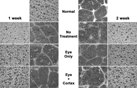
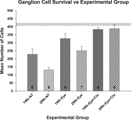
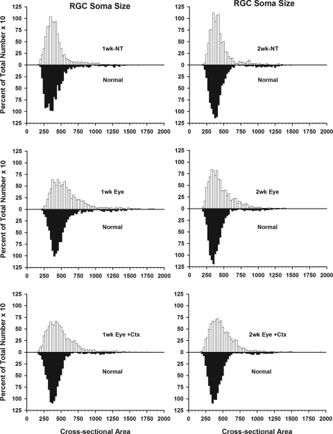
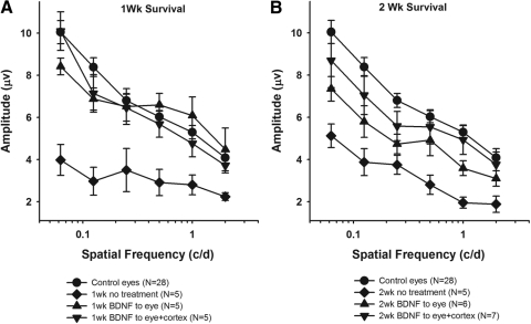
Methods: Retinal ganglion cell survival and pattern electroretinographic responses were compared in normal cat eyes and in eyes that received (1) a mild nerve crush and no treatment, (2) a single intravitreal injection of BDNF at the time of the nerve injury, or (3) intravitreal treatment combined with 1 to 2 weeks of continuous delivery of BDNF to the visual cortex, bilaterally.
Results: Relative to no treatment, administration of BDNF to the eye alone resulted in a significant increase in ganglion cell survival at both 1 and 2 weeks after nerve crush (1 week, 79% vs. 55%; 2 weeks, 60% vs. 31%). Combined treatment of the eye and visual cortex resulted in a modest additional increase (17%) in ganglion cell survival in the 1-week eyes, a further significant increase (55%) in the 2-week eyes, and ganglion cell survival levels for both that were comparable to normal (92%-93% survival). Pattern ERG responses for all the treated eyes were comparable to normal at 1 week after injury; however, at 2 weeks, only the responses of eyes receiving the combined BDNF treatment remained so.
Conclusions: Although treatment of the eye alone with BDNF has a significant impact on ganglion cell survival after optic nerve injury, combined treatment of the eye and brain may represent an even more effective approach and should be considered in the development of future optic neuropathy-related neuroprotection strategies.
Figures
Similar articles
-
BDNF enhances retinal ganglion cell survival in cats with optic nerve damage.Invest Ophthalmol Vis Sci. 2001 Apr;42(5):966-74. Invest Ophthalmol Vis Sci. 2001. PMID: 11274073
-
BDNF preserves the dendritic morphology of alpha and beta ganglion cells in the cat retina after optic nerve injury.Invest Ophthalmol Vis Sci. 2008 Jun;49(6):2456-63. doi: 10.1167/iovs.07-1325. Epub 2008 Feb 8. Invest Ophthalmol Vis Sci. 2008. PMID: 18263808
-
BDNF treatment and extended recovery from optic nerve trauma in the cat.Invest Ophthalmol Vis Sci. 2013 Oct 9;54(10):6594-604. doi: 10.1167/iovs.13-12683. Invest Ophthalmol Vis Sci. 2013. PMID: 23989190 Free PMC article.
-
Effects of optic nerve injury, glaucoma, and neuroprotection on the survival, structure, and function of ganglion cells in the mammalian retina.J Physiol. 2008 Sep 15;586(18):4393-400. doi: 10.1113/jphysiol.2008.156729. Epub 2008 Jun 19. J Physiol. 2008. PMID: 18565994 Free PMC article. Review.
-
Intravitreal injection worsens outcomes in a mouse model of indirect traumatic optic neuropathy from closed globe injury.Exp Eye Res. 2021 Jan;202:108369. doi: 10.1016/j.exer.2020.108369. Epub 2020 Nov 22. Exp Eye Res. 2021. PMID: 33238184 Free PMC article. Review.
Cited by
-
Advances in Neuroprotection in Glaucoma: Pharmacological Strategies and Emerging Technologies.Pharmaceuticals (Basel). 2024 Sep 25;17(10):1261. doi: 10.3390/ph17101261. Pharmaceuticals (Basel). 2024. PMID: 39458902 Free PMC article. Review.
-
Role of BDNF/TrkB pathway in the visual system: Therapeutic implications for glaucoma.Expert Rev Ophthalmol. 2017;12(1):69-81. doi: 10.1080/17469899.2017.1259566. Epub 2016 Nov 23. Expert Rev Ophthalmol. 2017. PMID: 28751923 Free PMC article.
-
Intrinsically photosensitive retinal ganglion cells in glaucoma.Front Cell Neurosci. 2022 Sep 23;16:992747. doi: 10.3389/fncel.2022.992747. eCollection 2022. Front Cell Neurosci. 2022. PMID: 36212698 Free PMC article. Review.
-
In Vivo Evaluation of White Matter Integrity and Anterograde Transport in Visual System After Excitotoxic Retinal Injury With Multimodal MRI and OCT.Invest Ophthalmol Vis Sci. 2015 Jun;56(6):3788-800. doi: 10.1167/iovs.14-15552. Invest Ophthalmol Vis Sci. 2015. PMID: 26066747 Free PMC article.
-
Neutralization of BDNF attenuates the in vitro protective effects of olfactory ensheathing cell-conditioned medium on scratch-insulted retinal ganglion cells.Cell Mol Neurobiol. 2011 Apr;31(3):357-64. doi: 10.1007/s10571-010-9626-5. Epub 2010 Nov 17. Cell Mol Neurobiol. 2011. PMID: 21082236 Free PMC article.
References
-
- Kalil RE. Removal of visual cortex in the cat: effects on the morphological development of the retino-geniculo-cortical pathway. In: Stone J, Dreher B, Rapaport D. eds. Development of Visual Pathways in Mammals: Proceedings of a satellite symposium of the XXIX International Congress of the Union of Physiological Sciences, Sydney, Australia, August 24–27, 1983 New York: Alan R. Liss; 1984: 257–274
-
- Pearson HE, Labar DR, Payne BR, Cornwell P, Aggarwal N. Transneuronal retrograde degeneration in the cat retina following neonatal ablation of visual cortex. Brain Res 1981; 212: 470–475 - PubMed
-
- Weber AJ, Kalil RE, Stanford LR. Morphology of single, physiologically identified Y retinogeniculate axons in the cat following damage to visual cortex at birth. J Comp Neurol 1989; 289: 446–455 - PubMed
-
- Pearson HE, Sonstein WJ, Stoffler DJ. Selectivity of kainic acid as a neurotoxin within the dorsal lateral geniculate nucleus of the cat: a model for transneuronal retrograde degeneration. J Neurocytol 1991; 20: 376–386 - PubMed
-
- Pearson HE, Stoffler DJ. Retinal ganglion cell degeneration following loss of postsynaptic target neurons in the dorsal lateral geniculate nucleus of the adult cat. Exp Neurol 1992; 116: 163–171 - PubMed
Publication types
MeSH terms
Substances
Grants and funding
LinkOut - more resources
Full Text Sources
Miscellaneous
