. 2009 Sep 15;183(6):3634-41.
doi: 10.4049/jimmunol.0900974. Epub 2009 Aug 26.

TLR3-stimulated dendritic cells up-regulate B7-H1 expression and influence the magnitude of CD8 T cell responses to tumor vaccination

Affiliations

TLR3-stimulated dendritic cells up-regulate B7-H1 expression and influence the magnitude of CD8 T cell responses to tumor vaccination

Vesna Pulko et al. J Immunol. .

Abstract

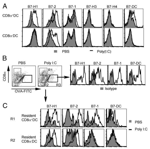
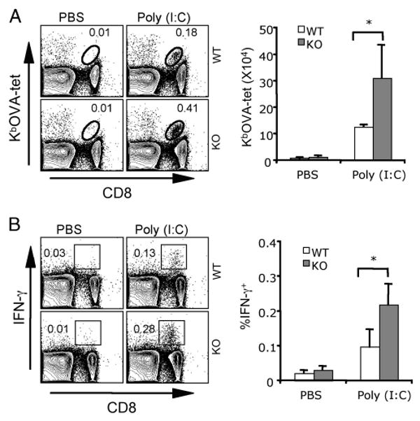
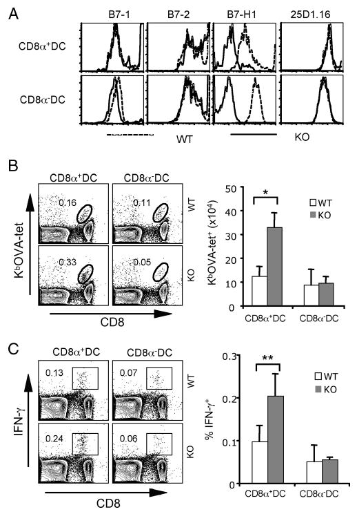
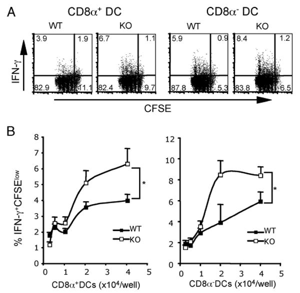
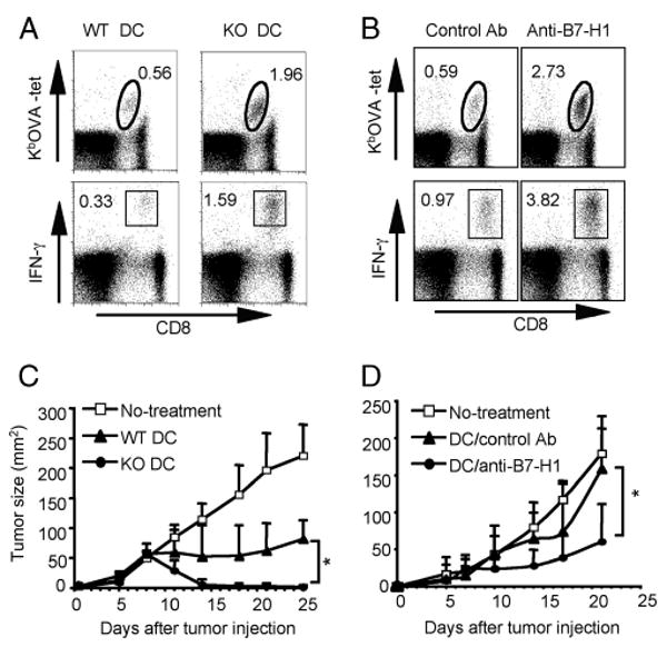
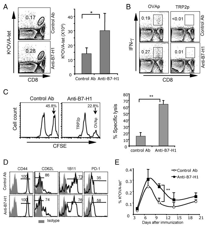
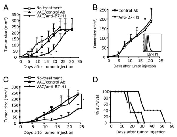
Agonists of TLR have been explored as vaccine adjuvants for tumor immunotherapy. However, their immunological consequences are not fully understood. Although TLR signaling increases the functional potential of dendritic cells (DCs) for priming T cells, coinduction of potentially negative immunoregulatory capacities may impair effector T cell generation. We examined the expression and function of B7 family costimulatory molecules on DCs after activation with the TLR3 agonist, polyinosinic:polycytidylic acid. We demonstrated that polyinosinic:polycytidylic acid consistently up-regulated both B7-2 and B7-H1 molecules on resident, migratory DCs from spleen and lymph nodes. Depletion or blockade of B7-H1 on activated DCs increased the magnitude of effector CD8 T cell expansion. DC-based or protein-based tumor vaccines, in combination with B7-H1 blockade, induced strong effector CD8 T cell responses, resulting in protective immunity against newly established tumors. Our studies suggest that TLR3 signaling has the potential to up-regulate both positive and negative coregulatory molecules on APCs. Selective blockade of negative regulatory molecules in combination with TLR3 agonist may be an effective strategy for increasing the efficacy of tumor vaccines.


Conflict of interest statement

Disclosures: Both E. D. Kwon and the Mayo Clinic have received royalties greater than the federal threshold for significant financial interest from the licensing to Medarex of technology related to B7-H1. Additionally, E. D. Kwon and the Mayo Clinic have contractual rights to receive future royalties from the licensing of this technology. In addition, some of the authors (E. D. Kwon, X. Frigola, and H. Dong) have filed patents for the potential use of B7-H1, B7-H3, and B7-H4 as prognostic markers for assessment of cancer.

Figures

FIGURE 1
FIGURE 1
Poly(I:C) up-regulated both B7-2 and B7-H1 on DCs. A, TLR3 agonist poly(I:C) (50 μg) or PBS (control) was injected i.p. into WT mice. B and C, FITC-OVA protein (20 μg) was injected s.c. with PBS or poly(I:C) (20 μg) in WT mice. One day later, CD11c+ DCs were purified from spleen (A) or draining lymph nodes (B and C), and stained with anti-CD11c, anti-CD8α, and other Abs, as indicated. Shaded histograms represent isotype controls (B). Migratory DCs are identified as CD8α FITC-OVA+ cells (R3), and resident CD8α+/− DCs as FITC-OVA (R1 and R2). Data show profiles of CD11c+ CD8α+/− DCs.
FIGURE 2
FIGURE 2
Immunization with poly(I:C) induced a more robust expansion of Ag-specific CD8 T cells in B7-H1-deficient mice. WT or B7-H1 KO mice were immunized i.p. with whole OVA protein (0.5 mg) and poly(I:C) (50 μg) or PBS. Seven days after immunization, splenocytes were isolated and analyzed for OVA-specific CD8 T cells by KbOVA tetramer staining (A). Functionality of OVA-reactive CD8 T cells was analyzed by intracellular staining for IFN-γ after 5-h restimulation with OVA peptide (B). Bar graphs show the average numbers or percentages ± SD of three mice per group. One representative of three experiments is shown; *, p < 0.05.
FIGURE 3
FIGURE 3
Immunization with B7-H1-deficient CD8α+ DCs increased Ag-reactive CD8 T cells. CD8α+ or CD8α DCs were purified from spleens of WT or B7-H1 KO Act-mOVA mice 20 h after poly(I:C) (50 μg/ml) injection. A, Histograms show phenotypes of splenic DC subsets. Presentation of OVA peptide within MHC I complex was identified by anti-Kb/OVA mAb (25D1.16). B and C, Immunization of naive WT mice with CD8α+ or CD8α DCs (1 × 10⁵, s.c.). One week after immunization, splenocytes were isolated and stained with KbOVA tetramer and anti-CD8 Ab (B). Effector function of CD8 T cells was analyzed by intracellular staining for IFN-γ after 5-h restimulation with OVA peptide (C). One representative of three experiments is shown. Bar graphs show the average numbers or percentages ± SD of three or four mice per group; *, p < 0.01; **, p < 0.05.
FIGURE 4
FIGURE 4
DC B7-H1 limited the expansion of effector CD8 T cells in vitro. CD8α+ or CD8α DCs were purified from spleens of WT or B7-H1 KO Act-mOVA mice 20 h after poly(I:C) injection (50 μg). A, CFDA-SE-labeled OT-1 CD8 T cells (1 × 10⁵/well) were incubated with WT or B7-H1 KO DC subsets (4 × 10⁴/well) for 60 h. Proliferation of CD8 T cells was analyzed by flow cytometry based on the dilution of CFSE. Production of IFN-γ was analyzed by intracellular staining 16 h after addition of brefeldin A. One of two independent experiments is shown. B, Percentages of IFN-γ+CFSElow OT-1 CD8 T cells in culture with graded DC subsets, as indicated. *, p < 0.05.
FIGURE 5
FIGURE 5
Immunization with B7-H1-deficient or blocked DCs increased Ag-specific effector CD8 T cells and suppressed tumor growth. WT mice were immunized i.v. with A, poly(I:C)-activated OVA peptide-pulsed WT or B7-H1 KO BM-derived DCs, or with B, poly(I:C)-activated OVA peptide-pulsed WT DCs preincubated with anti-B7-H1 blocking or control Ab. One week later, splenocytes were isolated and stained with KbOVA tetramer and anti-CD8 Ab (upper panels in A and B). Effector function of CD8 T cells was analyzed by intracellular staining for IFN-γ after 5-h restimulation with OVA peptide (lower panels in A and B). One representative of three experiments is shown. C and D, Immunotherapy with poly(I:C)-activated DC vaccines. B16-OVA tumor cells (5 × 10⁵) were injected s.c. into WT mice on day 0. Mice were treated with C, WT or B7-H1 KO DC vaccines (s.c.) on days 3, 6, and 10, or with D, WT DC vaccines combined with B7-H1 blocking Ab or control Ab (s.c.) on days 3, 6, and 10. Data show tumor sizes as mean ± SD of four or five mice per group. *, p < 0.05. One of two independent experiments is shown.
FIGURE 6
FIGURE 6
Immunization with protein/poly(I:C) combined with B7-H1 blockade increased Ag-specific effector CD8 T cells. WT mice were immunized i.p. with whole OVA protein (0.5 mg) and poly(I:C) (50 μg) on day 0. Anti-B7-H1 (10B5) or control Abs (hamster Ig) (200 μg) were injected (i.p.) on days 0 and 3. Eight days after immunization, splenocytes were isolated and analyzed for OVA-specific CD8 T cells by KbOVA tetramer staining (A). Bar graphs show the average numbers ± SD of three mice per group; *, p < 0.05. Functionality of OVA-reactive CD8 T cells was analyzed by intracellular staining for IFN-γ after 5-h restimulation with OVA peptide or control TRP2 peptide (B). One representative of three experiments is shown. C, In vivo cytotoxicity was measured in mice on day 7 after immunization with OVA/poly(I:C) combined with control Ab or B7-H1 blocking Ab. Histogram plots show the remaining target cells in spleen. The bar graph shows the average percentages of specific lysis in three mice; **, p < 0.01. D, Phenotype of OVA-specific CD8 T cells on day 8 after immunization. E, Kinetics of OVA-specific (KbOVA-tetramer+) CD8 T cell response to OVA/poly(I:C) with or without B7-H1 blockade. Data show the average percentages ± SD of three mice at each time point. One representative of two experiments is shown; **, p < 0.01.
FIGURE 7
FIGURE 7
Vaccination with B7-H1 blockade improved preventive and therapeutic antitumor immunity. A, B16-OVA tumor cells (1 × 10⁶) were injected s.c. into WT mice 14 days after immunization with OVA/poly(I:C) with or without B7-H1 blockade. Data show the average tumor size ± SD of five mice per group; *, p < 0.05, compared with control Ab groups. B, B16-OVA tumors were treated with anti-B7-H1 blocking Ab (10B5) or control Ab (200 μg, i.p.) on days 3, 6, 9, and 12 after tumor inoculation. Expression of B7-H1 by cultured B16-OVA tumor cells (inset, isotype staining is shown with shaded histogram). C and D, B16-OVA tumor cells (5 × 10⁵/mouse) were injected s.c. into WT mice on day 0. Mice were immunized i.p. with OVA/poly(I:C) and anti-B7-H1 (10B5) or control Ab (hamster Ig) (200 μg) every 2 days for a total of five times beginning on day 2 (A). C, Data show the average tumor size ± SD of five mice per group; *, p < 0.05. D, The survival of mice treated with vaccine combined with B7-H1 blockade; p < 0.05, compared with vaccine without B7-H1 blockade. One of two independent experiments is shown.

References

    1. Janeway CA, Jr, Medzhitov R. Innate immune recognition. Annu Rev Immunol. 2002;20:197–216. - PubMed
    1. Banchereau J, Steinman RM. Dendritic cells and the control of immunity. Nature. 1998;392:245–252. - PubMed
    1. Kawai T, Akira S. TLR signaling. Cell Death Differ. 2006;13:816–825. - PubMed
    1. Yang D, Chen Q, Su SB, Zhang P, Kurosaka K, Caspi RR, Michalek SM, Rosenberg HF, Zhang N, Oppenheim JJ. Eosinophil-derived neurotoxin acts as an alarmin to activate the TLR2-MyD88 signal pathway in dendritic cells and enhances Th2 immune responses. J Exp Med. 2008;205:79–90. - PMC - PubMed
    1. Kobayashi M, Kweon MN, Kuwata H, Schreiber RD, Kiyono H, Takeda K, Akira S. Toll-like receptor-dependent production of IL-12p40 causes chronic enterocolitis in myeloid cell-specific Stat3-deficient mice. J Clin Invest. 2003;111:1297–1308. - PMC - PubMed

Publication types