. 2009 Sep 15;183(6):3971-9.
doi: 10.4049/jimmunol.0901604. Epub 2009 Aug 26.

Deficiency of endothelial heparan sulfates attenuates allergic airway inflammation

Affiliations

Deficiency of endothelial heparan sulfates attenuates allergic airway inflammation

Riaz I Zuberi et al. J Immunol. .

Abstract

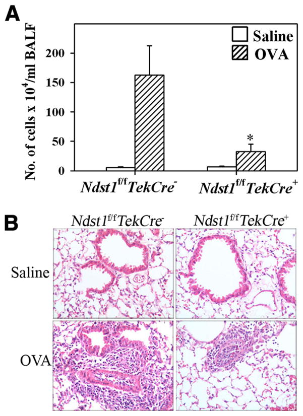
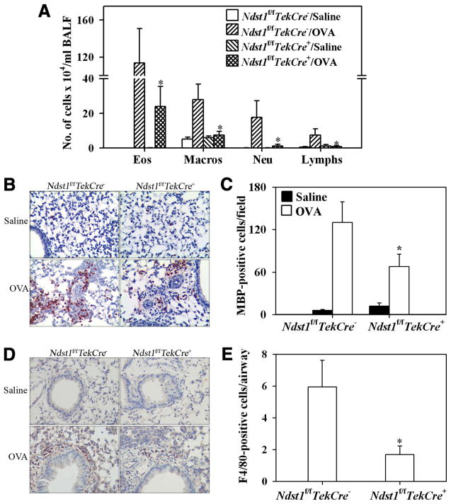
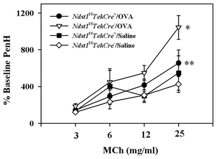
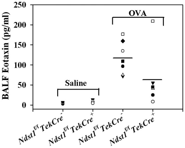
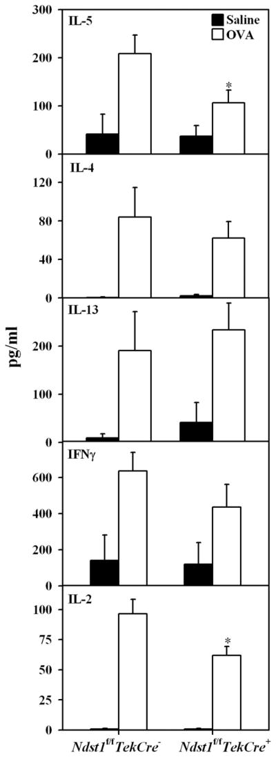
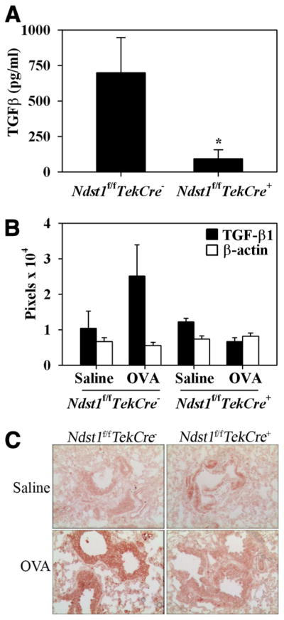
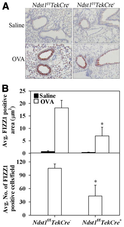
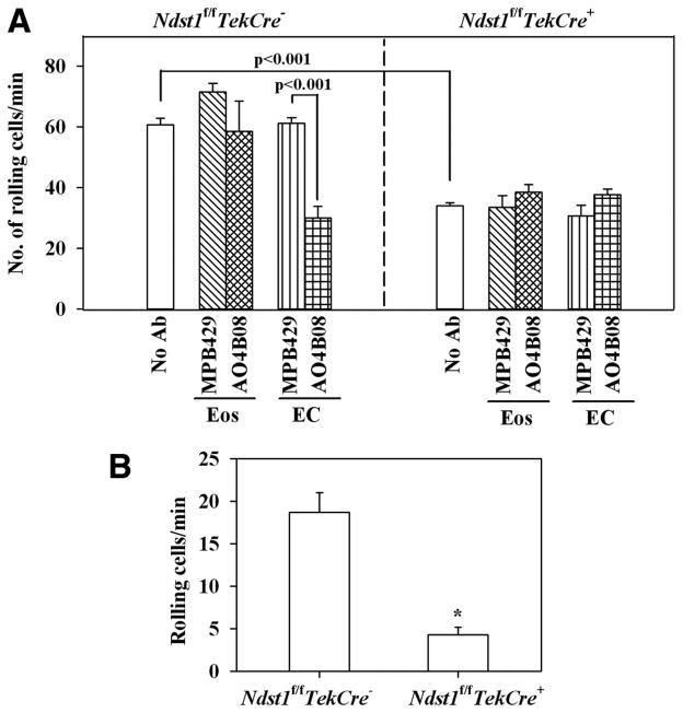
The effect of targeted inactivation of the gene encoding N-deacetylase/N-sulfotransferase-1 (Ndst1), a key enzyme involved in the biosynthesis of heparan sulfate (HS) chains, on the inflammatory response associated with allergic inflammation in a murine model of OVA-induced acute airway inflammation was investigated. OVA-exposed Ndst1(f/f)TekCre(+) (mutant) mice deficient in endothelial and leukocyte Ndst1 demonstrated significantly decreased allergen-induced airway hyperresponsiveness and inflammation characterized by a significant reduction in airway recruitment of inflammatory cells (eosinophils, macrophages, neutrophils, and lymphocytes), diminished IL-5, IL-2, TGF-beta1, and eotaxin levels, as well as decreased expression of TGF-beta1 and the angiogenic protein FIZZ1 (found in inflammatory zone 1) in lung tissue compared with OVA-exposed Ndst1(f/f)TekCre(-) wild-type littermates. Furthermore, murine eosinophils demonstrated significantly decreased rolling on lung endothelial cells (ECs) from mutant mice compared with wild-type ECs under conditions of flow in vitro. Treatment of wild-type ECs, but not eosinophils, with anti-HS Abs significantly inhibited eosinophil rolling, mimicking that observed with Ndst1-deficient ECs. In vivo, trafficking of circulating leukocytes in lung microvessels of allergen-challenged Ndst1-deficient mice was significantly lower than that observed in corresponding WT littermates. Endothelial-expressed HS plays an important role in allergic airway inflammation through the regulation of recruitment of inflammatory cells to the airways by mediating interaction of leukocytes with the vascular endothelium. Furthermore, HS may also participate by sequestering and modulating the activity of allergic asthma-relevant mediators such as IL-5, IL-2, and TGF-beta1.


Conflict of interest statement

Disclosures

The authors have no financial conflicts of interest.

Figures

FIGURE 1
FIGURE 1
Cellular infiltration of the airways in response to allergen challenge is inhibited in Ndst1f/fTekCre+ mice. Ndst1f/fTekCre+ mice and WT littermates were sensitized and challenged with OVA while control mice received only saline. BALF and lung tissue collected 3 h after the last allergen challenge were processed for evaluation of cellular infiltration. A, Cells recovered from the BALF were counted with a hemocytometer and expressed as the number of cells × 104/ml of BALF (n = 8–11 mice for control groups and 14–16 mice for OVA-challenged groups). *, p = 0.011. B, Cellular infiltration of lung tissue was evaluated by H&E staining of paraformaldehyde-fixed lung tissue sections. Representative images from each group at a magnification of × 20 are shown.
FIGURE 2
FIGURE 2
Allergen-challenged Ndst1f/fTekCre+ mice exhibit decreased airway recruitment of eosinophils and macrophages. A, BALF collected from control (saline) and OVA-sensitized and challenged Ndst1f/fTekCre+ mice and WT littermates was evaluated for differential cell counts by microscopic evaluation of cytocentrifuged slides (n = 5–11 mice for control groups and 11–16 mice for OVA-challenged groups). B, Lung tissue eosinophils in these mice were evaluated by immunohistochemical staining for eosinophil-specific MBP using rat mAbs specific for murine MBP. Representative images from each group at a magnification of × 20 are shown. C, MBP-positive cells in five randomly selected nonoverlapping microscopic fields were counted (× 40 magnification) and results were expressed as the average number of cells per field (n = 5 mice/group). D, Lung tissue macrophages in control and OVA-exposed Ndst1f/fTekCre+ mice and WT littermates were evaluated by immunohistology with rat anti-mouse F4/80. Representative images from each group at a magnification of × 20 are shown. E, Airways in a microscopic field were identified and the number of F4/80-positive cells around the airways was counted (× 40 magnification). Results were expressed as the number of F4/80-positive cells per airway, which was determined by dividing the total number of F4/80-positive cells by the total number of airways (n = 4 mice/group). *, p < 0.05.
FIGURE 3
FIGURE 3
AHR is diminished in allergen-challenged Ndst1f/fTekCre+ mice. Airway responsiveness in control and allergen-challenged Ndst1f/fTekCre+ mice and WT littermates was measured 3 h after the last challenge by whole body plethysmography. Mice were first exposed to saline followed by increasing concentrations of nebulized MCh. The enhanced pause (Penh) in breathing after each nebulization was monitored, and results for each MCh concentration were expressed as a percentage of the baseline Penh values following exposure to saline (n = 5 mice for the control groups and 10–12 mice for the OVA-challenged groups). *, p < 0.01; **, p = 0.028.
FIGURE 4
FIGURE 4
Allergen-challenged Ndst1f/fTekCre+ mice exhibit decreased eotaxin levels. BALF collected from control and allergen-challenged Ndst1f/fTekCre+ mice and WT littermates was used to evaluate eotaxin levels by ELISA. Concentration of eotaxin in the BALF was determined against a standard curve generated (range, 7.8–500 pg/ml) and expressed as pg/ml of BALF (n = 4–5 mice for the control groups and 6–7 mice for the OVA-challenged groups).
FIGURE 5
FIGURE 5
IL-5 and IL-2 levels are decreased in the BALF of allergen-challenged Ndst1f/fTekCre+ mice. IL-5, IL-13, IL-4, IFN-γ, and IL-2 levels in BALF from control and allergen-challenged Ndst1f/fTekCre+ mice and WT littermates were evaluated by ELISA using Ab pairs and recombinant cytokines for generation of standard curves. Cytokine levels were expressed as pg/ml of BALF (n = 4 mice for the control groups and 6–11 mice for the OVA-challenged groups). *, p < 0.01 for IL-5 and p < 0.05 for IL-2.
FIGURE 6
FIGURE 6
Allergen-challenged Ndst1f/fTekCre+ mice exhibit decreased TGF-β1 levels in BALF and lung tissue. A, TGF-β1 levels were measured in BALF from control and allergen-challenged Ndst1f/fTekCre+ mice and WT littermates by ELISA. TGF-β1 levels in OVA-challenged Ndst1f/fTekCre+ mice and WT littermates after subtracting TGF-β1 levels in corresponding control mice are shown as pg/ml of BALF (n = 7 each for control and OVA-challenged WT littermates and 8 each for control and OVA-challenged Ndst1f/fTekCre+ mice). *, p < 0.05. B, TGF-β1 levels in total lung tissue lysates were determined by Western blotting using rabbit Abs against TGF-β1. Blots were probed with HRP-conjugated anti-mouse β-actin to monitor levels of β-actin expression as an internal control. Bands were visualized on x-ray films and scanned and analyzed using ImageJ to quantitate the density (pixels) of the bands (n = 4 mice/group). C, Distribution of TGF-β1 in lung tissue from control and allergen-challenged Ndst1f/fTekCre+ mice and WT littermates was determined by immunohistochemistry with polyclonal Abs against TGF-β1. Representative images from each group at a magnification of × 20 are shown. *, p < 0.05.
FIGURE 7
FIGURE 7
Expression of FIZZ1 is inhibited in allergen-challenged Ndst1f/fTekCre+ mice. A, FIZZ1 expression in lung tissue of OVA-challenged Ndst1f/fTekCre+ mice and WT littermates as well as in unchallenged control mice was evaluated by immunohistochemistry using goat polyclonal Abs against murine FIZZ1. Representative images from each group at a magnification of ×20 are shown. B, Total FIZZ1 expression was quantitated from captured images (five fields per mouse) at ×10 magnification using ImageJ image analysis system and expressed as average FIZZ1-positive area per field. FIZZ1-positive type II alveolar epithelial cells in five randomly selected microscopic fields with no visible airways or blood vessels were counted (×40 magnification) and expressed as the average number of cells per field. A negligible number of FIZZ1-positive type II alveolar epithelial cells were present in saline-exposed control mice (n = 5 each for control and OVA-challenged WT littermates and 4 each for control and OVA-challenged Ndst1f/fTekCre+ mice). *, p < 0.05.
FIGURE 8
FIGURE 8
Rolling of murine eosinophils on MLECs and circulating leukocytes on vascular endothelium of Ndst1f/fTekCre+ mice. A, Single-cell suspensions of murine eosinophils (n = 2 mice in duplicate) were infused into a flow chamber holding coverslips coated with monolayers of TNF-α-stimulated lung EC from Ndst1f/fTekCre+ mice or WT littermates (n = 2 mice/group) that were treated with media alone, anti-HS Ab AO4B08, or control Ab MPB49 at a wall shear stress of ~1.0 dyn/cm2 for 5 min and the interactions of the injected cells with the coated coverslips were recorded. In some cases, the eosinophils were treated with AO4B08 or MPB49 before infusion into the flow chamber. Interaction of the injected cells with the MLEC-coated coverslips was recorded for subsequent offline analysis. Slow-flowing cells demonstrating multiple discrete interruptions in flow were considered as rolling cells. Results were expressed as the number of rolling cells per minute. B, The interaction of acridine orange-labeled circulating leukocytes with the endothelium of revascularized microvessels of lung allografts from allergen-challenged Ndst1f/fTekCre+ mice and WT littermates mice was evaluated by IVM. Leukocytes visibly interacting with the lung microvascular endothelium and passing at a slower rate than the main blood stream were considered as rolling cells and were quantitated by manually counting the number of rolling cells passing through a reference point in a 200-μm vessel segment and expressed as the number of rolling cells per minute (n = 4–8 lung microvessels/mouse, 2 mice/group). *, p < 0.001.

References

    1. Bishop JR, Schuksz M, Esko JD. Heparan sulphate proteoglycans fine-tune mammalian physiology. Nature. 2007;446:1030–1037. - PubMed
    1. Rostand KS, Esko JD. Microbial adherence to and invasion through proteoglycans. Infect Immun. 1997;65:1–8. - PMC - PubMed
    1. Kjellén L. Glucosaminyl N-deacetylase/N-sulphotransferases in heparan sulphate biosynthesis and biology. Biochem Soc Trans. 2003;31:340–342. - PubMed
    1. Grobe K, Inatani M, Pallerla SR, Castagnola J, Yamaguchi Y, Esko JD. Cerebral hypoplasia and craniofacial defects in mice lacking heparan sulfate Ndst1 gene function. Development. 2005;132:3777–3786. - PMC - PubMed
    1. Pallerla SR, Pan Y, Zhang X, Esko JD, Grobe K. Heparan sulfate Ndst1 gene function variably regulates multiple signaling pathways during mouse development. Dev Dyn. 2007;236:556–563. - PubMed

Publication types