. 2009 Nov;76(10):1081-8.
doi: 10.1038/ki.2009.321. Epub 2009 Aug 26.

Intra-tubular deposits, urine and stone composition are divergent in patients with ileostomy

Affiliations

Intra-tubular deposits, urine and stone composition are divergent in patients with ileostomy

Andrew P Evan et al. Kidney Int. 2009 Nov.

Abstract

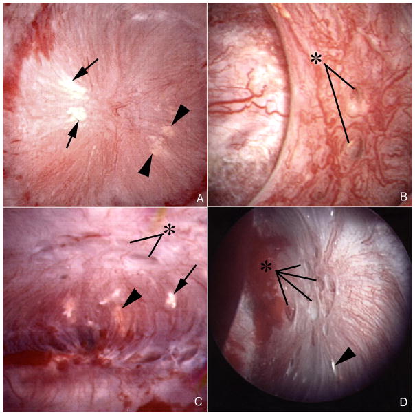
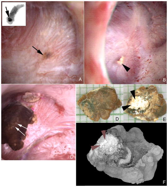
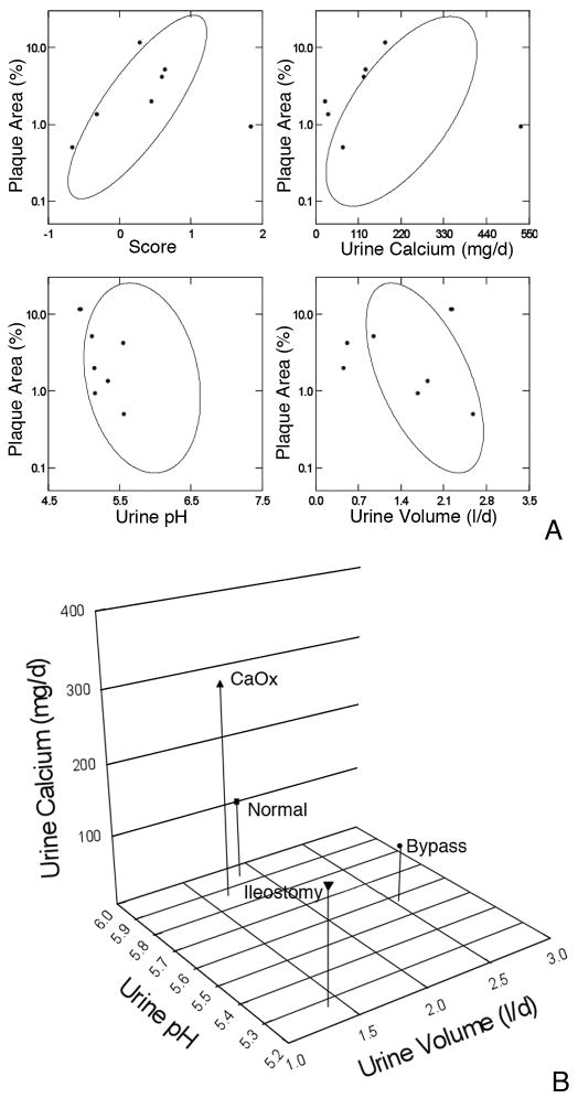
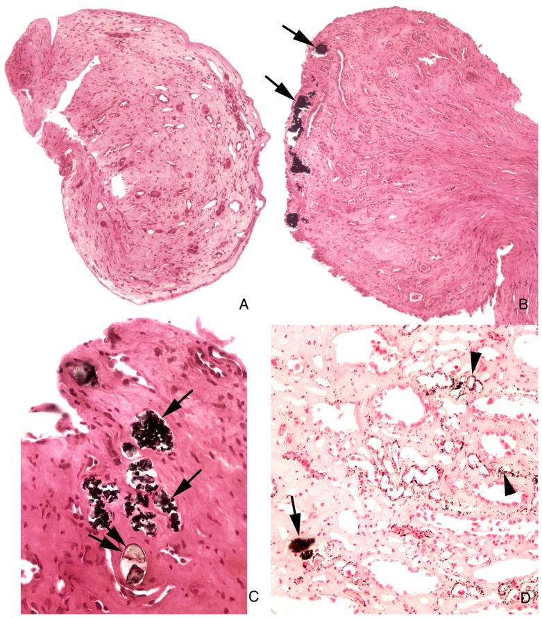
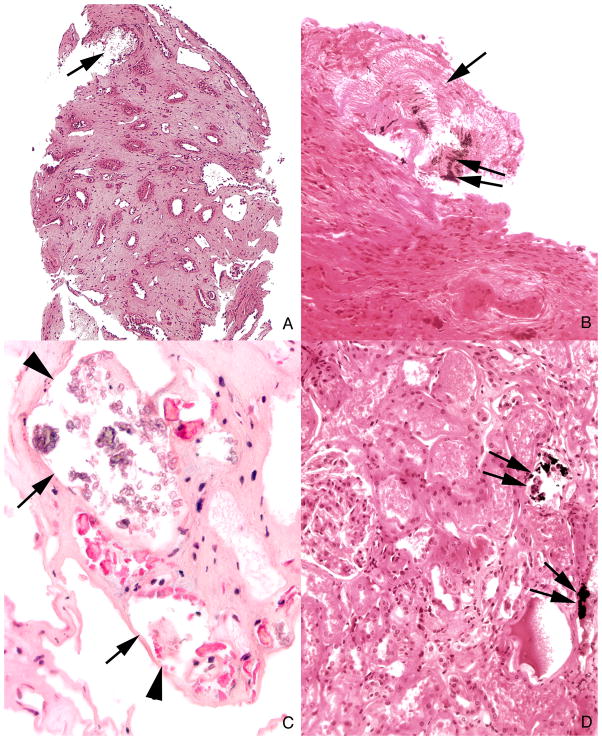
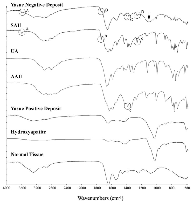
Patients with ileostomy typically have recurrent renal stones and produce scanty, acidic, sodium-poor urine because of abnormally large enteric losses of water and sodium bicarbonate. Here we used a combination of intra-operative digital photography and biopsy of the renal papilla and cortex to measure changes associated with stone formation in seven patients with ileostomy. Papillary deformity was present in four patients and was associated with decreased estimated glomerular filtration rates. All patients had interstitial apatite plaque, as predicted from their generally acid, low-volume urine. Two patients had stones attached to plaque; however, all patients had crystal deposits that plugged the ducts of Bellini and inner medullary collecting ducts (IMCDs). Despite acid urine, all crystal deposits contained apatite, and five patients had deposits of sodium and ammonium acid urates. Stones were either uric acid or calcium oxalate as predicted by supersaturation, however, there was a general lack of supersaturation for calcium phosphate as brushite, sodium, or ammonium acid urate because of the overall low urine pH. This suggests that local tubular pH exceeds that of bulk urine. Despite low urine pH, patients with an ileostomy resemble those with obesity bypass, in whom IMCD apatite crystal plugs are found. They are, however, unlike these bypass patients in having interstitial apatite plaque. IMCD plugging with sodium and ammonium acid urate has not been found previously and appears to correlate with formation of uric acid stones.


Conflict of interest statement

DISCLOSURE

The authors have no interests to disclose relevant to this publication.

Figures

Figure 1
Figure 1. Endoscopic images of papilla from ileostomy patients with kidney stones
Papillary morphology of ileostomy patients ranged from a normal conical shape (panels a–c) to flattened and retracted (panel d). Dilated openings (asterisks) to ducts of Bellini were common in both the normal and deformed papilla (panels b–d). Variable amounts of white (arrows) and yellow (arrowheads) plaque were seen separately (panel d) or on the same papilla (panels a and c).
Figure 2
Figure 2. Endoscopic images showing both intraluminal plugs and attached stone in ileostomy patients
A dark region on the papillary surface (panel a, arrow) marks a large intraluminal deposit deep to the urothelium. The insert at the upper left corner of panel a shows this same intraluminal deposit (arrow) by micro-CT in a biopsy sample removed at the site of the deposit. Mineral plugs protruding from dilated ducts of Bellini (panel b, arrowhead) were common. In addition, several stones were noted attached to sites of Randall’s plaque. Panel c shows such a stone (double arrows). This stone was removed and photographed by light microscopy (panels d and e) and scanned by micro-CT (panel f). The papillary surface of this stone (panels e and f, double arrowheads) reveals a whitish region that was determined to be hydroxyapatite by micro- CT. Magnification, x30 (d and e).
Figure 3
Figure 3. Comparison of present plaque data to our prior reports
3A: Percent plaque coverage of renal papillae gauged via intra-operative digital imaging (y axes of all 4 panels) varied in our prior reports (9) of normal, idiopathic calcium stone formers and obesity bypass patients as in the elliptical confidence bands shown on each panel. The present 7 patients are over plotted in grey symbols. In general, the patients fall along or within the ellipses. 3B: Compared to our prior study cases ileostomy patients have much lower pH as a predominant cause of plaque, and moderately lower urine volume and calcium excretion. Values are means.
Figure 4
Figure 4. Histologic images of Yasue stained sections from papillary biopsies from ileostomy patients
The morphology of the papillary biopsies ranged from a normal architecture without deposits (panel a) to an abnormal appearance characterized by numerous dilated inner medullary collecting ducts and ducts of Bellini filled with Yasue positive (panels b, c and d, arrows) and Yasue negative deposits (panel c double arrows) always associated with a loss of lining cells (arrows) and surrounded by interstitial fibrosis. Sites of interstitial plaque (Randall’s) were noted in the basement membrane of thin loops of Henle (panel d, arrowheads). Magnification, x100 (a and b); x300 (c and d).
Figure 5
Figure 5. Histologic images of tubules with Yasue negative deposits in inner medulla and Yasue positive deposits in cortex
Panels a and b each show a large deposit in a duct of Bellini (arrow) that is Yasue negative except for a very small region of Yasue positive staining (double arrow). These plugged ducts are generally very dilated, completely filled with mineral, surrounded by interstitial fibrosis and lack tubular lining cells. Several tubules filled with Yasue negative deposits (arrows) are seen at a higher magnification in panel c to show the loss of the tubular lining cells (arrowheads). Panel d shows two cortical collecting ducts stained with Yasue positive deposits (arrows). Magnification, x100 (a); x200 (b); x300 (c); x200 (d).
Figure 6
Figure 6. Micro-FTIR spectra of Yasue negative and positive crystal deposits in ileostomy patients
This figure illustrates a series of infrared spectra obtained for a set of standards (sodium acid urate (SAU), uric acid (UA), ammonium acid urate (AAU), and hydroxyapatite, for a site of a Yasue positive deposit in the tissue, for a site of a Yasue negative deposit in the tissue and for the normal tissue with embedding medium. The infrared spectrum of the Yasue negative deposit is consistent with SAU and AAU: Bands A, B and D highlighted by circles in the spectrum for the Yasue negative deposit correspond with bands a, b, and d, at wavenumbers 3598, 1738 and 1257 respectively in the spectrum for SAU. Band C in the spectra for the Yasue negative deposit (highlighted by a circle) corresponds with band c at wavenumber 1385 in the spectrum for AAU. A broad band at about 1100 (arrow) in the Yasue negative spectrum suggests phosphate or sulfate. The infrared spectrum of the Yasue positive deposit is consistent with hydroxyapatite as is obvious by the matching of the large band at about 1,000. Normal tissue has no bands that correspond with those for the standards.

References

    1. Bambach CP, Robertson WG, Peacock M, Hill GL. Effect of intestinal surgery on the risk of urinary stone formation. Gut. 1981;22:257–263. - PMC - PubMed
    1. Clarke AM, Chirnside A, Hill GL, et al. Chronic dehydration and sodium depletion in patients with established ileostonies. Lancet. 1967;2:740–743. - PubMed
    1. Worcester EM. Stones due to bowel disease. In: Coe FL, Favus MJ, Pak CY, et al., editors. Kidney Stones: Medical and Surgical Management. chap 39. Philadelphia: Lippincott-Raven; 1996. pp. 883–903.
    1. Parks JH, Worcester EM, O’Connor RC, Coe FL. Urine stone risk factors in nephrolithiasis patients with and without bowel disease. Kidney Int. 2003;63:255–265. - PubMed
    1. Randall RE. Urate nephropathy following chronic ileostomy acidosis. Am J Nephrol. 2002;22:372–375. - PubMed

Publication types

LinkOut - more resources