. 2009 Nov 1;517(1):87-104.
doi: 10.1002/cne.22112.

Arborization pattern of engrailed-positive neural lineages reveal neuromere boundaries in the Drosophila brain neuropil

Affiliations

Arborization pattern of engrailed-positive neural lineages reveal neuromere boundaries in the Drosophila brain neuropil

Abhilasha Kumar et al. J Comp Neurol. .

Abstract

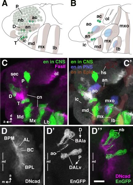
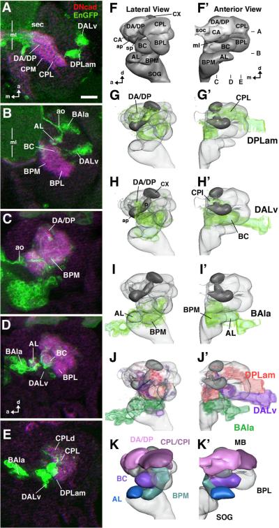
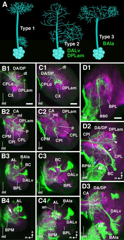
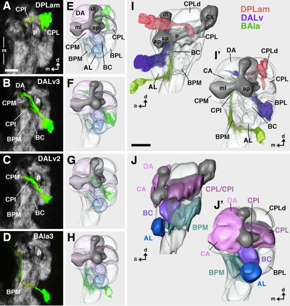
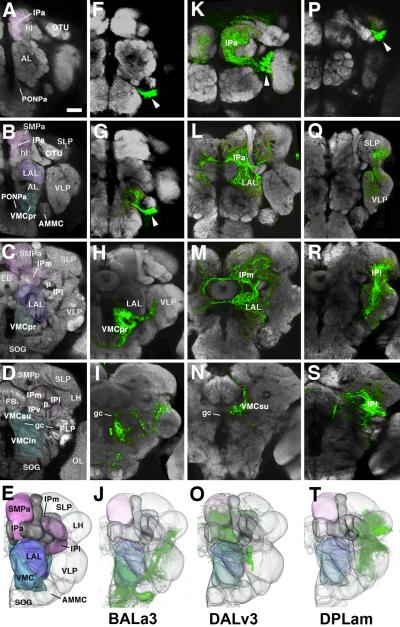
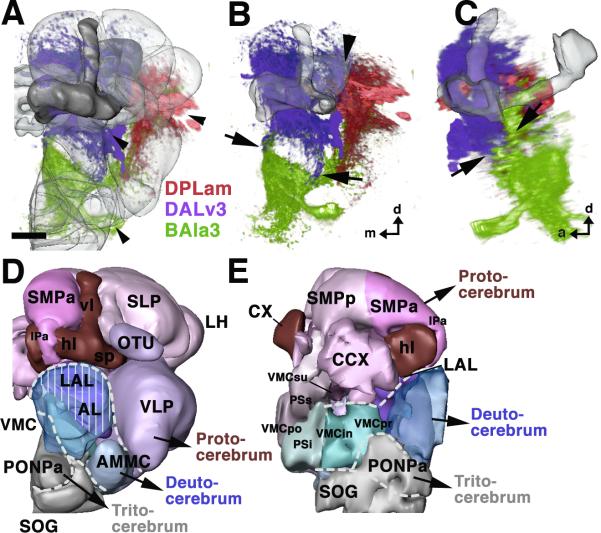
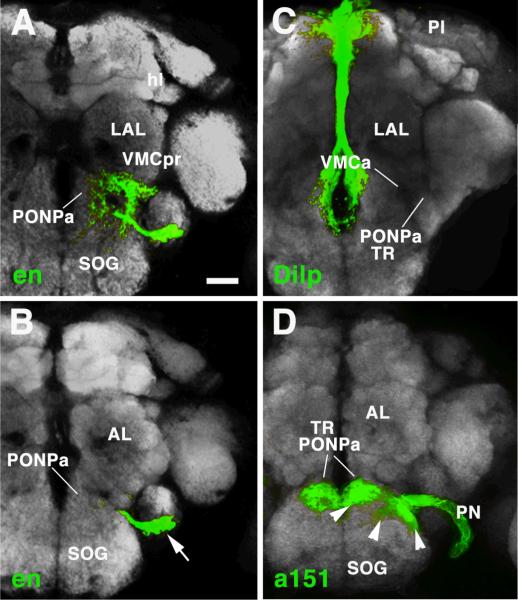
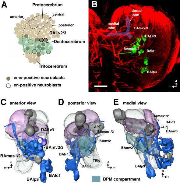
The Drosophila brain is a highly complex structure composed of thousands of neurons that are interconnected in numerous exquisitely organized neuropil structures such as the mushroom bodies, central complex, antennal lobes, and other specialized neuropils. While the neurons of the insect brain are known to derive in a lineage-specific fashion from a stereotyped set of segmentally organized neuroblasts, the developmental origin and neuromeric organization of the neuropil formed by these neurons is still unclear. In this study we used genetic labeling techniques to characterize the neuropil innervation pattern of engrailed-expressing brain lineages of known neuromeric origin. We show that the neurons of these lineages project to and form most arborizations, in particular all of their proximal branches, in the same brain neuropil compartments in embryonic, larval and adult stages. Moreover, we show that engrailed-positive neurons of differing neuromeric origin respect boundaries between neuromere-specific compartments in the brain. This is confirmed by an analysis of the arborization pattern of empty spiracles-expressing lineages. These findings indicate that arborizations of lineages deriving from different brain neuromeres innervate a nonoverlapping set of neuropil compartments. This supports a model for neuromere-specific brain neuropil, in which a given lineage forms its proximal arborizations predominantly in the compartments that correspond to its neuromere of origin.


Figures

Fig.1
Fig.1. Metameric engrailed expression in the embryonic head
A: Schematic of early (stage 11) embryonic head, lateral view. Ectodermal engrailed (en) stripes corresponding to posterior domains of head segments are shaded [lb labium; mx maxilla; md mandible; ic intercalary segment; an antennal segment; ac acron (“head spot”)]. Brain neuroblasts (nb) are shaded green. Dark green indicates sets of en-positive neuroblasts. It is notable that these neuroblasts spatially overlap with en expression domains. B: Schematic of mid-stage (stage 13) embryonic head, lateral view. Spatial relationship of en-positive neuroblasts and ectodermal en strips is maintained. Blue areas indicate primordia of head sensory complexes (labium, maxilla, antenna) which partially overlap with en stripes. C: Z-projections of confocal sections of stage 14 embryonic head labeled with antibody against En (green) and Fasciclin II (magenta); lateral view. Note metamerical clusters of neurons (lineages) derived from en-positive neuroblasts. The three posterior clusters (Md, Mx, Lb) demarcate the three neuromeres of the gnathal segments, which will later form the subesophageal ganglion. The anterior clusters correspond to the neuromeres of the supraesophageal ganglion (P protocerebrum, derived from acron; D deutocerebrum, derived from antennal segment; T tritocerebrum, derived from intercalary segment). C': Same Z-projection as in C. The different tissues contributing to en expression domains are shown in different colors. en-positive clusters of neurons are in green. en in the ectoderm (by that stage: epidermal primordium) is shaded brown; parts of sensory primordia expressing en (ao antennal organ; mxo maxillary organ) are in blue. Note that the en-positive clusters and the corresponding en stripes from which they derive are still in close proximity, except for the protocerebral cluster of neurons that, due to morphogenetic movements in the head, has moved away from the epidermal head spot (hs). D, D', D": Z-projection of confocal sections of stage 15 embryonic brain labeled with antibodies against DN-cadherin (D; magenta in D") and GFP expressed by an en-Gal4 driver (D'; green in D"); dorsal view; only right brain hemisphere is shown; vertical line indicates midline (ml). Note pattern of en-positive lineages (deutocerebral BAla3; protocerebral DALv) and their relationship to the primordia of brain compartments (AL Antennal lobe; BC basocentral; BPL baso-posterior lateral; BPM baso-posterior medial). Each lineage consists of the superficial neuroblast (white line and arrowhead indication, D') and a chain of primary neurons. Other abbreviations: cn cervical connective; ol optic lobe; sec supraesophageal commissure. Bar: 10μm (for C–D)
Fig.2
Fig.2. engrailed lineages in the late embryonic brain
A, B: Z-projection of confocal sections of stage 17 embryonic brain labeled with antibodies against DN-cadherin (magenta) and GFP expressed by an en-Gal4 driver (green); dorsal view; only right brain hemisphere is shown; vertical line indicates midline (ml). Dorso-ventral focal plane of A and B is indicated in panel F' to the right. C–E: Z-projections of parasagittal confocal sections of stage 17 embryonic brain prepared as the one shown in A/B. Anterior is to the left, dorsal up. Medio-lateral focal planes of C–E are indicated in panel F'. F, F': 3D digital models of stage 17 embryonic brain hemisphere in lateral view (F) and anterior view (F'), showing neuropile compartments (AL larval antennal lobe; ap anterior appendix of larval mushroom body; BC baso-central; BPL baso-posterior lateral; BPM baso-posterior medial; CA centro-anterior; CPL centro-posterior lateral; CX calyx; DA dorso-anterior; DP dorso-posterior; sec supraesophageal commissure; ml medial lobe of mushroom body; SOG subesophageal ganglion (=anterior ventral nerve cord); sp spur of mushroom body). G–I, G'–I': Digital brain models as in F/F', with volume renderings of the en lineages visible in stage 17 embryo (DPLam, DALv, BAla3; shaded green). J, J': Montage of the en lineages in one model (DPLam red; DALv purple; BAla3 green). K, K': Digital brain models as in F/F', with neuropile compartments innervated by en lineages shown in different colors. En expression in late embryo appears in three main clusters. The dorsal-most cluster corresponds to the DPLam lineage (A, E, G/G'), which arborizes in CPL, CPI, DP and DA (A, C–E, G/G'). The medial cluster (DALv; A, B, E, H/H') consists of two adjacent lineages, DALv2 and DALv3, with indistinguishable projections in the embryo and early larva. Proximal projections are in the BC compartment (B, D, H/H'); more distally, projections overlap with those of DPLam in CPI (see G' and H') and DA and CA (see G, H, F). In the deutocerebrum, one lineage (BAla3) is distinguished (B, E, I/I'). It projects to the BPM compartment (C, I/I'). The larval AL compartment (antennal lobe) is labeled by sensory afferents of en-positive antennal organ (ao; BD, I/I'). Bar: 10μm
Fig.3
Fig.3. engrailed lineages during larval development
A: Schematic representation of different types of lineages encountered in brain (type 1: separate proximal and distal arborization; type 2: continuous arborization; type 3: distal arborization; Larsen et al., personal communication). B1–B4, C1–C4, D1–D3: Z-projection of confocal sections of larval brains labeled with antibodies against DN-cadherin (magenta) and GFP expressed by an en-Gal4 driver (green); only right brain hemisphere is shown; vertical line indicates midline (ml). B1– B4: first larval instar, dorsal view, focal plane goes from dorsal (B1) to ventral (B4). C1–C4: second larval instar, dorsal view, focal plane goes from dorsal (C1) to ventral (C4). D1–D3: late third larval instar; anterior view; focal plane goes from anterior (D1) to posterior (D3). The arborization pattern of en lineages is similar as shown for late embryo in Fig.2. The protocerebral DPLam lineage (cell bodies shown in B2, C2, D2) arborizes widely in CPL, CPI, DP/DA, and CA. DALv (comprising two neighboring lineages; cell bodies in B3, C3, D3) has arborizations in BC, as well as CPI and DP/DA where they overlap with fibers of DPLam. BAla3 (cell bodies in B3, C4, D3) arborizes in BPM. en-Gal4 driven GFP labeling in larval antennal lobe (AL; B4, C4, D3) is due to en-positive afferents from antennal organ. At third instar, secondary neurons have been added to en lineages. They form distinctive secondary axon tracts (SATs) that project into territory innervated by primary neurons (white arrow head in D2: SAT of DPLam; arrow in D2: SAT of DALv2/3; blue arrow in D2: SAT of BAla3). Note that there are en–positive fibers ascending from the ventral cord (asc), as well as en-positive surface glia (sg). Other abbreviations: BPL baso-posterior lateral compartment; CPM centro-posterior medial compartment; CX calyx; dl dorsal lobe; ml medial lobe; p peduncle). Bars: 10μm (B1–B4; C1–C4); 25μm (D1–D3)
Fig.4
Fig.4. Secondary neurons of engrailed lineages visualized in late third instar larva by en-Gal4 driven MARCM clones
Heat shock to induce Flippase mediated recombination was given shortly after larval hatching, leading to labeling of secondary neurons. A–D: Z-projection of confocal sections of late larval brains labeled with antibodies against Nc82 (grey) and GFP expressed by an en-Gal4 driver (green); anterior view; only right brain hemisphere is shown; vertical line indicates midline (ml). E–H: Digital 3D models of third instar brains (anterior view) with volume renderings of secondary en lineages (green). The lineages correspond to that shown in A–D. In models, mushroom body is shown in dark gray for better orientation. Compartments are rendered transparent. I, I': Digital 3D model of third instar brain with montage of all four en lineages; lateral view (I) and anterior view (I'). J, J': Digital models showing neuropile compartments in lateral view (J) and anterior view (J'). Lineages depicted in I/I' are shown in different colors: DPLam red, DALv2/3 purple; BAla3 green. Compartments in E–J' are shaded in colors that reflect the lineages they are innervated by (dark blue: antennal organ; light blue: BAla3; purple: DALv2/3; burgundy/pink: DPLam plus DALv. Note that axon tracts of secondary lineages are associated with the same compartments that were innervated by primary neurons of corresponding lineages (see Fig.2/3). For abbreviations of compartments see legend of Fig.2. Bar: 25μm
Fig.5
Fig.5. Arborization of secondary engrailed lineages in adult brain
A–S: Upper four panels of each column show Z-projections of frontal confocal sections of adult brains labeled with Nc82 antibody (synapses, grey) which delineates neuropile compartment. Each panel represents a Z projection of optical sections (“brain slice”) of approximately 20 micron thickness. Rows of panels start anteriorly in the brain (top) and move posteriorly (second to bottom). Panels of the first row (A, F, K, P) show the neuropile right in front of the horizontal lobe of the mushroom body (hl); the second row (B, G, L, Q) right behind the horizontal lobe; third row (C, H, M, R) at the level of the ellipsoid body (EB); fourth row (D, I, N, S) at the level of the fan-shaped body. E–T: The bottom row presents 3D digital models of adult brain neuropile in anterior view. The first column identifies the compartments (AL antennal lobe; AMMC antennomechanosensory and motor center; CCX central complex; CX calyx of mushroom body; EB ellipsoid body; FB fan-shaped body; gc great commissure; hl horizontal lobes of mushroom body; IP inferior protocerebrum; IPa anterior domain of IP; IPl lateral domain of IP; IPm medial domain of IP; IPv ventral domain of IP; LAL lateral accessory lobe; LH lateral horn; OL optic lobe; OTU optic tubercle; p peduncle of mushroom body; PLP postero-lateral protocerebrum; PONPa anterior perioesophageal neuropile; PSi inferior posterior slope (VMCpo); PSs superior domain of posterior slope (=VMCpo); SLP superior lateral protocerebrum; SMP superior medial protocerebrum; SMPa anterior domain of SMP; SMPp posterior domain of SMP; SOG suboesophageal ganglion; vl vertical lobes of mushroom body; sp spur of mushroom body; VLP ventrolateral protocerebrum; VMC ventromedial cerebrum; VMCin infracommissural domain of VMC; VMCpo postcommissural domain of VMC; VMCpr precommissural domain of VMC; VMCsu supracommissural domain of VMC). Color scheme corresponds to that one of Figure 4: the VMC, whose larval forerunner (BPM) is innervated by en lineage BAla3, is shaded blue; LAL (larval precursor: BC), innervated by DALv2/3, is purple; IPa (larval precursor: CA), SMPa (larval precursor: DA) and IPm (larval precursor: CPI), all innervated by DALv and DPLam, are pink; IPl (larval precursor: CPL), innervated mostly by DPLam, is burgundy. The second, third and fourth column each shows the arborization pattern of a single en lineage (BAla3 (F–J), DALv3 (K–O), and DPLam (P–T), respectively). Clusters of cell bodies of the lineages are identified by arrowheads. Note that the compartments that contain arborizations of the secondary neurons largely correspond to those that had input from primary neurons of the corresponding lineage. An exception is DPLam, where secondary neurons contribute substantially to the VLP, which had no primary arborizations; moreover, secondary DPLam neurons do not project to the SMP and IPa, both of which did receive primary neuronal input. Bar: 25μm
Fig.6
Fig.6. Secondary engrailed lineages in the adult brain and suggested neuromere boundaries
A–C: montages of volume renderings of the three en lineages shown in different colors (DPLam: red; DALv3: purple; BAla3: green). A and B show anterior views; in A, neuropile compartments are shaded. C presents a lateral view. Arrowheads in A point at the cell body clusters of the three lineages. Arrows in B and C outline the sharp boundary between arborizations of DALv3 (above) and BAla3 (below). This boundary corresponds to the structurally distinct boundary between LAL compartment (above) and VMC (below). Large arrowhead in B demarcates another, less sharply demarcated boundary between the IPm and IPl (figure 5); DPLam neurites are largely confined to the IPl, DALv3 neurites to the IPm. D, E: 3D digital models of the neuropile compartments of the adult brain, presenting suggested neuromere boundaries as white hatched lines. D shows anterior view, E medial view. Color coding is similar to that used in Figures 4 and 5. Compartments of the protocerebrum are shown in shades of red and purple, deutocerebral compartments in blue, and the tritocerebrum in grey. Note that in D, the hatched line indicating the proto-deutocerebral boundary arches dorsally over the deutocerebral antennal compartment (AL), which is rendered transparent to let one see through to the VMC and LAL located behind. The LAL (vertical lines) belongs to the protocerebrum, while the VMC (no vertical lines) to the deutocerebrum. Also, note that the volume renderings in A–C do not reveal the fact that BAla3 terminal arbors are excluded from the PONPa; this is visible in Z-Projections shown in Fig.5F, G. For abbreviations of neuropile compartments, see Fig.5. Bar: 25μm
Fig.7
Fig.7. Tentative delineation of the tritocerebrum
A, B: Z-projections of frontal confocal sections of adult brain labeled with Nc82 antibody (grey; labels neuropile compartments). The en-positive BAla3 lineage is shown in green; arrow in B points at cell bodies. Panel A represents neuropile at level of mushroom body horizontal lobe (hl) and lateral accessory lobe (LAL); focal plane shown in B represents neuropile more anteriorly, at the level of the antennal lobe (AL). C, D: Z-projections of confocal sections at levels corresponding to those shown in A and B, respectively. In C, projection of Dilp-positive neurons of the Pars intercerebralis (PI) to the anterior perioesophageal neuropile (PONPa) is visualized by Dilp-Gal4, UAS-mcd8GFP (Rulifson et al., 2002). In D, the sensory afferents of the pharyngeal nerve (PN, labeled by Gal4-driver line a151; kindly provided by Dr.Julie Simpson, JFRC, USA), which carries axons of the sensory neurons that originate in the intercalary segment, are shown in the anterior-most tip of the perioesophageal compartment. Note that the anterior perioesophageal neuropile is devoid of arborizations of the deutocerebral BAla3 lineage (A, B). We take this finding to support the notion that the PONPa represents the tritocerebrum (TR). Bar: 25μm
Fig.8
Fig.8. Projection of ems-positive lineages in the late larval brain
A: Schematic map of embryonic brain neuroblasts (lateral view; after Urbach and Technau, 2003a, b; Sprecher et al., 2007). Neuroblasts that were classified as deutocerebral based on their spatial relationship to en and other markers are shaded green. ems (shaded in brown) is expressed in one tritocerebral neuroblast, four deutocerebral neuroblasts, and four neuroblasts along the posterior border of the protocerebrum. Two of these are the DALv2/3 neuroblasts that also turn on en (shown as black-frame neuroblasts). B: Z-projection of confocal section of a late larval brain labeled with anti-Neurotactin (red; visualizes secondary lineages), and anti-Ems (green). The Ems-positive lineages of interest are pointed out by labels; they include the protocerebral DALv3, and the deutocerebral BAmv2/3, BAlc1/2 and BAlp3. Green lines connect the clusters of somata to the belonging, BP106-positive axon tracts. C–E: Digital 3D models of late larval brains with neuropile compartments rendered semi-transparent and ems-positive lineages rendered blue (deutocerebral) and purple (protocerebral). Three deutocerebral lineages, BAmv3, BAlc1, and BAlp3 connect the deutocerebral antennal lobe with the protocerebrum; two deutocerebral lineages, BAmv2/3, project to the BPM compartment (D, E). BAmas1/2 are most likely tritocerebral, since they have proximal arbors in this compartment (Lichtneckert et al., 2007). Bar: 25μm.

Similar articles

Cited by

References

    1. Baba Y, Comer CM. Antennal motor system of the cockroach, Periplaneta americana. Cell Tissue Res. 2008;331:751–62. - PubMed
    1. Beaster-Jones L, Kaltenbach SL, Koop D, Yuan S, Chastain R, Holland LZ. Expression of somite segmentation genes in amphioxus: a clock without a wavefront? Dev Genes Evol. 2008;218:599–611. - PubMed
    1. Bello BC, H. F, Gould AP. A pulse of the Drosophila Hox protein Abdominal-A schedules the end of neural proliferation via neuroblast apoptosis. Neuron. 2003;37:209–19. - PubMed
    1. Bossing T, Udolph G, Doe CQ, Technau GM. The embryonic central nervous system lineages of Drosophila melanogaster. I. Neuroblast lineages derived from the ventral half of the neuroectoderm. Dev Biol. 1996;179:41–64. - PubMed
    1. Boyan G, Williams L. A single cell analysis of engrailed expression in the early embryonic brain of the grasshopper Schistocerca gregaria: ontogeny and identity of the secondary headspot cells. Arthropod Struct Dev. 2002;30:207–18. - PubMed

Publication types

MeSH terms

LinkOut - more resources